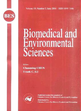
Volume 19 Issue 3
Jun.  2006
Turn off MathJax
Article Contents

RI-AN YU, CHENG-FENG YANG, XUE-MIN CHEN. DNA Damage, Apoptosis and C-myc, C-fos, and C-jun Overexpression Induced by Selenium in Rat Hepatocytes[J]. Biomedical and Environmental Sciences, 2006, 19(3): 197-204.
Citation: RI-AN YU, CHENG-FENG YANG, XUE-MIN CHEN. DNA Damage, Apoptosis and C-myc, C-fos, and C-jun Overexpression Induced by Selenium in Rat Hepatocytes[J]. Biomedical and Environmental Sciences, 2006, 19(3): 197-204.

DNA Damage, Apoptosis and C-myc, C-fos, and C-jun Overexpression Induced by Selenium in Rat Hepatocytes

Funds:  国家自然科学基金(30271110;30471500)
  • Objective To study the effects of selenium on DNA damage, apoptosis and c-myc, c-fos, and c-jun expression in rat hepatocytes. Methods Sodium selenite at the doses of 5, 10, and 20 μmol/kg was given to rats by i.p. and there were 5 male SD rats in each group. Hepatocellular DNA damage was detected by single cell gel electrophoresis (or comet assay).Hepatocellular apoptosis was determined by TUNEL (TdT-mediated dUTP nick end labelling) and flow cytometry. C-myc,c-fos, and c-jun expression in rat hepatocytes were assayed by Northern dot hybridization. C-myc, c-fos, and c-jun protein were detected by immunohistochemical method. Results At the doses of 5, 10, and 20 μmol/kg, DNA damage was induced by sodium selenite in rat hepatocytes and the rates of comet cells were 34.40%, 74.80%, and 91.40% respectively. Results also showed an obvious dose-response relationship between the rates of comet cells and the doses of sodium selenite (r=0.9501,P<0.01). Sodium selenite at the doses of 5, 10, and 20 μmol/kg caused c-myc, c-fos, and c-jun overexpression obviously. The positive brown-yellow signal for proteins of c-myc, c-fos, and c-jun was mainly located in the cytoplasm of hepatocytes with immunohistochemical method. TUNEL-positive cells were detected in selenium-treated rat livers. Apoptotic rates (%) of selenium-treated liver cells at the doses of 5, 10, and 20 μmol/kg were (3.72±1.76), (5.82±1.42), and (11.76±1.87) respectively, being much higher than those in the control. Besides an obvious dose-response relationship between apoptotic rates and the doses of sodium selenite (r=0.9897, P<0.01), these results displayed a close relationship between DNA damage rates and apoptotic rates, and the relative coefficient was 0.9021, P<0.01. Conclusion Selenium at 5-20 μmol/kg can induce DNA damage, apoptosis, and overexpression of c-myc, c-fos, and c-jun in rat hepatocytes.
  • 加载中
  • 加载中
通讯作者: 陈斌, bchen63@163.com
  • 1. 

    沈阳化工大学材料科学与工程学院 沈阳 110142

  1. 本站搜索
  2. 百度学术搜索
  3. 万方数据库搜索
  4. CNKI搜索

Article Metrics

Article views(1129) PDF downloads(9) Cited by()

Proportional views
Related

DNA Damage, Apoptosis and C-myc, C-fos, and C-jun Overexpression Induced by Selenium in Rat Hepatocytes

Funds:  国家自然科学基金(30271110;30471500)

Abstract: Objective To study the effects of selenium on DNA damage, apoptosis and c-myc, c-fos, and c-jun expression in rat hepatocytes. Methods Sodium selenite at the doses of 5, 10, and 20 μmol/kg was given to rats by i.p. and there were 5 male SD rats in each group. Hepatocellular DNA damage was detected by single cell gel electrophoresis (or comet assay).Hepatocellular apoptosis was determined by TUNEL (TdT-mediated dUTP nick end labelling) and flow cytometry. C-myc,c-fos, and c-jun expression in rat hepatocytes were assayed by Northern dot hybridization. C-myc, c-fos, and c-jun protein were detected by immunohistochemical method. Results At the doses of 5, 10, and 20 μmol/kg, DNA damage was induced by sodium selenite in rat hepatocytes and the rates of comet cells were 34.40%, 74.80%, and 91.40% respectively. Results also showed an obvious dose-response relationship between the rates of comet cells and the doses of sodium selenite (r=0.9501,P<0.01). Sodium selenite at the doses of 5, 10, and 20 μmol/kg caused c-myc, c-fos, and c-jun overexpression obviously. The positive brown-yellow signal for proteins of c-myc, c-fos, and c-jun was mainly located in the cytoplasm of hepatocytes with immunohistochemical method. TUNEL-positive cells were detected in selenium-treated rat livers. Apoptotic rates (%) of selenium-treated liver cells at the doses of 5, 10, and 20 μmol/kg were (3.72±1.76), (5.82±1.42), and (11.76±1.87) respectively, being much higher than those in the control. Besides an obvious dose-response relationship between apoptotic rates and the doses of sodium selenite (r=0.9897, P<0.01), these results displayed a close relationship between DNA damage rates and apoptotic rates, and the relative coefficient was 0.9021, P<0.01. Conclusion Selenium at 5-20 μmol/kg can induce DNA damage, apoptosis, and overexpression of c-myc, c-fos, and c-jun in rat hepatocytes.

RI-AN YU, CHENG-FENG YANG, XUE-MIN CHEN. DNA Damage, Apoptosis and C-myc, C-fos, and C-jun Overexpression Induced by Selenium in Rat Hepatocytes[J]. Biomedical and Environmental Sciences, 2006, 19(3): 197-204.
Citation: RI-AN YU, CHENG-FENG YANG, XUE-MIN CHEN. DNA Damage, Apoptosis and C-myc, C-fos, and C-jun Overexpression Induced by Selenium in Rat Hepatocytes[J]. Biomedical and Environmental Sciences, 2006, 19(3): 197-204.

Catalog

    /

    DownLoad:  Full-Size Img  PowerPoint
    Return
    Return