


-
Cerebrovascular disease has been the leading cause of mortality in the world[1]. In China alone, 1.5–2 million new patients are diagnosed with cerebrovascular disease ever year, and 80%–85% of them suffer from acute cerebral ischemia[2]. The acute cerebral ischemia can lead to anoxia and ischemia of brain tissue and subsequent necrosis or encephalomalacia, resulting in focal or global cerebral injury. At present, the treatment differs by patients and mortality remains to be high. Hence, further study on the molecular mechanism of the disease is essential to further improve the prognosis of the patients.
Studies have shown that DNA methylation can lead to the occurring of arteriosclerosis, and results in subsequent morbidity by regulating the arteriosclerosis gene expression[3].
In addition, it has been found that Wnt/β-catenin pathway, regulated by factors, such as Skip, SFRPs (secreted frizzled related proteins) and SOST (Sclerostin), plays an important role in angiogenesi[4]. Register et al.[5] discovered that the level of SOST is negatively correlated with the level of carotid artery plaque calcification. In addition, hypermethylation of SOST DNA promoter occurred in patients with aneurysm of aorta[6], which leads to the aberrant activation of the Wnt/β-catenin pathway, further promotes the occurring of atherosclerosis.
Whether the methylation of SOST gene is different between patients with acute cerebral ischemic and those without has not been reported. We speculate that abnormal methylation of SOST gene may occur more frequently in stroke patients, which, if true, could indicate an important mechanism of stroke. Here, we studied whether there is methylation of SOST gene promoter (Supplementary Table S1, available in www.besjournal.com) in patients with acute ischemic stroke accompanied by atherosclerosis, and whether there is difference in the level of the methylation (Supplementary Figure S1 and Supplementary Table S2, available in www.besjournal.com), and SOST gene methylation between stroke patients and controls.
Gene identity: 50964 (2,000 bp up and downstream in transcription start site): 1 agagcctgtg ctactggaag gtggcgtgcc ctcctctggc tggtaccatg cagctcccac 61 tggccctgtg tctcgtctgc ctgctggtac acacagcctt ccgtgtagtg gagggccagg 121 ggtggcaggc gttcaagaat gatgccacgg aaatcatccc cgagctcgga gagtaccccg 181 agcctccacc ggagctggag aacaacaaga ccatgaaccg ggcggagaac ggagggcggc 241 ctccccacca cccctttgag accaaaggta tggggtggag gagagaattc ttagtaaaag 301 atcctgggga ggttttagaa acttctcttt gggaggcttg gaagactggg gtagacccag 361 tgaagattgc tggcctctgc cagcactggt cgaggaacag tcttgcctgg aggtggggga 421 agaatggctc gctggtgcag ccttcaaatt caggtgcaga ggcatgaggc aacagacgct 481 ggtgagagcc cagggcaggg aggacgctgg ggtggtgagg gtatggcatc agggcatcag 541 aacaggctca ggggctcaga aaagaaaagg tttcaaagaa tctcctcctg ggaatatagg 601 agccacgtcc agctgctggt accactggga agggaacaag gtaagggagc ctcccatcca 661 cagaacagca cctgtggggc accggacact ctatgctggt ggtggctgtc cccaccacac 721 agacccacat catggaatcc ccaggaggtg aacccccagc tcgaagggga agaaacaggt 781 tccaggcact cagtaacttg gtagtgagaa gagctgaggt gtgaacctgg tttgatccaa 841 ctgcaagata gccctggtgt gtgggggggt gtgggggaca gatctccaca aagcagtggg 901 gaggaaggcc agagaggcac ccctgcagtg tgcattgccc acggcctgcc cagggagctg 961 gcacttgaag gaatgggagt tttcggcaca gttttagccc ctgacatggg tgcagctgag 1021 tccaggccct ggaggggaga gcagcatcct ctgtgcagga gtagggacat ctgtcctcag 1081 cagccacccc agtcccaacc ttgcctcatt ccaggggagg gagaaggaag aggaaccctg 1141 ggttcctggt caggcctgca cagagaagcc caggtgacag tgtgcatctg gctctataat 1201 tggcaggaat cctgaggcca tgggggcgtc tgaaatgaca cttcagacta agagcttccc 1261 tgtcctctgg ccattatcca ggtggcagag aagtccactg cccaggctcc tggaccccag 1321 ccctccccgc ctcacaacct gttgggacta tggggtgcta aaaagggcaa ctgcatggga 1381 ggccagccag gaccctccgt cttcaaaatg gaggacaagg gcgcctcccc ccacagctcc 1441 ccttctaggc aaggtcagct gggctccagc gactgcctga agggctgtaa ggaacccaaa 1501 cacaaaatgt ccaccttgct ggactcccac gagaggccac agcccctgag gaagccacat 1561 gctcaaaaca aagtcatgat ctgcagagga agtgcctggc ctaggggcgc tattctcgaa 1621 aagccgcaaa atgccccctt ccctgggcaa atgcccccct gaccacacac acattccagc 1681 cctgcagagg tgaggatgca aaccagccca cagaccagaa agcagcccca gacgatggca 1741 gtggccacat ctcccctgct gtgcttgctc ttcagagtgg gggtgggggg tggccttctc 1801 tgtcccctct ctggtttggt cttaagacta tttttcattc tttcttgtca cattggaact 1861 atccccatga aacctttggg ggtggactgg tactcacacg acgaccagct atttaaaaag 1921 ctcccaccca tctaagtcca ccataggaga catggtcaag gtgtgtgcag gggatcaggc 1981 caggcctcgg agcccaatct ctgcctgccc agggagtatc accatgaggc gcccattcag 2041 ataacacaga acaagaaatg tgcccagcag agagccaggt caatgtttgt ggcagctgaa 2101 cctgtaggtt ttgggtcaga gctcagggcc cctatggtag gaaagtaacg acagtaaaaa 2161 gcagccctca gctccatccc ccagcccagc ctcccatgga tgctcgaacg cagagcctcc 2221 actcttgccg gagccaaaag gtgctgggac cccagggaag tggagtccgg agatgcagcc 2281 cagccttttg ggcaagttct tttctctggc tgggcctcag tattctcatt gataatgagg 2341 gggttggaca cactgccttt gattcctttc aagtctaatg aattcctgtc ctgatcacct 2401 ccccttcagt ccctcgcctc cacagcagct gccctgattt attaccttca attaacctct 2461 actcctttct ccatcccctg tccacccctc ccaagtggct ggaaaaggaa tttgggagaa 2521 gccagagcca ggcagaaggt gtgctgagta cttaccctgc ccaggccagg gaccctgcgg 2581 cacaagtgtg gcttaaatca taagaagacc ccagaagaga aatgataata ataatacata 2641 acagccgacg ctttcagcta tatgtgccaa atggtatttt ctgcattgcg tgtgtaatgg 2701 attaactcgc aatgcttggg gcggcccatt ttgcagacag gaagaagaga gaggttaagg 2761 aacttgccca agatgacacc tgcagtgagc gatggagccc tggtgtttga accccagcag 2821 tcatttggct ccgaggggac agggtgcgca ggagagcttt ccaccagctc tagagcatct 2881 gggaccttcc tgcaatagat gttcaggggc aaaagcctct ggagacaggc ttggcaaaag 2941 cagggctggg gtggagagag acgggccggt ccagggcagg ggtggccagg cgggcggcca 3001 ccctcacgcg cgcctctctc cacagacgtg tccgagtaca gctgccgcga gctgcacttc 3061 acccgctacg tgaccgatgg gccgtgccgc agcgccaagc cggtcaccga gctggtgtgc 3121 tccggccagt gcggcccggc gcgcctgctg cccaacgcca tcggccgcgg caagtggtgg 3181 cgacctagtg ggcccgactt ccgctgcatc cccgaccgct accgcgcgca gcgcgtgcag 3241 ctgctgtgtc ccggtggtga ggcgccgcgc gcgcgcaagg tgcgcctggt ggcctcgtgc 3301 aagtgcaagc gcctcacccg cttccacaac cagtcggagc tcaaggactt cgggaccgag 3361 gccgctcggc cgcagaaggg ccggaagccg cggccccgcg cccggagcgc caaagccaac 3421 caggccgagc tggagaacgc ctactagagc ccgcccgcgc ccctccccac cggcgggcgc 3481 cccggccctg aacccgcgcc ccacatttct gtcctctgcg cgtggtttga ttgtttatat 3541 ttcattgtaa atgcctgcaa cccagggcag ggggctgaga ccttccaggc cctgaggaat 3601 cccgggcgcc ggcaaggccc ccctcagccc gccagctgag gggtcccacg gggcagggga 3661 gggaattgag agtcacagac actgagccac gcagccccgc ctctggggcc gcctaccttt 3721 gctggtccca cttcagagga ggcagaaatg gaagcatttt caccgccctg gggttttaag 3781 ggagcggtgt gggagtggga aagtccaggg actggttaag aaagttggat aagattcccc 3841 cttgcacctc gctgcccatc agaaagcctg aggcgtgccc agagcacaag actgggggca 3901 actgtagatg tggtttctag tcctggctct gccactaact tgctgtgtaa ccttgaacta 3961 cacaattctc cttcgggacc tcaatttcca ctttgtaaaa tgagggtgga ggtgggaata 4021 ggatctcgag gagactattg gcatatgatt ccaaggactc cagtgccttt tgaatgggca Note. SOST, sclerostin. Table S1. SOST promoter sequencing
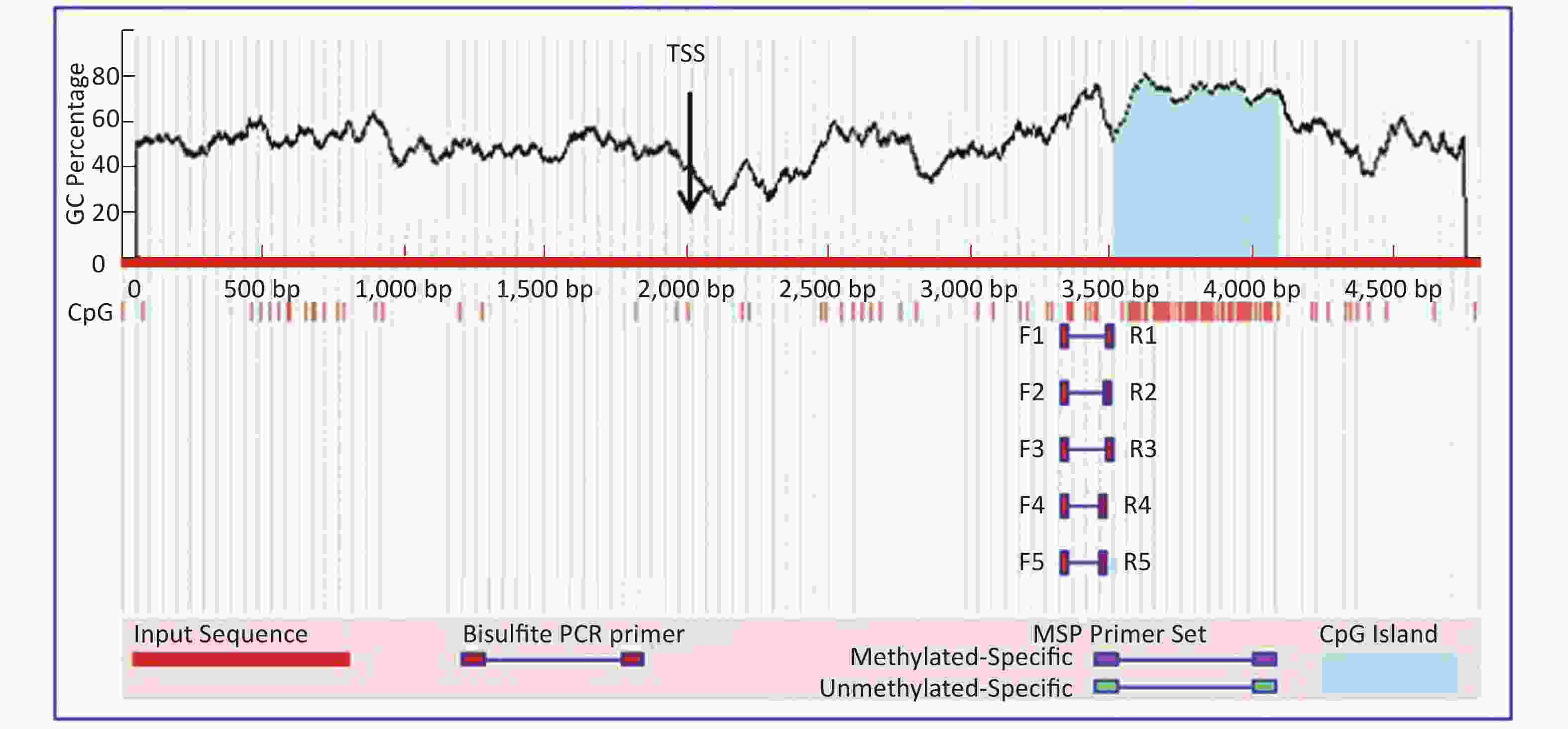
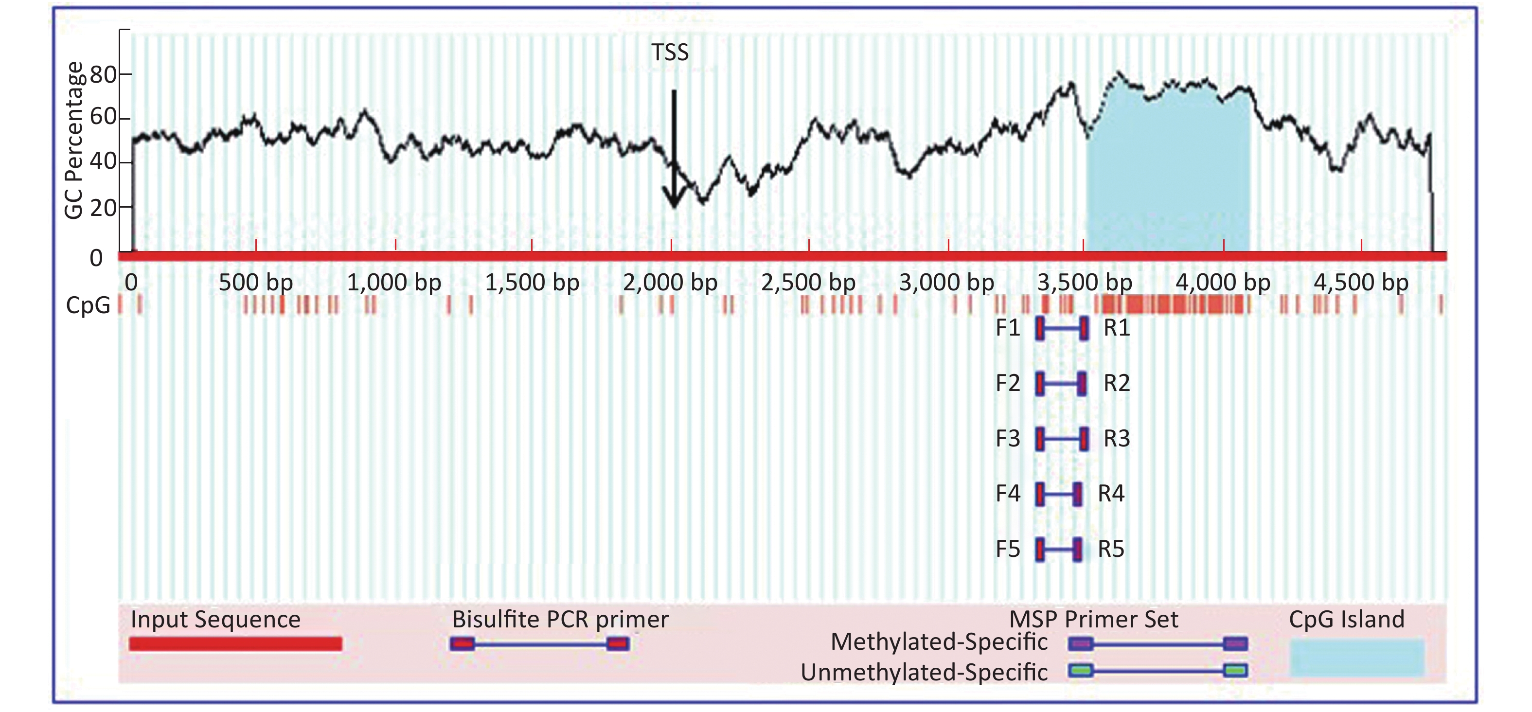
Figure S1. The CpG island predication of SOST promoter. 2,000 bp in the Upstream and 2,800 bp (including 1st exon) downstream of TSS were include, respectively. From http://itsa.ucsf.edu/~urolab/methprimer. CpG island: a region abundant of C and G nucleic acid base. TSS: transcription start site. SOST, sclerostin.
No. Size (bp) Start and end Island 1 576 3,514–4,089 Note. SOST, sclerostin. CpG sites: a region abundant of C and G nucleic acid base. Table S2. Predication of cpg sites around the SOST promoter
Based on our preliminary study results and the estimation of required sample size (using formula of hypothesis testing for mean: (n = [(tα + tβ)2Ơ2]/§2, tα = 1.645, tβ = 1.298; Ơ: standard deviation; §: mean difference), we collected the peripheral blood samples from 41 patients diagnosed with acute ischemic stroke and 41 healthy volunteers matched on the frequency of age and gender for this case-control study. The patients were recruited consecutively from February 2017 to May 2017 in Anhui provincial hospital (China), and the controls were stratified by age and gender and randomly selected from the physical examination center of Anhui Provincial Hospital. All patients and controls were Han nationality and had no kinship with each other.
The inclusion criteria were: patients who were diagnosed with acute ischemic stroke (confirmed by magnetic resonance imaging)[7], meanwhile, patients had no consanguinity with each other. The exclusion criteria were as follows: patients were receiving anticoagulation or thrombolysis treatment, patients with atrial fibrillation or a history of atrial fibrillation, patients with cranio-cerebral trauma, idiopathic cerebral infarction, a history of MI (myocardial infarction), hemopathy, tissue-damaging infection or took antithrombotics, patients with osteoporosis or who took medications that have an impact on DNA methylation, such as oxytetracycline, dimethyl sulfoxide, curcumin, quinolone. The inclusion criteria of control group were: volunteers who received routine physical examination in heath management center of the same institute. The exclusion criteria were: patients diagnosed with stroke and acute coronary syndrome; patients receiving anticoagulation or thrombolysis treatment, patients with atrial fibrillation or a history of atrial fibrillation.
Detection of DNA Methylation We first mixed 200 μL blood, 800 μL SDS lysate and 10 μL (20 mg/μL) PK together. The DNA was extracted using Phenol extraction method and Gel electrophoresis was used to detect the DNA product. DNA methylation detection kit (CW2140, CWBIO) was used to detect the methylation of SOST gene. The DNA solution was preserved under −20 ℃.
PCR Process PCR Amplifier was used to amplify the DNA fragment. The amplification system included 1 μL DNA template, 0.8 μL primers (5 μmol/L), 2 μL 10×PCR Buffer, 1.6 μL Dntp (2.5 mmol/L), 0.2 μL Taq enzyme. DdH2O supplement was added to reach the total volume of 20 μL. The primers [designed using Methprimer (http://www.urogene.org/methprimer/index1.html)] were SOST (Amplicon size: 332 bp): hSOST-Bf: 5’-TGGTATGAAGTAGAGAGGGGTTTTA-3’, hSOST-Br: 5’-CCACCCCATACCTTTAATCTCA-3’. The reaction protocol was 5 min at 95 ℃ activation, 10 cycles of 30 s at 95 ℃, 30 s at 60 ℃ to 50 ℃ Δ1 ℃ and 30 s at 72 ℃. Then 30 Cycles of 30 s at 95 ℃, 30 s at 50 ℃, and 30 s at 72 ℃. 30 min for 60 ℃ at last.
Sequencing We confirmed the DNA using gel electrophoresis (2% agarose) and purified the DNA with DNA purify and recycle kit (CW0524, CWBIO). We then finished the process of transformation and colony authentication using T vector connection kit (CW0801, CWBIO), selecting the positive samples for sequencing. The results of DNA methylation were obtained at last.
Statistical Analysis SPSS19.0 was used for statistical analysis. The formula of C/(C+T) *100 (C: cytosine, T: thymine) was adopted to calculate the rate of methylation. Student T test was used to analyze the difference in SOST gene methylation between patients and controls; chi-square test was used to analyze the categorical data. Logistic regression was applied with P < 0.05 defined as being statistically significant. Stratified analysis by gender and status of menopause were also conducted.
The characteristics of the patients and controls were shown in Table 1. Blood glucose, total cholesterol levels, the percentages of subjects with IMT ≥ 0.9 mm, and subjects with plaque were significantly different between two groups (P = 0.0499, P = 0.0312, P = 0.0000, respectively).
Item Cerebral ischemic patients (n = 41) Controls (n = 41) P value Gender 0.506a Male (%) 24 (58.54) 21 (51.22) Female (%) 17 (41.46) 20 (48.78) Ages (years) 55.71 ± 8.11 56.32 ± 5.82 0.6968b Hypertension (n) No (11) No (36) 0.000c I stage (3) I stage (5) II stage (0) II stage (0) III stage (27) III stage (0) Blood glucose (mmol/L) 6.94 ± 3.76 5.58 ± 1.54 0.0499b* Triglyceride (mmol/L) 1.84 ± 1.18 1.55 ± 1.05 0.2586b Homocysteine (mmol/L) 13.17 ± 5.02 − Total cholesterol (mmol/L) 4.41 ± 1.21 5.03 ± 0.95 0.023b* Carotid artery Subjects with IMT ≥ 0.9 mm 29 13 0.000a* Subjects with plaque 26 15 0.000a* Note. aTwo sides χ2 test; bTwo sides students T test; cMann-Whitney U test; *Indicating a significant difference. IMT, intima-media thickness. Table 1. General characteristics of the patients and controls
Overall there was no significant difference in SOST DNA methylation between the patients and controls (P = 0.27, Supplementary Table S3 available in www.besjournal.com), neither was any difference when stratified by gender (P = 0.94 for female, Supplementary Table S4 available in www.besjournal.com, P = 0.21 for male, Supplementary Table S5 available in www.besjournal.com).
No Gender Age (year) Methylation rate (%) Cases 1 M 64 95.6 2 M 57 96.3 3 M 54 96.3 4 M 55 98.1 5 M 58 91.9 6 M 61 96.9 7 M 62 91.9 8 M 49 95.0 9 M 45 95.0 10 M 52 93.1 11 M 62 89.4 12 M 44 91.9 13 M 47 90.0 14 M 52 85.6 15 M 43 96.3 16 M 41 91.9 17 F 46 93.1 18 F 50 91.9 19 F 61 88.1 20 F 64 92.5 21 F 53 89.4 22 F 60 83.1 23 F 64 95.6 24 F 60 86.3 25 F 66 95.6 26 F 48 91.9 27 F 60 92.5 28 F 38 94.4 29 F 48 92.5 30 F 57 93.8 31 F 71 93.1 32 M 72 88.7 33 M 61 93.1 34 M 61 90.6 35 F 56 91.2 36 M 65 97.5 37 M 57 95.6 38 F 55 95.0 39 M 55 89.4 40 M 63 93.1 41 M 47 90.6 Controls C1 M 64 96.3 C2 M 56 88.1 C3 M 54 78.7 C4 M 53 88.7 C5 M 59 93.8 C6 M 60 93.8 C7 M 62 94.4 C8 M 50 91.9 C9 M 46 91.2 C10 M 52 93.1 C11 M 62 93.1 C12 M 50 94.4 C13 M 46 78.1 C14 M 50 97.5 C15 M 50 85.6 C16 M 51 91.2 C17 F 51 94.4 C18 F 50 86.9 C19 F 61 90.0 C20 F 62 93.8 C21 F 53 98.8 C22 F 60 94.4 C23 F 64 95.6 C24 F 57 82.5 C25 F 58 96.3 C26 F 64 90.6 C27 F 52 95.0 C28 F 62 93.8 C29 F 43 95.6 C30 F 50 87.5 C31 F 64 93.1 C32 M 64 96.3 C33 M 57 93.8 C34 M 56 92.5 C35 F 57 86.3 C36 M 58 94.4 C37 M 63 92.5 C38 F 58 90.6 C39 F 61 91.9 C40 F 63 93.1 C41 F 56 83.1 P value (T Test 2) 0.27 Note. M, male; F, female. Table S3. Methylation rates between groups
No Gender Age (year) Methylation rate (%) Cases 1 F 46 93.1 2 F 50 91.9 3 F 61 88.1 4 F 64 92.5 5 F 53 89.4 6 F 60 83.1 7 F 64 95.6 8 F 60 86.3 9 F 66 95.6 10 F 48 91.9 11 F 60 92.5 12 F 38 94.4 13 F 48 92.5 14 F 57 93.8 15 F 71 93.1 16 F 56 91.2 17 F 55 95.0 Controls C1 F 51 94.4 C2 F 50 86.9 C3 F 61 90.0 C4 F 62 93.8 C5 F 53 98.8 C6 F 60 94.4 C7 F 64 95.6 C8 F 57 82.5 C9 F 58 96.3 C10 F 64 90.6 C11 F 52 95.0 C12 F 62 93.8 C13 F 43 95.6 C14 F 50 87.5 C15 F 64 93.1 C16 F 57 86.3 C17 F 58 90.6 C18 F 61 91.9 C19 F 63 93.1 C20 F 56 83.1 P value (T Test 2) 0.94 Note. F, female. Table S4. Methylation rates in women
No Gender Age (year) Methylation rate (%) Cases 1 M 64 95.6 2 M 57 96.3 3 M 54 96.3 4 M 55 98.1 5 M 58 91.9 6 M 61 96.9 7 M 62 91.9 8 M 49 95.0 9 M 45 95.0 10 M 52 93.1 11 M 62 89.4 12 M 44 91.9 13 M 47 90.0 14 M 52 85.6 15 M 43 96.3 16 M 41 91.9 17 M 72 88.7 18 M 61 93.1 19 M 61 90.6 20 M 65 97.5 21 M 57 95.6 22 M 55 89.4 23 M 63 93.1 24 M 47 90.6 Control C1 M 64 96.3 C2 M 56 88.1 C3 M 54 78.7 C4 M 53 88.7 C5 M 59 93.8 C6 M 60 93.8 C7 M 62 94.4 C8 M 50 91.9 C9 M 46 91.2 C10 M 52 93.1 C11 M 62 93.1 C12 M 50 94.4 C13 M 46 78.1 C14 M 50 97.5 C15 M 50 85.6 C16 M 51 91.2 C17 M 64 96.3 C18 M 57 93.8 C19 M 56 92.5 C20 M 58 94.4 C21 M 63 92.5 P value (T Test 2) 0.21 Note. M, male. Table S5. Methylation rates in males
Since most patients also had hypertension, we conducted a subgroup analysis by the status of hypertension. There was a significant difference between the patients and controls (P = 0.00, Table 1) and hypertension was a risk factor of acute cerebral ischemia. Logistic analysis adjusting for ages and gender also did not show significant association between gender or age with the methylation.
HTML
-
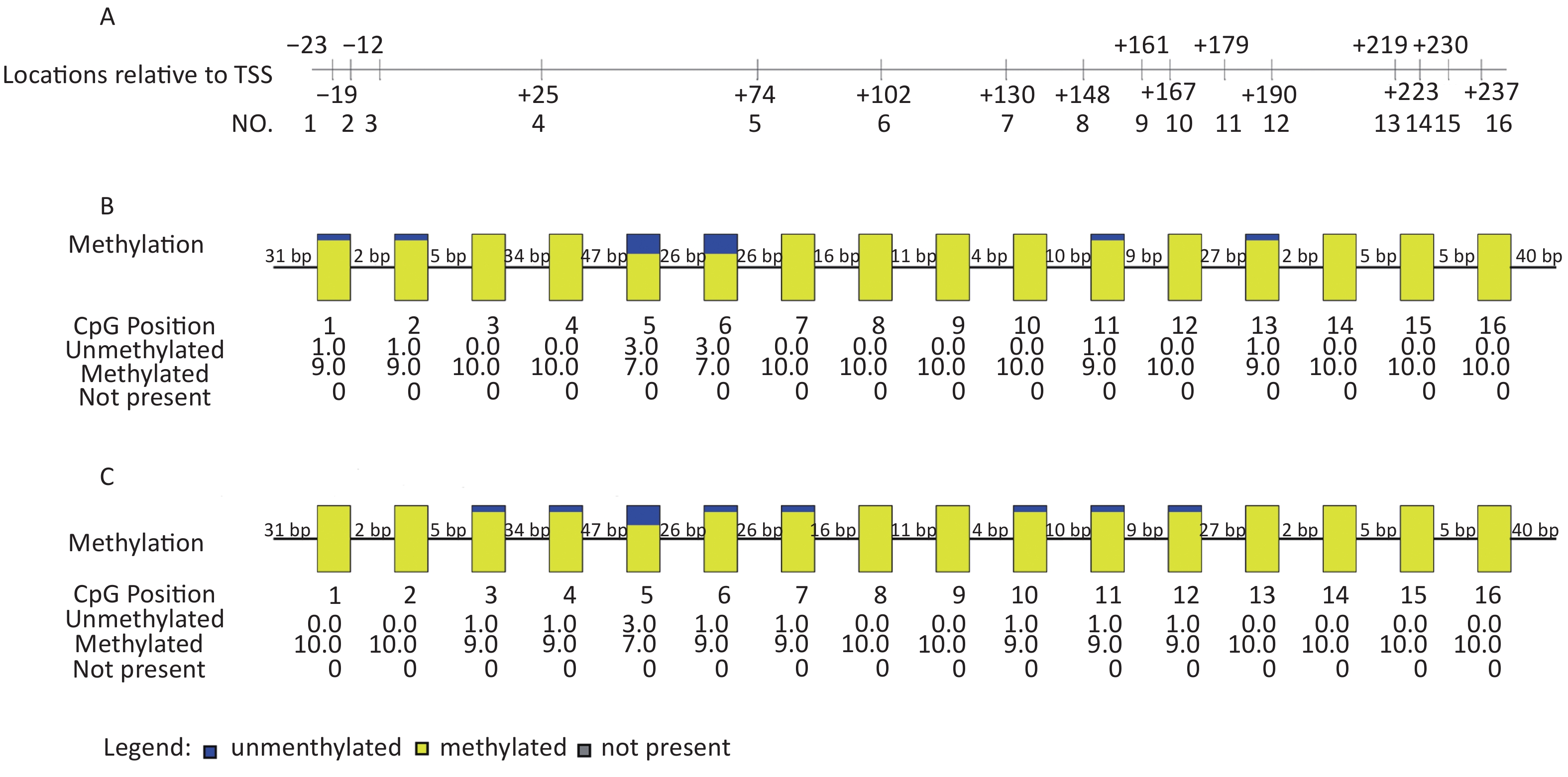
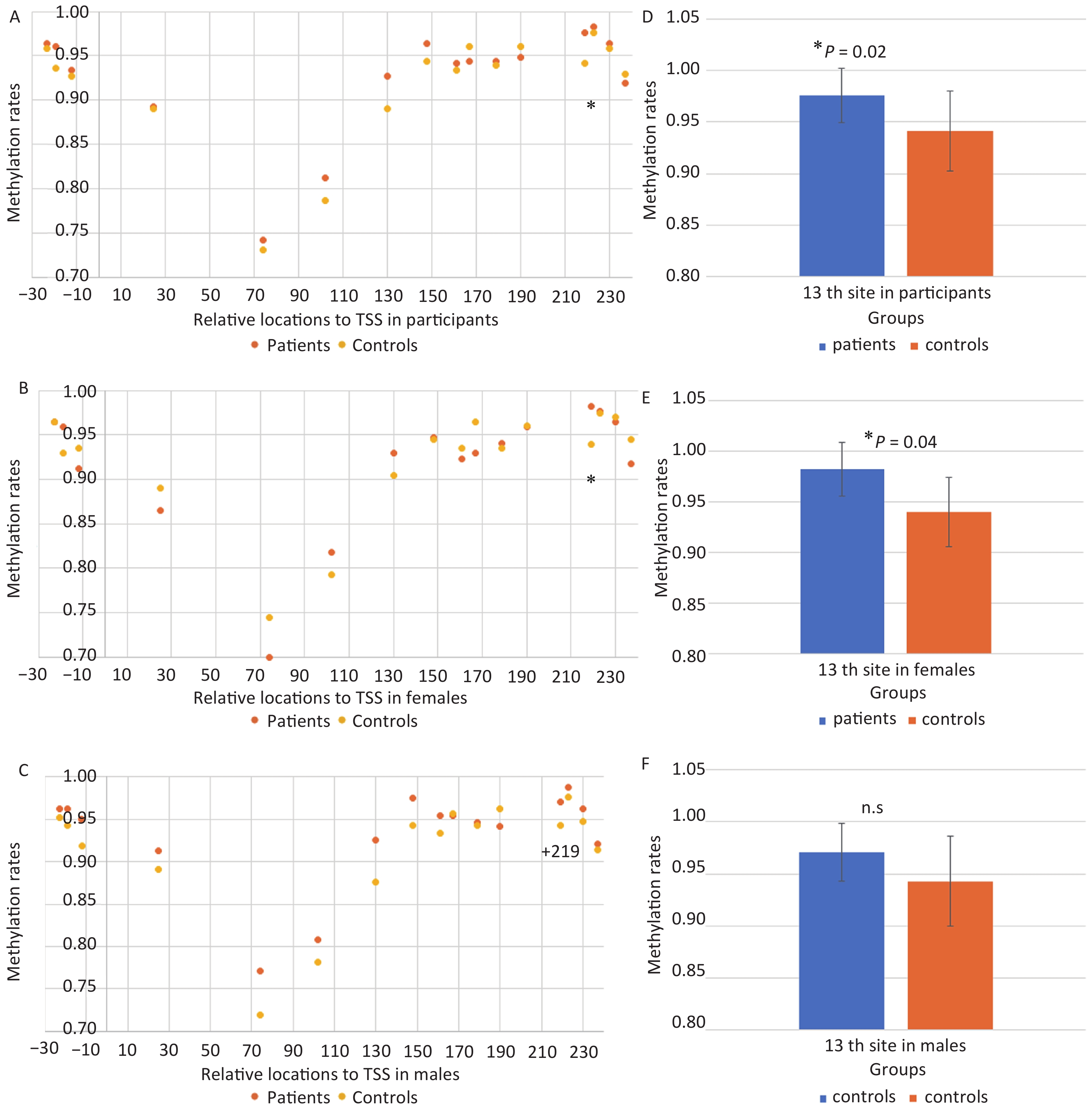
Next, we found that 16 CpG sites were located in the SOST DNA sequence (260 bp) from −23 to +237, including −23, −19, −12, +25, +74, +102, +130, +148, +161, +167, +179, +190, +219, +223, +230, +237 sites (Before and after the transcription initiation, Supplementary Figure S2). We found that the mean methylation level at +219 CpG site was significantly higher in stroke patients than controls (0.98 vs. 0.94, P = 0.023, Figure 1A–B) . In addition, we compared the level of SOST methylation at +219 CpG site between two groups stratified by gender. Interestingly, we found that the mean methylation level +219 CpG site was significant higher for stroke patients in females (0.98 vs. 0.94, P = 0.04, Figure 3B and 3E), but not in males (P = 0.20, Figure 1D and 1F). This indicated that the association between +219 CpG methylation and acute cerebral ischemic stroke might vary by gender.
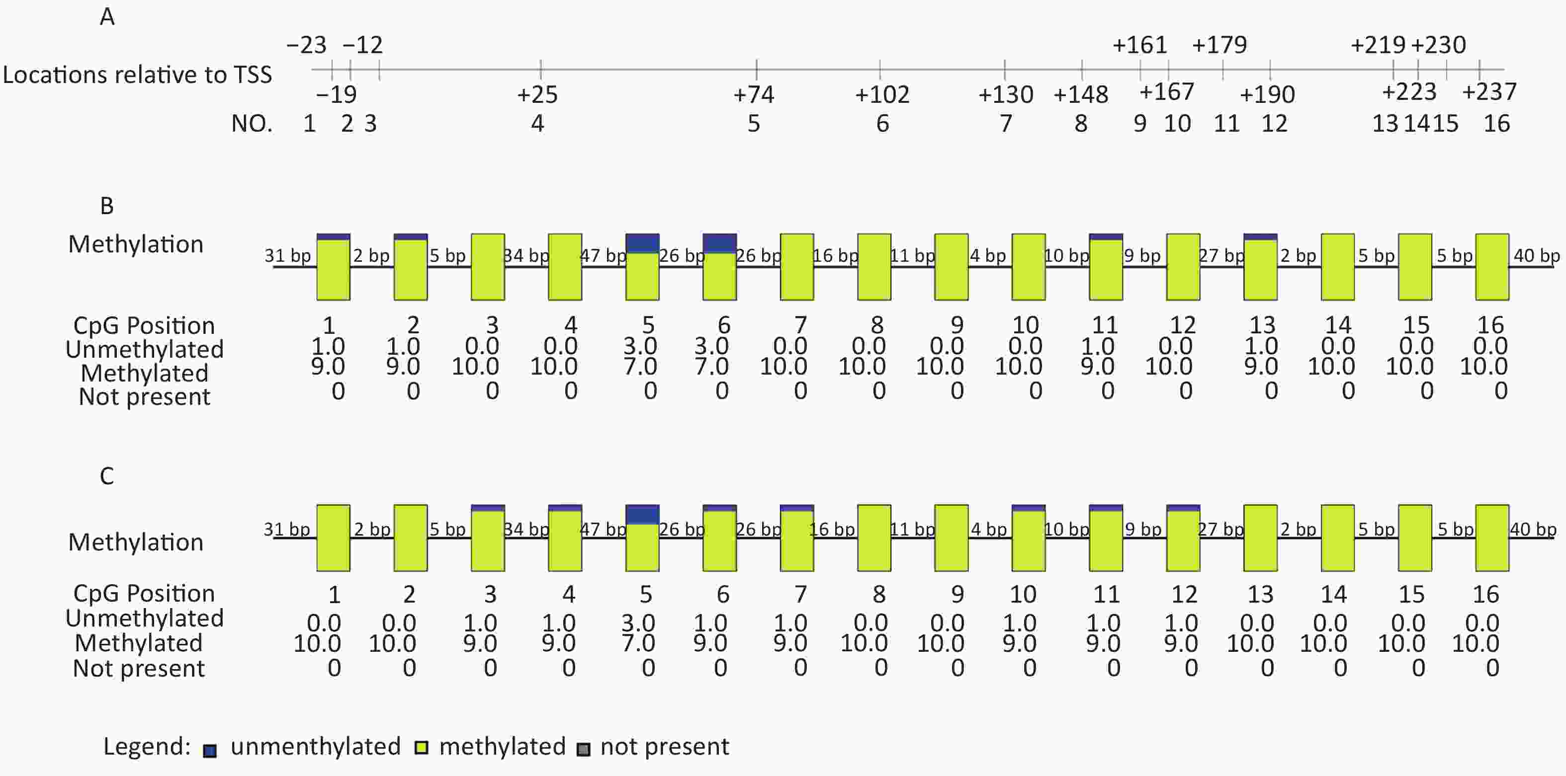
Figure S2. SOST CpG sites analysis and methylation rates of one case. (A) Sequencing of the amplified DNA fragments. sixteen islands were analyzed. (B) Sixteen island methylation rates of 17th patient (Female, 46 years). The whole methylation rate of 17th patient is 93.1% and rate of the +219 site (13th) is 100%. (C) The island methylation rates of 17th control (Female, 51 years). The whole methylation rate of 17th control is 94.4% and rate of the +219 site (13th) is 90.0%. Each rectangle is a CpG site. The numbers under the CpG positions represent the amounts of methylated and unmethylated colons. SOST, sclerostin. CpG island, a region abundant of C and G nucleic acid base. DNA, deoxyribonucleic acid. TSS, transcription start site.

Figure 1. Methylation Level of +219 Site. (A)(D) sixteen sites were analyzed in 41 patients and 41 controls. +219 site (*) level in patients is higher than controls. (B)(E) in female (17 patients vs. 20 controls) a significant difference was identified at +219 site. (C)(F) in male (24 patients vs. 21 controls) no significant differences was identified by +219 site. The results are from Students’ Test. n.s, no significant. *P < 0.05, indicated a significant difference.
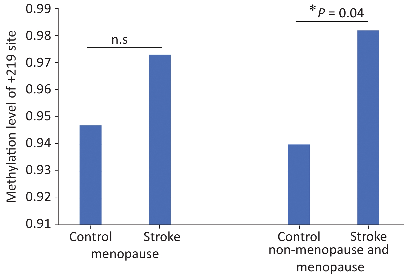
Furthermore, we analyzed the level of +219 CpG methylation in menopause female and found that there was no significant difference between patients and controls (Figure 2 and Supplementary Table S6 available in www.besjournal.com).
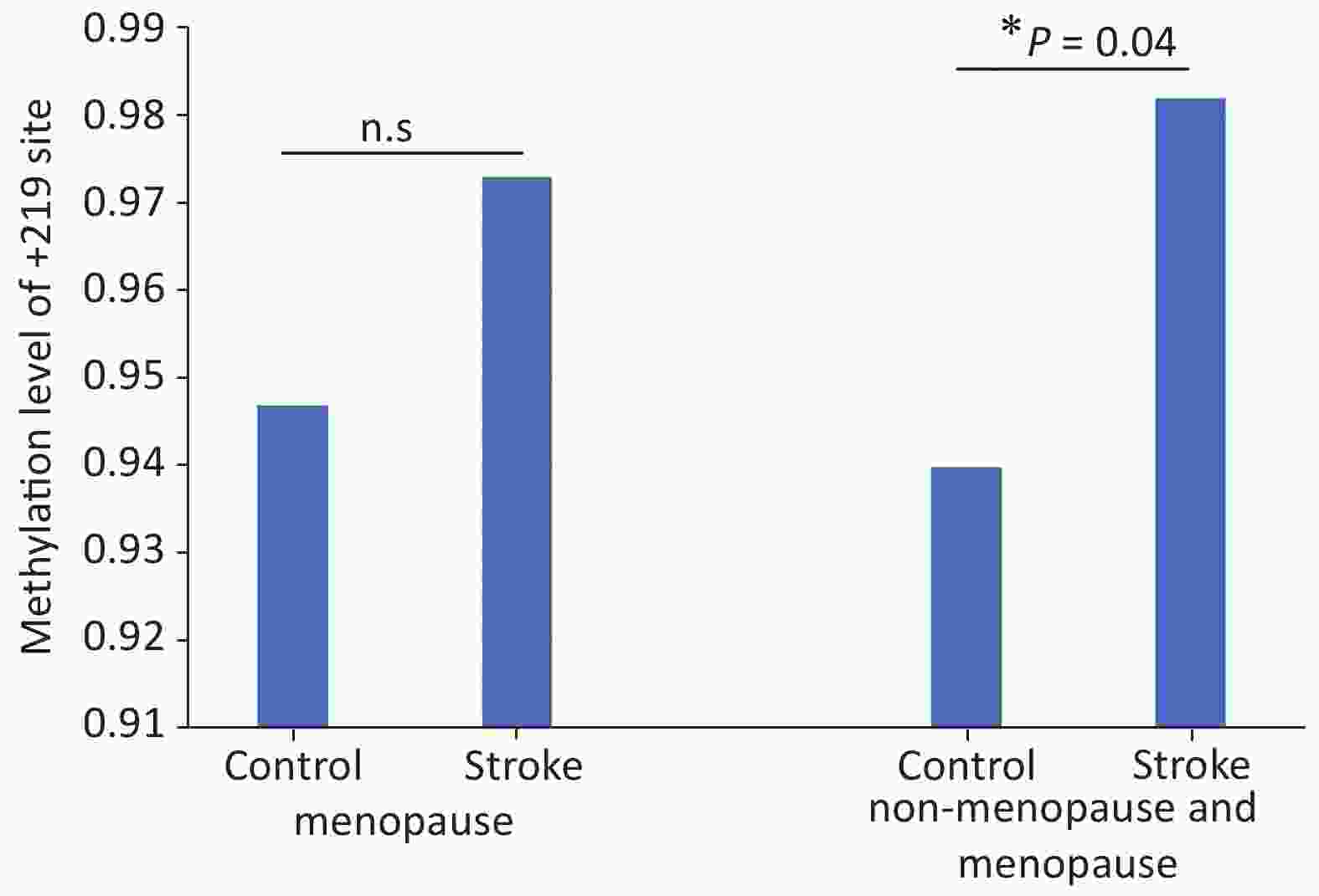
Figure 2. Mean methylation status of +219 sites in female by menopause. In menopause (11 patients vs. 15 controls) no significant difference was identified. Before the menopause (17 patients vs. 20 controls) a significant difference was identified. The results are from Students’ Test. *P < 0.05, indicated a significant difference; n.s: no significant.
Status Group Number Mean STD P menopause Control 15 0.947 0.074 0.367 Stroke 11 0.973 0.065 non-menopause Control 20 0.94 0.681 0.04* Stroke 17 0.982 0.053 Note. STD, standard deviation. *Significant differences. Table S6. +219 Site methylation levels in menopause
We found that the difference in methylation level between patients and control were all 0.05 at the +74 site in females and +74/+130 sites in male, which is higher than 0.03 and 0.04 at +219 site between patients and controls in general and female patients and female controls, respectively. However, the differences were not statistically significant (P values are 0.418, 0.194, and 0.202, respectively). (Supplementary Table S7 available in www.besjournal.com)
Gender Site Group Number Mean ± STD Difference P value female +74 controls 20 0.745±0.216 0.05 0.418 patients 17 0.700±0.162 male +74 controls 20 0.719±0.140 0.05 0.194 patients 17 0.771±0.123 +130 controls 20 0.876±0.160 0.05 0.202 patients 17 0.925±0.846 Note. STD, standard deviation. Table S7. Methylation levels at +74 and +130 sites in female and male
In our study, we confirmed that methylation rate at +219 site of SOST gene was higher in acute cerebral ischemic patients in this Chinese population. We found a significant difference in methylation level at +219 site of SOST gene between patients with acute cerebral ischemic stroke and controls in female subjects, which might be the most important finding of our study.
Interestingly, we discovered that the methylation of +219 site was different by gender in patients with cerebral arterial thrombosis. Most of female subjects in the study (26/37, 70.3%) already had menopause, and there was no significant difference between the patients and controls (Supplementary Table S8 available in www.besjournal.com). Limited to those subjects who had menopause, there was no difference in methylation level of +219 site between patients and controls (Figure 2). Therefore, we speculated that the +219 site methylation might be affected by hormone, and estrogen and androgen level might influence the +219 site methylation in patients with cerebral arterial thrombosis. Reports have shown that estrogen has a protective effect on ischemic brain injury, and its neuroprotective mechanisms present in many ways[8]. However, the impact of hormones on the methylation level of individual CpG site remains unclear. These data suggested that decreased estrogen and increased androgen may play a role in the reduced methylation level at +219 site in menopause women.
Group Menopause Non-menopause Sum P* Control 15 5 20 0.495 Stroke 11 6 17 Sum 26 11 37 Note. *Chi-square test. Table S8. Menopause in stroke and control
In previous study, HF lin[6] found that the methylation levels of MMP-2 (Matrix metalloproteinase-2) in the peripheral blood of the patients with stroke were lower than controls in its eight CpG sites, and also proved that a significant association was found only in men but not in women. That study also reported that the cerebral ischemic stroke was regulated by several genes, and a complex mechanism was present; and interestingly the significant difference between men and women was consistent with what we found in our study. Shan Jiang et al.[9] also showed that ER-α (estrogen receptor-α) gene promoter methylation rate was significant higher in patients with cerebral ischemic stroke than controls. They suggested that gene methylation was associated with cerebral ischemic stroke, but no report has studied the relationship between SOST gene and the disease. In fact, the relationship between SOST DNA methylation and many diseases have been reported in the literature[10], but no study about its association with cerebral ischemic stroke, especially the individual CpG site methylation and stroke, has been published. To explore the relationship between SOST and artery plaque type disease, we studied the association between SOST and cerebral ischemic stroke, and these findings will help further illuminate the mechanism of the disease.
Moreover, we also found that the differences in methylation level between patients and controls at +74 site in females and +74/+130 sites in males are all 0.05, which is higher than 0.03 and 0.04 at +219 site between patients and controls overall, between female patients and female controls. But the difference was not significant, suggesting large variations in methylation at these sites.
This study has several limitations: first, our study did not detect the SOST mRNA expression. Secondly, the sub-group analysis (by gender, menopause status) was based on small number of subjects. Finally, since it is a case-control study, the results are only for hypothesis-generating.
Further studies are needed to confirm the molecular mechanism, especially the impact of hormone level on +219 methylation, and to test the mRNA level. A larger study is needed to explore the methylation levels of +74 and +130 sites in the sub-group subjects.
Anyhow, we found higher methylation level of +219 CpG site in patients with cerebral ischemia comparing to controls in female subjects. The +219 CpG site partially elucidated the molecular mechanism of acute cerebral ischemic disease, and provided a molecular target for further studying and possibly developing new treatment for patients with acute cerebral ischemia.
Ethics approval and consent to participate: The study was approved by the ethics committee of Anhui Provincial Hospital (NO. P-012), and all objects signed the informed consent.
Conflicts of interest The authors declare that they have no competing interests.
Availability of data and materials The datasets used and/or analyzed during the current study are available from the corresponding author.
Authors’ contributions YIN Yi Hong designed the protocol, collected samples, analyzed the data, performed the examination, and wrote the manuscript. HU Wei was a major contributor in collecting samples, analyzing the data, and writing the manuscript. ZHU Zheng Yu was a major contributor in enrolling cases in groups. HAN Min was a major contributor in collecting samples and data. XIE Zhao Hong supervised the designing of protocol and collecting of data. BI Jian Zhong supervised the designing of protocol and writing of the manuscript. All authors read and approved the final manuscript.

 Quick Links
 DownLoad: