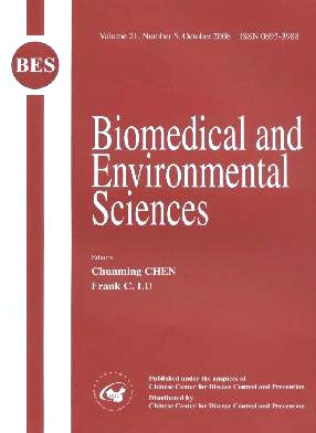
Volume 21 Issue 5
Oct.  2008
Turn off MathJax
Article Contents

QIAN WEN, LIMA, YAN-PING CHEN, LIN YANG, WEI LUO, XIAO-NING WANG. A Rabbit Model of Hormone-induced Early Avascular Necrosis of the Femoral Head[J]. Biomedical and Environmental Sciences, 2008, 21(5): 398-403.
Citation: QIAN WEN, LIMA, YAN-PING CHEN, LIN YANG, WEI LUO, XIAO-NING WANG. A Rabbit Model of Hormone-induced Early Avascular Necrosis of the Femoral Head[J]. Biomedical and Environmental Sciences, 2008, 21(5): 398-403.

A Rabbit Model of Hormone-induced Early Avascular Necrosis of the Femoral Head

Funds:  国家高技术研究发展计划(863计划)(2007AA02Z458)%Science and Technology Program of Guangdong Province(2005B34001004)%Science and Technology Program of Guangzhou(2005Z3-E0151)%Medical Science Research Fund of Guangdong Provice(B2007107)
  • Objective To establish an experimental model of early stage avascular necrosis of the femoral head (ANFH) caused by corticosteroid in adult rabbits and to observe the pathological changes with various imaging techniques. Methods ANFH was induced by a combination of hypersensitivity vasculitis caused by injection of horse serum and subsequent administration of a high dose of corticosteroid. The pathological changes were detected with digital radiography (DR), computed tomography (CT), magnetic resonance imaging (MRI), ink artery infusion angiography, hematoxylin--eosin staining, and mmunohistochemistry. Results The imageological and athological changes corresponded to the clinical characteristics of early stage ANFH. DR showed bilaterally increased bone density, an unclear epiphyseal line, and blurred texture of cancellous bone. CT showed spot-like low-density imaging of cancellous bone, thinner cortical bone, osteoporosis, and an unclear epiphyseal line. MRI showed bone marrow edema and spot-like high signals in T2-weighted imaging in cancellous bone. Ink artery infusion angiography showed fewer obstructed blood vessels in the femoral head. HE staining of pathological sections showed fewer trabeculae and thin bone, an increased proportion of empty osteocyte lacunae, decreased hematopoiesis, thrombosis, and fat cell hypertrophy. Lmmunohistochemistry showed attenuated expression of vascular endothelial growth factor in osteoblasts and chondrocytes, and on the inner membrane of blood vessels. Conclusion Experimental rabbit model of early stage ANFH caused by corticosteroid can be successfully established and provide the foundation for developing effective methods to treat early stage ANFH.
  • 加载中
  • 加载中
通讯作者: 陈斌, bchen63@163.com
  • 1. 

    沈阳化工大学材料科学与工程学院 沈阳 110142

  1. 本站搜索
  2. 百度学术搜索
  3. 万方数据库搜索
  4. CNKI搜索

Article Metrics

Article views(1037) PDF downloads(19) Cited by()

Proportional views
Related

A Rabbit Model of Hormone-induced Early Avascular Necrosis of the Femoral Head

Funds:  国家高技术研究发展计划(863计划)(2007AA02Z458)%Science and Technology Program of Guangdong Province(2005B34001004)%Science and Technology Program of Guangzhou(2005Z3-E0151)%Medical Science Research Fund of Guangdong Provice(B2007107)

Abstract: Objective To establish an experimental model of early stage avascular necrosis of the femoral head (ANFH) caused by corticosteroid in adult rabbits and to observe the pathological changes with various imaging techniques. Methods ANFH was induced by a combination of hypersensitivity vasculitis caused by injection of horse serum and subsequent administration of a high dose of corticosteroid. The pathological changes were detected with digital radiography (DR), computed tomography (CT), magnetic resonance imaging (MRI), ink artery infusion angiography, hematoxylin--eosin staining, and mmunohistochemistry. Results The imageological and athological changes corresponded to the clinical characteristics of early stage ANFH. DR showed bilaterally increased bone density, an unclear epiphyseal line, and blurred texture of cancellous bone. CT showed spot-like low-density imaging of cancellous bone, thinner cortical bone, osteoporosis, and an unclear epiphyseal line. MRI showed bone marrow edema and spot-like high signals in T2-weighted imaging in cancellous bone. Ink artery infusion angiography showed fewer obstructed blood vessels in the femoral head. HE staining of pathological sections showed fewer trabeculae and thin bone, an increased proportion of empty osteocyte lacunae, decreased hematopoiesis, thrombosis, and fat cell hypertrophy. Lmmunohistochemistry showed attenuated expression of vascular endothelial growth factor in osteoblasts and chondrocytes, and on the inner membrane of blood vessels. Conclusion Experimental rabbit model of early stage ANFH caused by corticosteroid can be successfully established and provide the foundation for developing effective methods to treat early stage ANFH.

QIAN WEN, LIMA, YAN-PING CHEN, LIN YANG, WEI LUO, XIAO-NING WANG. A Rabbit Model of Hormone-induced Early Avascular Necrosis of the Femoral Head[J]. Biomedical and Environmental Sciences, 2008, 21(5): 398-403.
Citation: QIAN WEN, LIMA, YAN-PING CHEN, LIN YANG, WEI LUO, XIAO-NING WANG. A Rabbit Model of Hormone-induced Early Avascular Necrosis of the Femoral Head[J]. Biomedical and Environmental Sciences, 2008, 21(5): 398-403.

Catalog

    /

    DownLoad:  Full-Size Img  PowerPoint
    Return
    Return