



-
Iodine is an essential trace element crucial for the neurological development of fetuses and infants[1]. Pregnant women, as a special physiological group, experience increased iodine requirements to support thyroid hormone synthesis for themselves and their fetuses. Multiple indicators can be used to assess iodine levels. Urinary iodine is an indicator of overall iodine status; however, it varies significantly among individuals and is susceptible to dietary influences[2]. Serum is the principal medium for iodine transport and its physiological function. Serum protein-bound iodine (PBI) reflects circulating thyroid hormone levels and represents a “stock” measure of thyroid hormone synthesis and secretion[3]. Free serum iodine is a dynamic and sensitive indicator of iodine supply and metabolic balance in real time[4].
To overcome the limitations of monitoring iodine during pregnancy, we introduced serum free-to-protein-bound iodine ratio (FPIR). FPIR uses a mathematical ratio to mitigate fluctuations in maternal binding proteins during pregnancy caused by plasma volume expansion, while reflecting the kinetic balance between acute inorganic iodine influx and long-term metabolic stores[5]. FPIR is a novel indicator of iodine nutritional status during pregnancy to compare with 24-h urine iodine (UI) concentration and investigate its association with maternal thyroid function and nodules.
This cross-sectional study was conducted from July 2021 to December 2022 across three nutritionally distinct regions of China to assess the iodine nutritional status of pregnant women. Participants were recruited from Yuncheng County (Shandong Province), a water-borne iodine-excess area; Longxi County (Gansu Province), an iodine-deficient region; and Tianjin, an area with adequate iodine intake. A random cluster sampling method was used to recruit the participants. All pregnant women registered for routine prenatal care within randomly selected clinic clusters were invited to participate.
All eligible participants had been local residents for at least 5 years. Pregnant women who were taking iodine-containing dietary supplements (such as multivitamins, kelp supplements, or potassium iodide tablets) or consumed foods high in iodine (such as kelp or nori) within the previous week were excluded. Additionally, a questionnaire was used to gather information on other health conditions, leading to the exclusion of women with serious diseases, such as hyperemesis gravidarum, hypertension, and anemia. Women with multiple pregnancies were excluded. A flowchart of this study is shown in Supplementary Figure S1.
On the day of the prenatal examination, pregnant women provided 5 mL of fasting venous blood between 08:00 and 11:00. After completing the prenatal examination, the participants collected 24-h urine samples at home, spanning from 08:00 on the first day to 08:00 on the next day, in iodine-free polyethylene bottles. After 24-h of urine collection, two 5-mL aliquots were obtained from each sample. A total of 2,106 24-h urine samples were obtained.
All urine and serum samples were stored at −80 °C for further analysis. The iodine concentrations in 24-h urine and serum samples were measured using inductively coupled plasma mass spectrometry (ICP-MS; iCAP Q, Thermo Fisher Scientific, Frankfurt am Main, Germany). For urine samples, the total inter-assay and intra-assay coefficients of variation for the UI measurements were 1.4%–3.2% and 0.6%–1.8%, respectively. Serum total iodine (tSI) levels were determined using tellurium for mass bias correction. The mean concentration of freeze-dried human serum (Ref. 201,405, Seronorm, Norway) was 71.7 ± 2.2 μg/L when using this method, whereas the certified value was 71.8 μg/L. Serum non-protein-bound iodine (nSI) was detected after protein precipitation using acetonitrile and distillation of 50 μL supernatant into 1 mL of 7 mmol/L ammonia solution.
Thyroid function measurements, including serum thyroid-stimulating hormone (TSH), free triiodothyronine (FT3), free thyroxine (FT4), thyroid peroxidase antibody, and thyroglobulin antibody (TgAb) levels, were performed using an ADVIA Centaur automatic chemiluminescence immunoassay kit (Siemens Healthcare Diagnostics).
The serum protein-bound iodine concentration was calculated using the following equation: PSI (μg/L) = tSI (μg/L) − nSI (μg/L). On thyroid ultrasonography, any spherical or ellipsoidal lesion with a length of at least 5 mm on at least one side was defined as a thyroid nodule. The diagnostic criteria for thyroid function are presented in Supplementary Table S1.
R software (version 4.5.1) and EmpowerStats (version 2.0) were used for all statistical analyses. Continuous data, tested for normality using the Kolmogorov-Smirnov test, were analyzed using one-way analysis of variance (with Tukey’s HSD) or the Kruskal-Wallis test (with Bonferroni-corrected Dunn's test), as appropriate. Categorical data were compared using chi-square tests. Generalized Additive and two-piecewise linear regression models were used to examine the nonlinear relationship between FPIR and logarithm of TSH (lnTSH) or thyroid nodules. Logistic regression analysis was performed to evaluate the association between FPIR and thyroid nodules. Missing data were addressed using multiple imputations (m = 5). Statistical significance was set at P < 0.05 (two-tailed).
A total of 3,375 pregnant women were included in the final analysis. Supplementary Table S2 shows the characteristics, iodine status, and thyroid function of pregnant women stratified by trimester. The median 24-h UI concentration was 192.38 μg/L, indicating adequate iodine nutrition in this population. A decreasing trend was observed in FT3 and FT4 levels from the first to third trimester (P < 0.001), whereas TSH levels increased (P < 0.001). The prevalence of thyroid nodules was 17.3%, which differed significantly between trimesters (P < 0.001).
The distributions and trends of FPIR and UI across trimesters are presented in Supplementary Figure S2. The FPIR showed a steady downward trend as the number of trimesters increased. In contrast, the UI exhibited a highly skewed distribution with considerable interindividual variability, particularly during the second and third trimesters. Compared to the UI, the FPIR demonstrated more stable homeostatic characteristics throughout pregnancy.
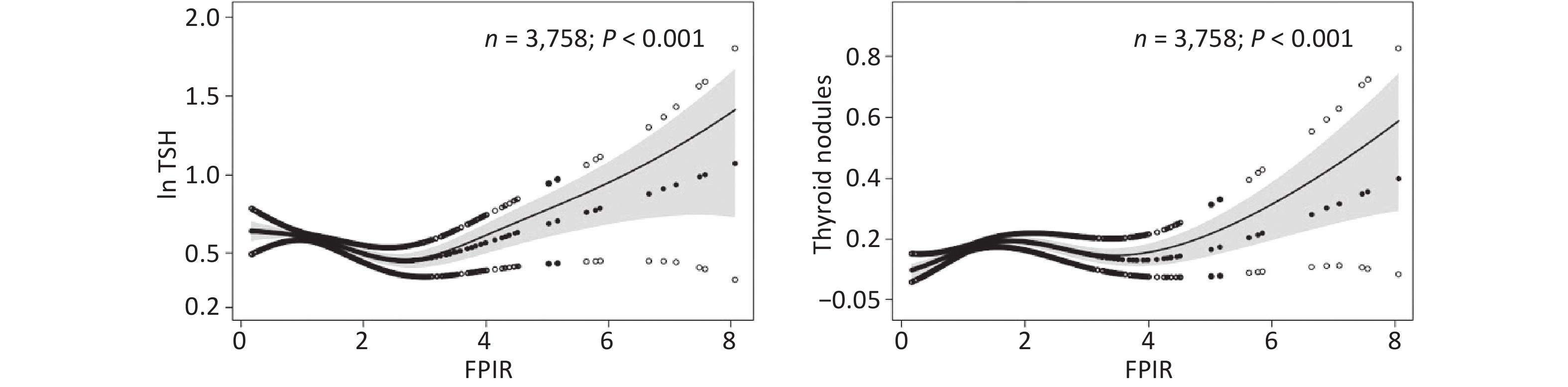
A nonlinear relationship between serum FPIR on both TSH and the presence of thyroid nodules is shown in Figure 1, exhibiting an "inverted S-shape," where solid lines represent segmented associations and shaded areas denote 95% confidence interval (CI). Table 1 shows that the inflection point for the association between FPIR and the logarithm of TSH (lnTSH) was 2.397. We adjusted for gestational age to account for physiological fluctuations in TSH levels during pregnancy. When the FPIR exceeded 2.397, a significant positive correlation was observed with lnTSH (β = 0.105, 95% CI: 0.014–0.196). When FPIR < 2.397, a significant negative correlation with lnTSH was observed (β = −0.119, 95% CI: −0.187 to −0.050). The prevalence of thyroid nodules increased with increasing FPIR values and gradually decreased after the inflection point.
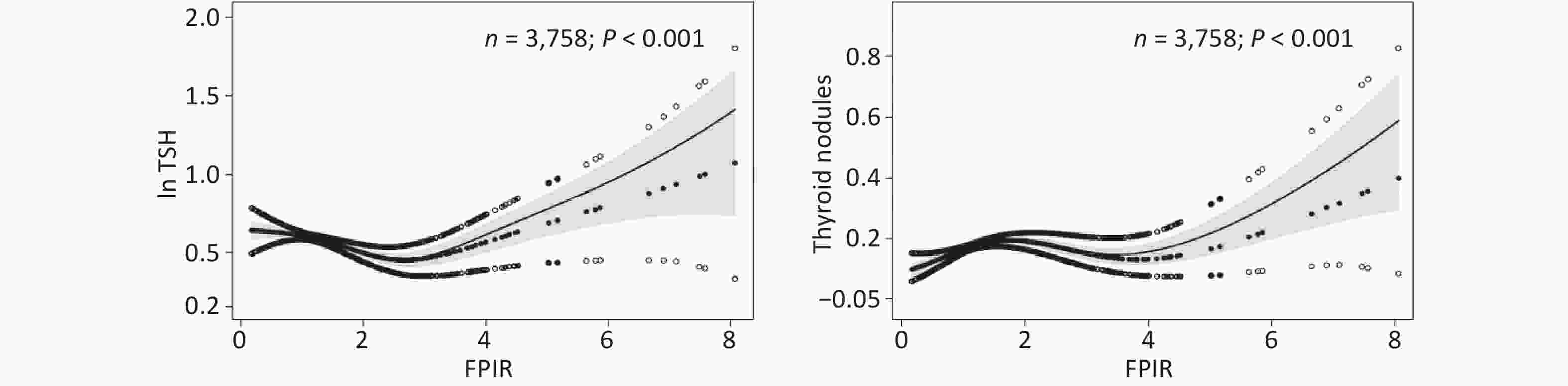
Figure 1. Analysis of the relationship between the FPIR and TSH or thyroid nodules in pregnant women. Adjusted for age, height, weight, income, gestational diabetes mellitus, educational parity, and gestational trimester. FPIR, serum free-to-protein-bound iodine ratio; lnTSH, natural logarithm of thyroid-stimulating hormone.
FPIR groups N β (95% CI) P-valuea lnTSH FPIR < 2.397 3,099 −0.119 (−0.187, −0.050) < 0.001 FPIR ≥ 2.397 276 0.105 (0.014, 0.196) 0.024 Thyroid nodules FPIR < 0.932 655 6.664 (2.081, 21.341) < 0.001 FPIR ≥ 0.932 2,720 1.002 (0.866, 1.158) 0.980 Note. aA 2-piece-wise linear regression model was used. The turning point of the FPIR was 2.397 for TSH and 0.932 for thyroid nodules. FPIR, serum free-to-protein-bound iodine ratio; lnTSH, natural logarithm of thyroid-stimulating hormone; CI, confidence interval. Table 1. Analysis of the threshold effect of FPIR on thyroid function indicators and thyroid nodules using a segmented linear regression model
As shown in Table 2, logistic regression was used to test whether an abnormal FPIR <1 or ≥ 2 predicted iodine status or thyroid nodules during pregnancy. A low ratio (FPIR < 1) was independently associated with iodine deficiency (odds ratio [OR] = 1.38, 95% CI: 1.08–1.76). A high ratio (FPIR ≥ 2) was inversely linked to TSH in both models (OR = −0.15, 95% CI: 0.33 to −0.02).
Outcome Unadjusted Adjusteda OR (95% CI) P-value OR (95% CI) P-value When FPIR ≤ 1 UI < 150 μg/L 1.38 (1.08,1.76) 0.001 1.38 (1.08,1.76) 0.001 TSH 0.10 (−0.02,0.22) 0.101 0.10 (−0.01,0.22) 0.085 Thyroid nodules 0.79 (0.63,0.97) 0.003 0.82 (0.66,1.02) 0.076 When FPIR ≥ 2 UI > 500 μg/L 0.42 (0.07,1.38) 0.237 0.42 (0.07,1.39) 0.238 TSH −0.14 (−0.32,0.03) 0.013 −0.15 (−0.33,0.02) 0.009 Thyroid nodules 0.97 (0.70,1.31) 0.029 0.96 (0.69,1.30) 0.077 Note. Adjusteda for age, height, weight, income, gestational diabetes mellitus, educational parity, and gestational trimester. FPIR, serum free-to-protein-bound iodine ratio; UI, 24-h urinary iodine concentration; TSH, thyroid-stimulating hormone; OR, odds ratio; CI, confidence interval. Table 2. Analysis of FPIR ≤ 1 or FPIR ≥ 2 as a risk factor of iodine status and thyroid function
This study was conducted across three regions in China with distinct iodine nutritional statuses to capture the comprehensive spectrum of iodine exposure since the implementation of the universal salt iodization program. Leveraging this natural gradient, we characterized FPIR as a new metric of iodine status and examined its relationship with both TSH profile and thyroid nodule prevalence during gestation.
Although urinary iodine is a baseline population metric, it captures only volatile short-term dietary intake and exhibits massive intra-individual variability[2]. During pregnancy, the diagnostic fidelity of the UI is fundamentally weakened by fluctuating maternal fluid intake, which triggers acute urine output shifts and distorts the concentration data via mechanical dilution[6]. Serum iodine concentration directly reflects bioavailable iodine in the thyroid gland and indicates the actual iodine level in the body. It accurately reflects the recent iodine nutritional status of the body and is not immediately affected by variations in dietary iodine intake[7].
Previous studies have focused on single investigations of serum iodine application across different populations and have lacked in-depth research on free iodine and protein-bound iodine in the serum. Serum-free iodine is the most direct raw material for thyroid hormone synthesis and reflects the dynamic equilibrium of iodine metabolism[4]. PBI is the primary form of iodine storage and transport in the body and reflects the long-term nutritional status of iodine[3]. This study demonstrated that prolonged iodine imbalance leads to abnormalities in the serum iodine spectrum, including bound iodine, which trigger thyroid dysfunction.
Our study indicates that lower FPIR values are negatively correlated with TSH levels. This finding is consistent with that of Sun et al., suggesting that excessively low serum iodine levels may be associated with thyroid dysfunction[8]. These observations underscore the importance of maintaining adequate iodine nutrition, particularly balanced serum-free and bound iodine levels, for maternal and fetal health during pregnancy[9]. Markedly low FPIR levels may predispose pregnant women to the development of thyroid nodules.
This study confirmed a significant dose-response relationship between the risk of thyroid nodules in pregnant women and FPIR. This may reflect persistent iodine exposure or metabolic abnormalities associated with structural alterations in the thyroid gland, highlighting the sensitivity of this physiological stage during pregnancy[10].
This cross-sectional study precluded the establishment of a causal relationship between FPIR values and the risk of incident thyroid nodules. Additionally, we did not collect detailed dietary information on iodine intake, such as food frequency questionnaires and/or 24-h dietary recalls. The generalizability of our findings to other populations requires further validation.
In conclusion, FPIR is a stable steady-state biomarker that provides a more consistent reflection of iodine nutritional status during pregnancy. A significant dose-response relationship was observed between FPIR and thyroid nodule risk.
HTML
Competing Interest None of the authors declare any conflicts of interest.
Ethics This study was approved by the Tianjin Medical University Medical Ethics Committee (approval no. TMUhMEC2020033). All participants were informed about the study objectives, required information, and biological samples to be collected, and written informed consent was obtained. This study was conducted in accordance with the principles of the Declaration of Helsinki.
Authors’ Contributions Z.J. and W.G. designed the study; Q.M., Q.J., H.Z., D.L., Y.W., P.L., F.L., M.J., and Y.L. participated in the investigation; Z.J. analyzed the data and wrote the first draft. W.Z. and W.G. contributed to the preparation of final manuscript.
Data Sharing The supplementary materials will be available in www.besjournal.com.
Reference

 Quick Links
 DownLoad: