



-
Despite major advances in antiretroviral therapy (ART), newly diagnosed human immunodeficiency virus (HIV) patients present with a wide range of clinical profiles, from asymptomatic to advanced disease[1]. Early markers of disease severity are needed, and serum albumin (ALB), a low-cost biomarker of nutritional status, inflammation, and liver function[2], is a strong candidate. Hypoalbuminemia (< 35 g/L) predicts poor outcomes in people living with HIV (PLWH), including opportunistic infections and mortality[3]. Prior studies link it to low CD4+ T-cell count and high viral load (HIV VL)[4], yet its role at diagnosis, before ART confounders, remains unclear. Gaps persist regarding its association with CD8+ T-cell count, the CD4+/CD8+ ratio, and liver enzymes, as well as whether these associations vary by age (< 50 vs. ≥ 50 years) or care setting (outpatient vs. inpatient). To address this, we conducted a retrospective cross-sectional study to examine the prevalence of hypoalbuminemia and its associations with demographic, immunovirological, and hepatic parameters, stratified by age and admission status, to evaluate ALB as an integrative biomarker for early risk stratification.
This retrospective cross-sectional study was approved by the ethics committee of Chongqing Public Health Medical Center (approval number: 2025-038-02-KY; date of approval: 25 December 2025; Chongqing, China), which waived the requirement for written informed consent. This study followed the recommendations for research involving human participants and the guidelines of the Declaration of Helsinki. Confidentiality and privacy were rigorously upheld in strict alignment with established clinical protocols. The study population comprised adults (men and women aged ≥ 18 years) who were newly diagnosed with HIV who presented to either the outpatient or inpatient departments of our hospital between January 2020 and October 2025. Our institution serves as the designated HIV voluntary counseling, testing, diagnosis, treatment and therapy quality control center for the municipality, receiving patients both through direct presentation and referral from other healthcare facilities across the municipality. HIV-specific antibodies were initially detected using two immunoassays. Positive screening results were subsequently confirmed by Western blot assays for the detection of HIV proteins, and plasma HIV RNA levels in peripheral blood were quantified using qRT-PCR. Patients with positive test results were subsequently verified as newly diagnosed after checking through the national case reporting system. Individuals not previously registered in this system were classified as newly diagnosed and ART-naïve.
Clinical data from individuals who meet the preceding inclusion/exclusion criteria were extracted from the hospital’s electronic medical record system. Specifically, the first laboratory-confirmed HIV positive report for each treatment-naive patient living with HIV was retrieved. Thus, clinical records from blood testing (immunological and biochemical assessments) requested by the attending physicians prior to the initiation of ART were reviewed to obtain baseline data. Generally, the interval between HIV diagnosis and baseline laboratory measurements was less than five days. The collected variables comprised patient ID, age, confirmed western blot result, HIV VL, CD4+ T-cell count, CD8+ T-cell count, CD4+/CD8+ ratio, serum ALB level, the admission type, and liver function markers including aspartate aminotransferase (AST) and alanine aminotransferase (ALT) levels. Individuals (1) whose HIV infection was not confirmed at our hospital (2) with prior confirmed HIV-positive results, (3) who had already initiated ART, (4) receiving concomitant medications before the first blood test, and (5) with a history of organ transplantation were initially excluded from the study. Patients with missing data for at least one of the preceding variables was excluded. Furthermore, those with reported HBV or HCV infection proven by recorded data were also excluded.
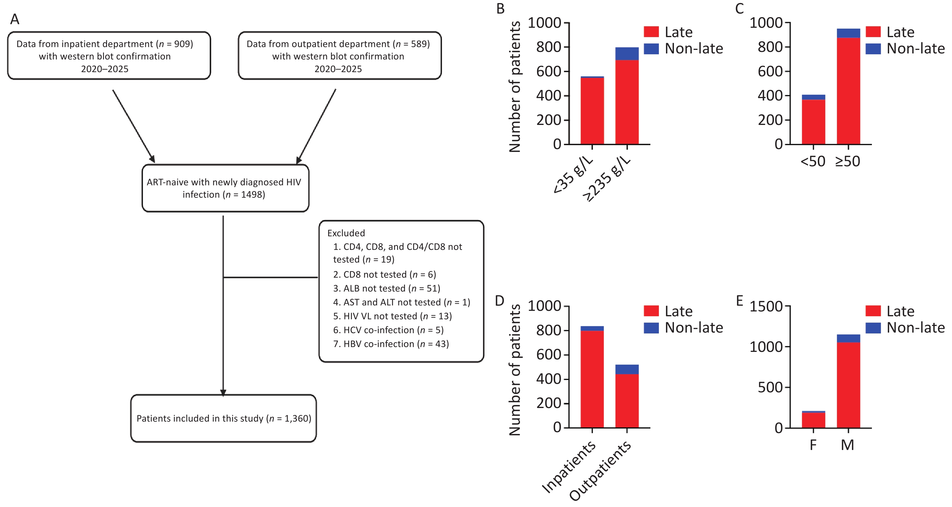
As described in Figure 1, the data were obtained from both inpatient and outpatient departments and were also stratified according to serum ALB levels and age. Based on ALB concentrations, patients were categorized into two groups: those with hypoalbuminemia (< 35 g/L) and those with ALB levels ≥ 35 g/L, in accordance with prior studies[4]. Additionally, participants were stratified by age into two groups: an older group comprising people living with HIV (PLWH) aged ≥ 50 years and a younger group comprising PLWH aged <50 years, consistent with previous literature[5].
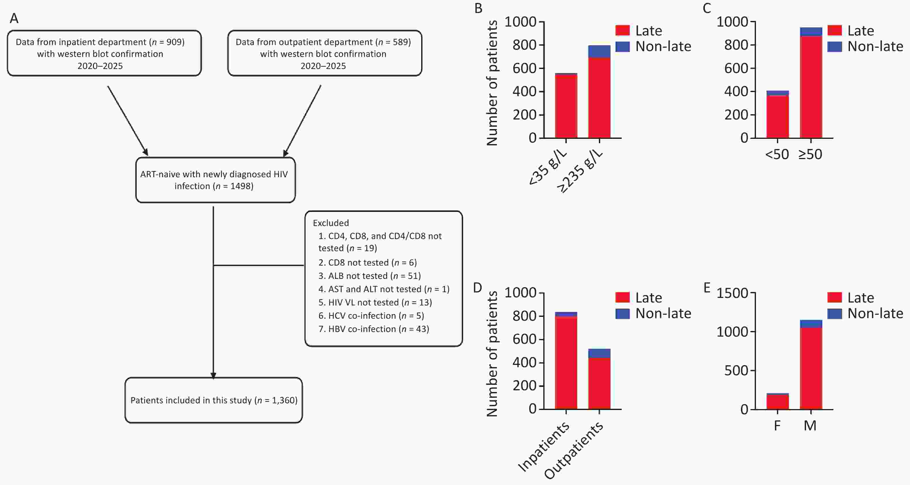
Figure 1. Study population and landscape of late HIV presentation among ART-naïve newly diagnosed PLWH. Panel A presents the study flow diagram. Panels B, C, D, and E illustrate late presentation categories stratified by ALB levels, age, admission type, and sex, respectively. M: male; F: female. Late refers to patients with CD4+ T-cell counts below 350 cells/µL; Non-late refers to patients with CD4+ T-cell counts of 350 cells/µL or more.
All data were initially recorded in Microsoft Excel and subsequently imported into GraphPad Prism (version 10, GraphPad Software, San Diego, CA, USA) for analysis. Data integrity and consistency were verified before statistical testing. The distribution of continuous variables was assessed using the Shapiro-Wilk test. Continuous variables were summarized using medians and interquartile ranges (IQRs) or means and standard deviations (SDs), as appropriate based on data distribution. Categorical variables were presented as frequencies and percentages. Serum ALB levels were analyzed both as a continuous variable and as a categorical variable [hypoalbuminemia (< 35 g/L) and ALB levels ≥ 35 g/L]. Comparisons between participants with hypoalbuminemia and those with ALB levels ≥ 35 g/L were performed using the Mann–Whitney U test or Student’s t-test for continuous variables, and the Chi square test or Fisher’s exact test for categorical variables, as appropriate. Bivariate associations between serum ALB levels and demographic (age, gender), admission type, immunovirological (HIV VL, CD4+ and CD8+ T-cell counts, CD4+/CD8+ ratio), and biochemical (ALT, AST) variables were evaluated using Spearman or Pearson correlation coefficients, depending on data distribution. To identify independent predictors of hypoalbuminemia, a multivariable logistic regression model was constructed. For this purpose, variables associated with hypoalbuminemia in bivariate analyses at a P value < 0.20, as well as clinically relevant covariates (age and gender), were included in the multivariable model. Collinearity was evaluated using variance inflation factors. Pre-specified subgroup analyses were conducted by admission type (inpatient vs. outpatient) and age group (<50 vs. ≥ 50 years) to assess potential effects of hypoalbuminemia on immunological profiles (CD4+ and CD8+ T-cell counts, CD4+/CD8+ ratio). All tests were two-sided, and a p value <0.05 was considered statistically significant.
From January 2020 to October 2025, 1498 ART-naïve, newly diagnosed PLWH were identified; 138 were excluded, leaving 1360 for analysis (Figure 1A). Most participants were late presenters (CD4+ T-cell count < 350 cells/µL) (Figure 1B-E). The median age was 58 (20) years; 84.6% were male, 61.6% were inpatients, and 38.4% were outpatients. The median HIV VL was 327, 000 (1, 055, 150) copies/mL, with median CD4+ and CD8+ T-cell counts of 119 (181) cells/µL and 528.5 (513) cells/µL, respectively. The median CD4+/CD8+ ratio was 0.21 (0.25). Median serum ALB, ALT, and AST levels were 36.5 (10.9) g/L, 22 (21) U/L, and 27 (20) U/L, respectively (Table 1).
Overall analysis Variables Total
(n = 1,360)ALB <35g/L (n = 561) ALB ≥35 g/L
(n = 799)P Sex Male 1,150 504 (89.84) 646 (80.85) < 0.0001 Female 210 57 (10.16) 153 (19.15) Age < 50 409 116 (20.68) 293 (36.67) < 0.0001 ≥ 50 951 445 (79.32) 506 (63.33) Admission Inpatient (n = 838) 838 526 (93.76) 312 (39.05) < 0.0001 Outpatient (n = 522) 522 35 (6.24) 487 (60.95) CD4+ T-cell count [median (IQR)] 119 (181) 59 (97.5) 175 (178.5) < 0.0001 CD8+ T-cell count [median (IQR)] 528.5 (513) 357 (402) 631 (522.5) < 0.0001 CD4+/CD8+ ratio [median (IQR)] 0.21 (0.25) 0.16 (0.21) 0.26 (0.27) < 0.0001 ALB [median (IQR)] 36.5 (10.9) 30.7 (5.09) 41.4 (7.48) < 0.0001 AST* [median (IQR)] 27 (20) 34 (28) 25 (13) <0.0001 ALT* [median (IQR)] 22 (21) 24 (23) 21 (18.5) 0.0056 HIV VL [median (IQR)] 327,000 (1,055,150) 669,000 (1,708,000) 166,000 (558,400) < 0.0001 *Multivariate analysis Variables Crude OR (95% CI) p Adjusted OR (95% CI) p Admission type Outpatient 1.00 1.00 Inpatient 23.46 (16.42–34.52) <0.0001 14.95 (10.32–22.26) <0.0001 Age range <50 1.00 1.00 ≥50 2.221 (1.734–2.859) <0.0001 2.147 (1.556–2.972) <0.0001 CD4+ T-cell counts 1.008 (1.007–1.010) <0.0001 1.007 (1.005–1.009) <0.0001 CD8+ T-cell counts 1.001 (1.001–1.002) <0.0001 1.000 (0.9996–1.000) 0.8618 CD4+/CD8 ratio 3.316 (2.026–5.600) <0.0001 0.3687 (0.1539–0.8238) 0.0196 HIV VL 1.00 0.2014 - - AST 0.9891 (0.9851–0.9928) <0.0001 0.9951 (0.9911–0.9987) 0.0110 ALT 0.9996 (0.9976–1.001) 0.6325 - - Note *The assessment of multicollinearity indicated no significant collinearity among the independent variables included in the multivariable logistic regression model (VIF ranges between 1.027 and 2.732). Table 1. Baseline characteristics, ALB status comparison, and regression analysis of factors associated with hypoalbuminemia in newly diagnosed PLWH (n = 1,360)
Hypoalbuminemia was present in 41.25% of participants at HIV diagnosis, which is lower than the 76.40% reported in Tanzania[3], and was significantly more common in inpatients than in outpatients (62.76% vs. 6.70%, P < 0.0001). The marked difference between inpatients and outpatients suggests that hypoalbuminemia is closely associated with disease severity and acute clinical decompensation at presentation, rather than merely reflecting demographic characteristics. In our study, hypoalbuminemia reflects a synergistic combination of both acute systemic inflammation at the time of presentation and advanced HIV disease requiring hospitalization. This dual etiology identifies a high-risk population needing multidisciplinary care combining treatment of acute opportunistic infections, nutritional support, timely ART, and holistic management. Thus, serum ALB on admission represents a simple biomarker for guiding intensified care. In addition, older individuals exhibited a higher prevalence of hypoalbuminemia compared with younger age groups (46.79% vs. 28.36%, P < 0.0001), indicating that older age is a significant correlate of hypoalbuminemia. Accordingly, clinicians should have a lower threshold for nutritional assessment and intervention in older newly diagnosed PLWH, as hypoalbuminemia may serve as a marker of frailty, inflammation, and poorer prognosis[6].
Individuals with hypoalbuminemia were older (79.32% vs. 63.33%), more frequently male (89.84% vs. 80.85%), and more likely to be hospitalized at diagnosis (93.76% vs. 39.05%) compared to those with ALB ≥ 35 g/L (P < 0.0001 for all, Table 1). Immunovirologically, they had significantly lower T-cell (CD4+ and CD8+) counts and CD4+/CD8+ ratios, alongside higher HIV VL (P < 0.0001), reinforcing the link between nutritional/inflammatory status and immune competence in ART-naïve PLWH. Indeed, ALB is a negative acute-phase reactant and a marker of systemic inflammation, nutritional status, and hepatic synthetic capacity[2]. Evidence suggests that HIV infection fosters a milieu of chronic immune activation and exhaustion, microbial translocation, and cytokine-mediated inflammation[7]. These features may, in turn, suppress ALB synthesis in hepatocytes[8] while simultaneously driving CD4+ T-cell depletion. Biochemically, hypoalbuminemia was associated with elevated ALT and AST levels compared with those in participants with ALB levels ≥ 35 g/L (p <0.001, Table 1), though both remained within normal limits. Hypoalbuminemia likely reflects systemic immune exhaustion and inflammation, not isolated liver dysfunction.
Serum ALB levels demonstrated a positive correlation with CD4+ T-cell counts (r = 0.5205, P < 0.0001), CD8+ T-cell counts (r =0.3907, P < 0.0001), and the CD4+/CD8+ ratio (r =0.3218, P < 0.0001). Conversely, serum ALB levels showed a negative correlation with HIV VL (r =−0.4045, P < 0.0001), ALT levels (r =−0.06678, P = 0.0138) and AST levels (r =−0.3113, P < 0.0001). In the multivariable analysis (Table 1), inpatient admission, older age, lower CD4+ T-cell count, a reduced CD4+/CD8+ ratio, and elevated AST levels were independent predictors of hypoalbuminemia. CD8+ T-cell count, HIV VL and ALT levels were not significant predictors. Our results suggest that viral replication affects ALB indirectly via immune and inflammatory pathways (e.g., CD4+ T-cell depletion, immune dysregulation, and comorbidities or opportunistic infections). Additionally, ALT was not an independent predictor, whereas AST remained significant, possibly reflecting systemic inflammation, mitochondrial dysfunction, or extrahepatic injury, all of which are linked to advanced HIV severity[9].
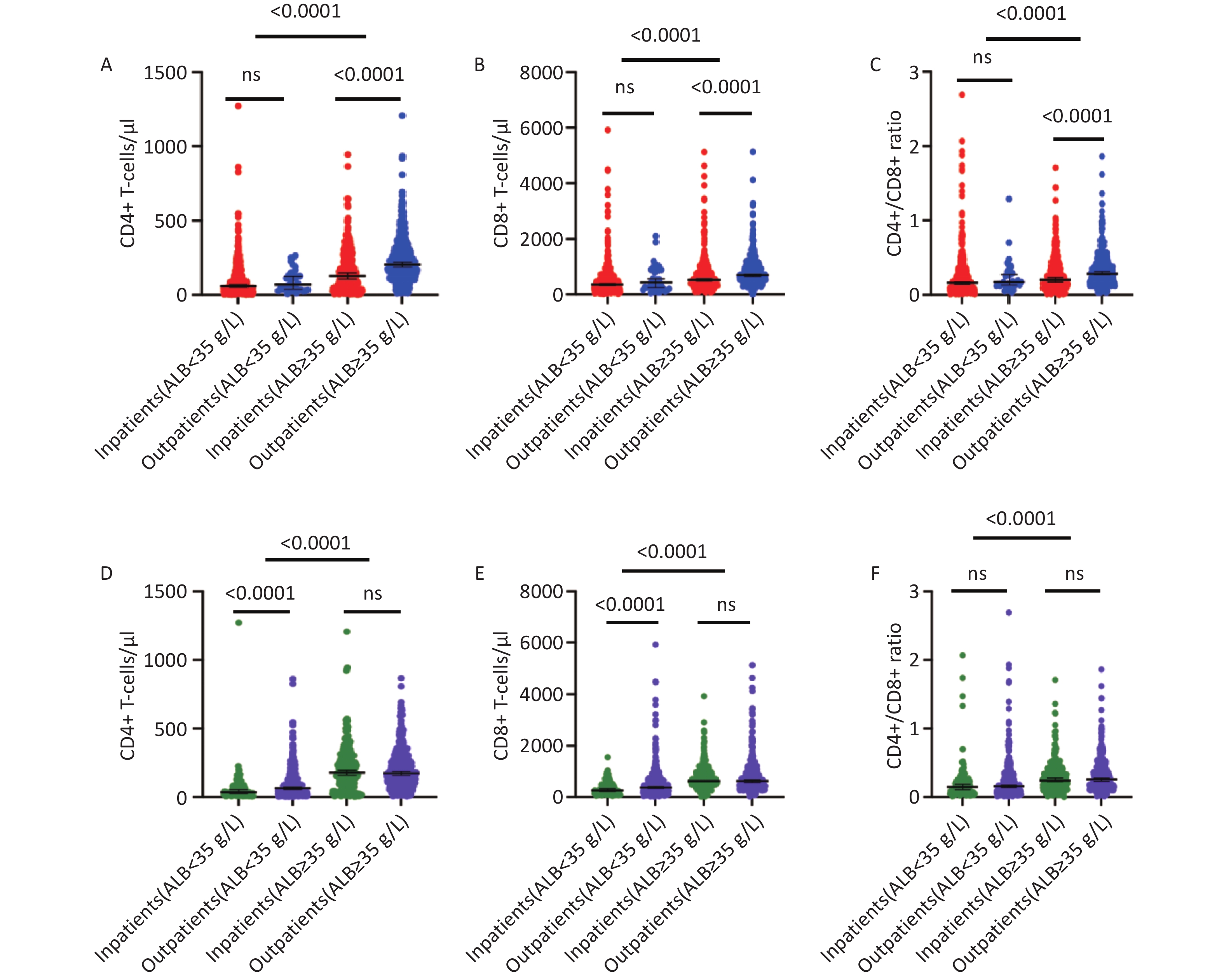
In the inpatients setting, those with serum ALB ≥35 g/L had significantly higher median (IQR) CD4+ T-cell counts [126 (186.5) vs. 58 (92.75) cells/µL], CD8+ T-cell counts [531 (509.5) vs. 356.5 (396.75) cells/µL], and CD4+/CD8+ ratios [0.20 (0.27) vs. 0.16 (0.21)] compared with inpatients with hypoalbuminemia (all P <0.001, Figure 2A-C). Similarly, in the outpatient setting, participants with ALB levels ≥35 g/L exhibited higher CD4+ T-cell counts [204 (169.5) vs. 68 (105) cells/µL], CD8+ T-cell counts [703 (513) vs. 437 (661.5) cells/µL], and CD4+/CD8+ ratios [0.28 (0.23) vs. 0.17 (0.18)] relative to those with hypoalbuminemia (all P < 0.001, Figure 2A-C). Notably, immunological profiles were broadly comparable between inpatients and outpatients with hypoalbuminemia, with similar median (IQR) CD4+ T-cell counts [58 (92.75) vs. 68 (105) cells/µL, P = 0.148] (Figure 2A), CD8+ T-cell counts [356.5 (396.75) vs. 437 (661.5) cells/µL, P = 0.446] (Figure 2B), and CD4+/CD8+ ratios [0.16 (0.21) vs. 0.17 (0.18), P = 0.295] (Figure 2C), suggesting that ALB levels may capture disease severity more accurately than admission status alone. From a clinical standpoint, these findings highlight serum ALB as a readily available and objective biomarker for early risk stratification, particularly in outpatient settings. Consequently, patients with low ALB levels, including those who do not meet criteria for hospital admission, may carry a disease burden comparable to that of hospitalized individuals. As such, they could benefit from closer monitoring, more intensive therapeutic interventions, or earlier escalation of care. Prognostically, hypoalbuminemia may help identify a subset of patients who appear to have lower acuity but are at heightened risk for clinical deterioration, complications, or unfavorable outcomes. Integrating ALB measurement into routine outpatient assessments has the potential to refine risk prediction models, support timely clinical decision-making, and ultimately improve patient outcomes by enabling more personalized and proactive management strategies.
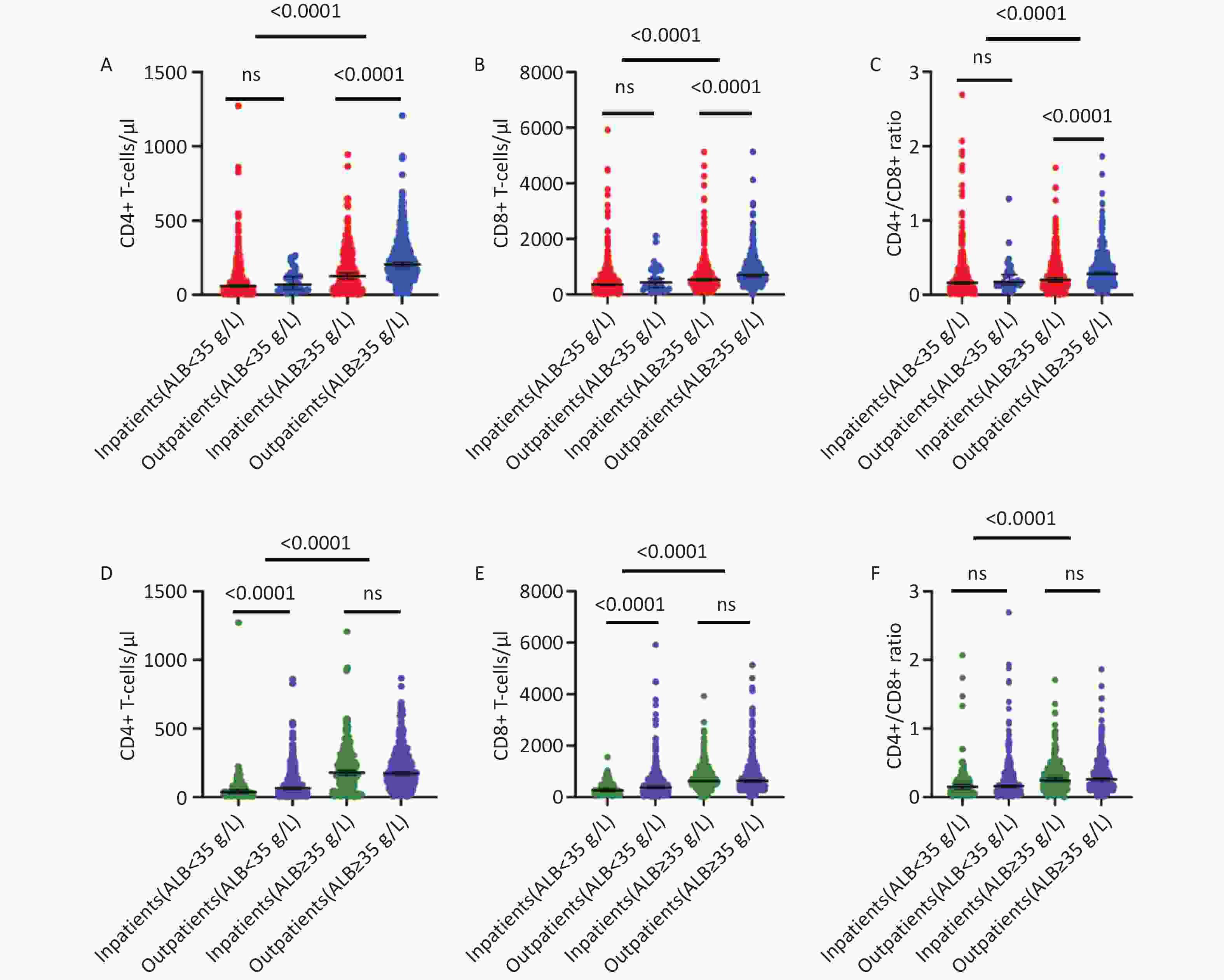
Figure 2. Impact of hypoalbuminemia on immunological profiles stratified by admission status and age group. Red indicates inpatient participants, blue indicates outpatient participants, green represents individuals younger than 50 years, and purple represents individuals aged 50 years or older. Black lines represent the median values of CD4+ T-cell counts, CD8+ T-cell counts, or CD4+/CD8+ ratio.
When stratified by age (50 years), younger participants with serum ALB levels ≥35 g/L exhibited significantly higher median (IQR) CD4+ T-cell counts [178 (203) vs.. 37 (65) cells/µL], CD8+ T-cell counts [632 (483.5) vs.. 266 (318) cells/µL], and CD4+/CD8+ ratios [0.24 (0.265) vs.. 0.15 (0.18)] compared with younger individuals with hypoalbuminemia (all P < 0.001). A similar pattern was observed among older participants, in whom those with ALB levels ≥ 35 g/L had higher CD4+ T-cell counts [174 (173) vs.. 66 (111) cells/µL], CD8+ T-cell counts [628 (539) vs.. 376 (417) cells/µL], and CD4+/CD8+ ratios [0.26 (0.27) vs.. 0.16 (0.22)] than their hypoalbuminemic counterparts (all P < 0.001). Notably, immunological profiles were comparable between younger and older individuals with serum ALB ≥ 35 g/L (P > 0.05 for all). Interestingly, younger individuals with hypoalbuminemia displayed a significantly altered immunological profile compared with other individuals (Figure 2D, E, and F). With further investigation in future studies, this pattern may suggest a phenomenon of accelerated immunological aging among young ART-naïve PLWH, particularly in the presence of hypoalbuminemia. Chronic inflammation, nutritional deficiencies, and persistent immune activation may synergistically accelerate immune senescence in this subgroup. Importantly, a declining CD4+/CD8+ ratio is increasingly recognized a being associated with aging and serves as an indicator of immunosenescence[10]. In clinical practice, elderly individuals commonly exhibit a CD4+/CD8+ ratio below 1, reflecting age-associated immunosenescence[10]. Based on our findings, hypoalbuminemia may therefore serve not only as a marker of disease severity but also as an indicator of premature immune aging, identifying young individuals at particularly high risk of adverse outcomes if ART initiation is delayed. Nevertheless, alternative explanations may account for the preceding observation, including delayed diagnosis in younger patients leading to greater immune decline before recognition. Additionally, a more aggressive disease phenotype, nutritional vulnerability signaled by hypoalbuminemia, and social determinants such as limited healthcare access or socioeconomic barriers could drive accelerated immune dysfunction independent of age-related factors.
We acknowledge that this study has some limitations. Its cross-sectional design precludes any inference regarding temporal or causal relationships between hypoalbuminemia, immune dysfunction, and immune aging. Data on nutritional status, inflammatory markers, opportunistic infections, and comorbidities were not always available and may have contributed to hypoalbuminemia. Practically, nutritional status is not routinely documented in newly diagnosed HIV patients, and inflammatory markers are not consistently available as part of the initial evaluation; therefore, we were unable to assess these potential confounding factors. Similarly, data on opportunistic infections, renal involvement, and comorbidities were only available for inpatients. In the outpatient setting, such information is difficult to obtain systematically, which limited our ability to rigorously control for these variables. This study highlights an important opportunity to encourage health policy makers to systematically incorporate the aforementioned data for newly diagnosed PLWH. Doing so would facilitate more robust and comprehensive research, ultimately enhancing the delivery of healthcare services for PLWH. Therefore, future studies using a prospective cohort design should explore the mechanistic pathways linking albumin metabolism and immune aging in HIV, as they may unveil a novel realm of exploration.
HTML
Competing Interests The authors declare no conflicts of interest. The funders had no role in the design of the study; in the collection, analyses, or interpretation of data; in the writing of the manuscript; or in the decision to publish the results.
Ethics The study protocol was approved by the ethics committee of the Chongqing Public Health Medical Center (approval number: 2025-038-02-KY; date of approval: 25 Decembre 2025; Chongqing, China). The requirement for written informed consent was waived by the institutional review board.
Author Contributions Conceptualization: Silvere D Zaongo and Yaokai Chen. Methodology: Silvere D Zaongo, and Mei Han. Formal analysis and investigation: Silvere D Zaongo and Mei Han. Supervision: Yaokai Chen. Writing review and editing: Silvere D Zaongo, Mei Han, Zhihua Ai, and Yaokai Chen. All authors commented on previous versions of the manuscript. All authors read and approved the final manuscript.
Data Sharing Upon request, and subject to review, the corresponding author will provide the data that support the findings of this study.
&These authors contributed equally to this work.
Reference

 Quick Links
 DownLoad: