





-
Sleep disorders, which are classified into seven major categories by the International Classification of Sleep Disorders, Third Edition (ICSD-3), including insomnia, affect more than one-third of the global population[1]. Chronic insomnia has emerged as a significant contributor to cognitive impairment and is considered a modifiable risk factor for Alzheimer's disease. However, the specific neural mechanisms linking sleep disruption to cognitive decline remain incompletely understood. This study employed multimodal magnetic resonance imaging (MRI) and graph-theoretical analysis to investigate the functional and structural network alterations in patients with chronic insomnia and their relationship with cognitive performance. We enrolled 31 patients with chronic insomnia according to the ICSD-3 criteria (excluding those with restless legs syndrome, mild obstructive sleep apnea, or unclear diagnoses) and 31 healthy controls matched for age and education. This study was approved by the hospital's Ethics Committee (2023ZDSYLL138-P01). All participants provided written informed consent. The inclusion criteria were as follows: age 40–80 years; right-handedness; absence of other sleep disorders; education of more than 8 years; no medical or neurological conditions; no brain abnormalities on T2-weighted fluid attenuated inversion recovery (T2-FLAIR) imaging; and no contraindications to MRI.
All participants underwent sleep assessments, including collection of sleep history and administration of the Athens Insomnia Scale (AIS), Pittsburgh Sleep Quality Index (PSQI), Epworth Sleepiness Scale (ESS), and Insomnia Severity Index (ISI). Mood was evaluated using the Hamilton Anxiety Rating Scale (HAMA) and Hamilton Depression Rating Scale (HAMD). Cognitive function was assessed by trained neurologists using domain-specific tests, including episodic memory (Auditory Verbal Learning Test–Delayed Recall, Rey–Osterrieth Complex Figure Test–Delayed Recall, Logical Memory Test–Delayed Recall); visuospatial function (Clock Drawing Test, Rey–Osterrieth Complex Figure Test–Copy); information processing speed (Digit Symbol Modalities Test, Stroop Color–Word Test, Parts A and B, Trail Making Test Part A); and executive function (Digit Span Test–Backward, Stroop Color-Word Test Part C, Semantic Similarity Test, Trail Making Test Part B, Verbal Fluency Test).
Multimodal MRI was performed using a 3.0-T Siemens Vida scanner and included resting-state blood oxygen level-dependent (BOLD) imaging, diffusion tensor imaging (DTI), T1-weighted three-dimensional magnetization-prepared rapid gradient-echo (3D-MPRAGE), and T2-FLAIR sequences. Participants were instructed to lie supine with their eyes closed while remaining awake. Earplugs and foam padding were used to minimize head motion. The BOLD data were processed using SPM8 and DPARSFA. Preprocessing included removal of the first 10 time points; exclusion of participants with excessive head motion (> 3 mm translation or > 3° rotation); spatial normalization to the Montreal Neurological Institute (MNI) space; linear detrending; band-pass filtering; and regression of head motion, white matter, global signal, and cerebrospinal fluid (CSF) signals. Four patients with chronic insomnia and two healthy controls (HCs) were excluded because of excessive head motion. An additional three patients with chronic insomnia and one with HC were excluded because T1-weighted images were unavailable. DTI data were processed using PANDA, including skull stripping, motion and eddy current correction, tensor reconstruction, and deterministic tractography using a termination criterion of a turning angle > 45° or fractional anisotropy (FA) < 0.2.
Both the functional and structural networks were parcellated into 90 brain regions using the Automated Anatomical Labeling (AAL) atlas. For functional networks, Pearson correlation coefficients calculated between the mean time series of each pair of regions were used to generate 90 × 90 connectivity matrices, which were subsequently thresholded based on sparsity (S). For structural networks, the number of white matter fibers (fiber number [FN]) connecting each pair of regions was used to define the edge weights, with connections retained only when FN was ≥3. Two patients with insomnia were excluded because of registration failure, whereas three patients with chronic insomnia and one HC were excluded because of poor T1-weighted images.
Graph-theoretical network analysis provides a powerful framework for characterizing the topological organization of whole-brain networks[2]. For each participant, topological properties of both structural and functional brain networks were calculated. These included global metrics, such as small-world parameters (σ, Cp, γ, Lp, λ)[3] and network efficiency measures (Eglob, Eloc)[4], as well as nodal metrics (NCp, Ne, NLe, DC, and BC) and edge properties.
Cognitive scores were standardized using Z-score transformation, and composite domain scores were calculated as the mean of the corresponding neuropsychological test scores. Group differences in demographic and clinical measures were assessed using independent-samples t-tests or chi-square tests, as appropriate. Neurocognitive scores were compared using analysis of covariance (ANCOVA), with age, sex, education, smoking, alcohol use, hypertension, diabetes, and hyperlipidemia as covariates (P < 0.05). Group differences in network metrics were assessed using permutation tests (10,000 permutations) with the same covariates. To control for multiple comparisons, the Benjamini-Hochberg false discovery rate (FDR) correction (q < 0.05) was applied to all nodal-level analyses. Network-based statistics (NBS) identified between-group differences in network connectivity using an initial threshold of P < 0.05, followed by 5,000 permutations and family-wise error (FEW)-corrected significance at P < 0.05.
No significant group differences were observed in age, education, or vascular risk factors (all P > 0.05). However, the insomnia group had a significantly higher proportion of females (7/24) than the HC group (18/13; P = 0.010). As expected, patients with chronic insomnia showed significantly higher scores on all sleep- and mood-related scales (all P < 0.001; Supplementary Table S1A).
After controlling for age, sex, and education, patients with insomnia showed significantly lower performance in executive function (P = 0.021, Cohen’s d = 0.58) and visuospatial function (P = 0.036) than HC. Episodic memory showed a nonsignificant trend toward poorer performance (P = 0.066, Cohen’s d = 0.42), whereas information processing speed did not differ significantly between the groups (P = 0.052; Supplementary Table S1B). At the individual test level, significant between-group differences were observed in the Digit Symbol Modalities Test (P = 0.011, Cohen’s d = 0.53), Semantic Similarity test (P = 0.006, Cohen’s d = 0.62), and Clock Drawing Test (P = 0.022, Cohen’s d = 0.54). Other neuropsychological tests showed no significant differences between the groups (all P > 0.05; Supplementary Table S1C).
Functional network analysis revealed that both groups retained small-world network architecture, with no significant between-group differences in global topological metrics (all P > 0.05). These findings suggest that the global efficiency and overall organizational properties of functional brain networks are largely preserved in patients with chronic insomnia. Exploratory nodal-level analyses revealed nominal alterations in functional network topology, including increased degree centrality and betweenness centrality in the right orbitofrontal cortex (Frontal_Sup_Orb_R; P = 0.010 and P = 0.047, respectively) and increased nodal metrics in the left inferior parietal lobule (Parietal_Inf_L; P = 0.016–0.037). The right orbitofrontal cortex is involved in decision-making and emotional regulation, whereas the inferior parietal lobule plays a critical role in attention and visuospatial processing. However, none of the observed nodal differences remained significant after FDR correction (all q > 0.05; Supplementary Table S2).
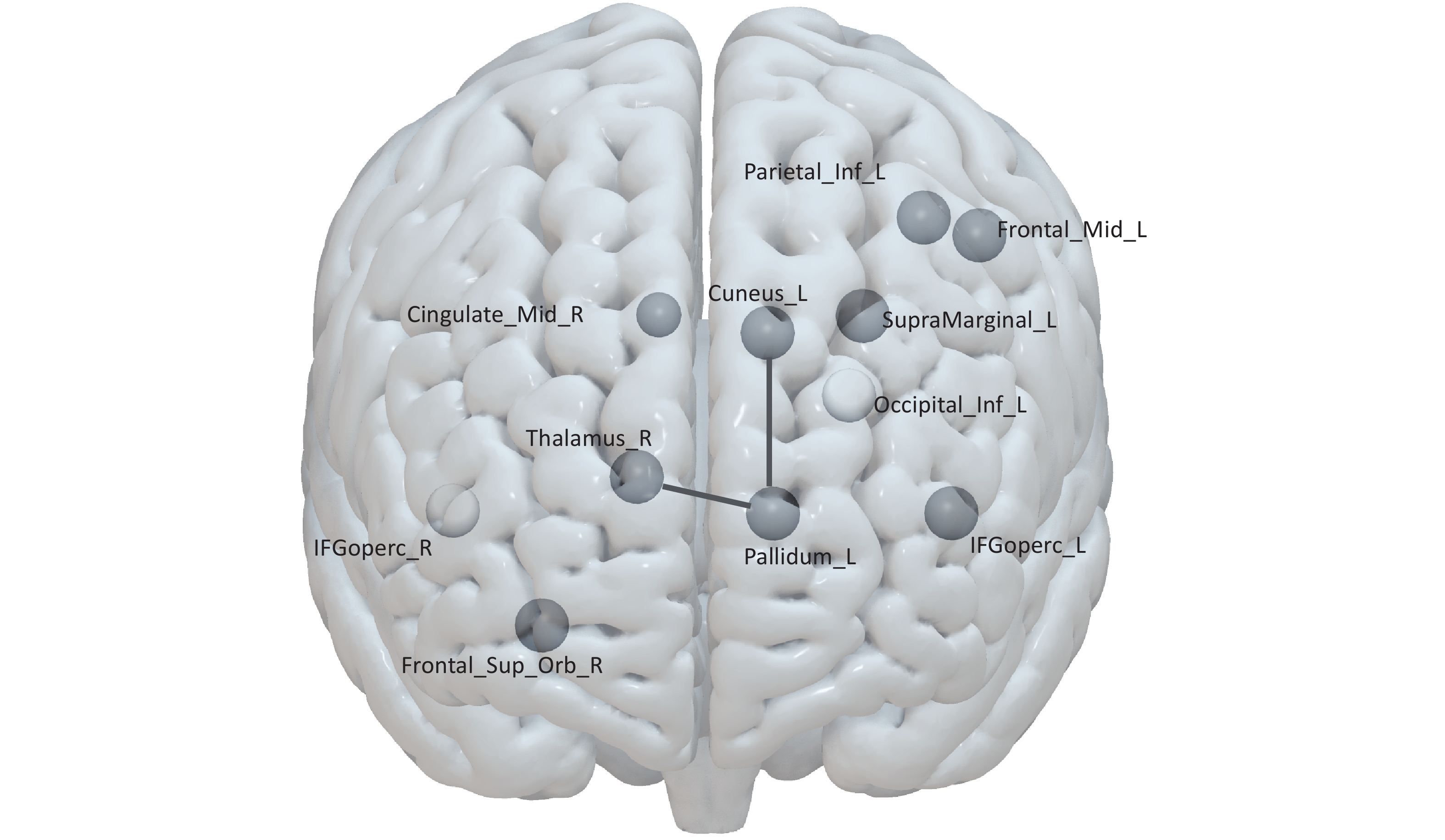
For the structural networks, both groups exhibited the characteristic small-world architecture, with no significant differences in the global network metrics. Exploratory nodal-level analyses revealed nominal alterations, including increased betweenness centrality in the left inferior frontal gyrus (IFGoperc. L; P = 0.011) and decreased betweenness centrality in the right inferior frontal gyrus (IFGoperc. R; P = 0.005), as well as increased nodal clustering coefficient and nodal local efficiency in several frontal and parietal regions (all P < 0.05). The inferior frontal gyrus is a key node of the executive control network, and its altered centrality may reflect compensatory reorganization in chronic insomnia. However, none of these nodal-level differences remained significant after FDR correction (all q > 0.05; Supplementary Table S3).Using deterministic tractography, network-based statistics (NBS) identified a significant structural connectivity component that differed between patients with chronic insomnia and HCs (initial threshold P < 0.05; 5,000 permutations; family-wise error-corrected P < 0.001). The affected connections primarily involved the left pallidum, left cuneus, and right thalamus (Figure 1 and Supplementary Table S4). The pallidum is a critical component of the basal ganglia-thalamocortical circuit involved in regulating sleep-wake transitions, whereas the cuneus is primarily involved in visual processing, and the thalamus serves as a relay station for sensory information. To determine whether these structural alterations were accompanied by corresponding functional changes, the functional connectivity of the affected pathways was further evaluated. No significant between-group differences were observed in the corresponding functional connectivity (Pallidum_L-Cuneus_L: P = 0.362; Pallidum_L-Thalamus_R: P = 0.533), suggesting that structural alterations were not paralleled by detectable changes in functional connectivity within these pathways. This finding underscores the complex and potentially non-linear relationships between structural and functional brain networks in chronic insomnia. Given the well-recognized limitations of deterministic tractography in resolving crossing fibers[5,6], these findings should be interpreted with caution.
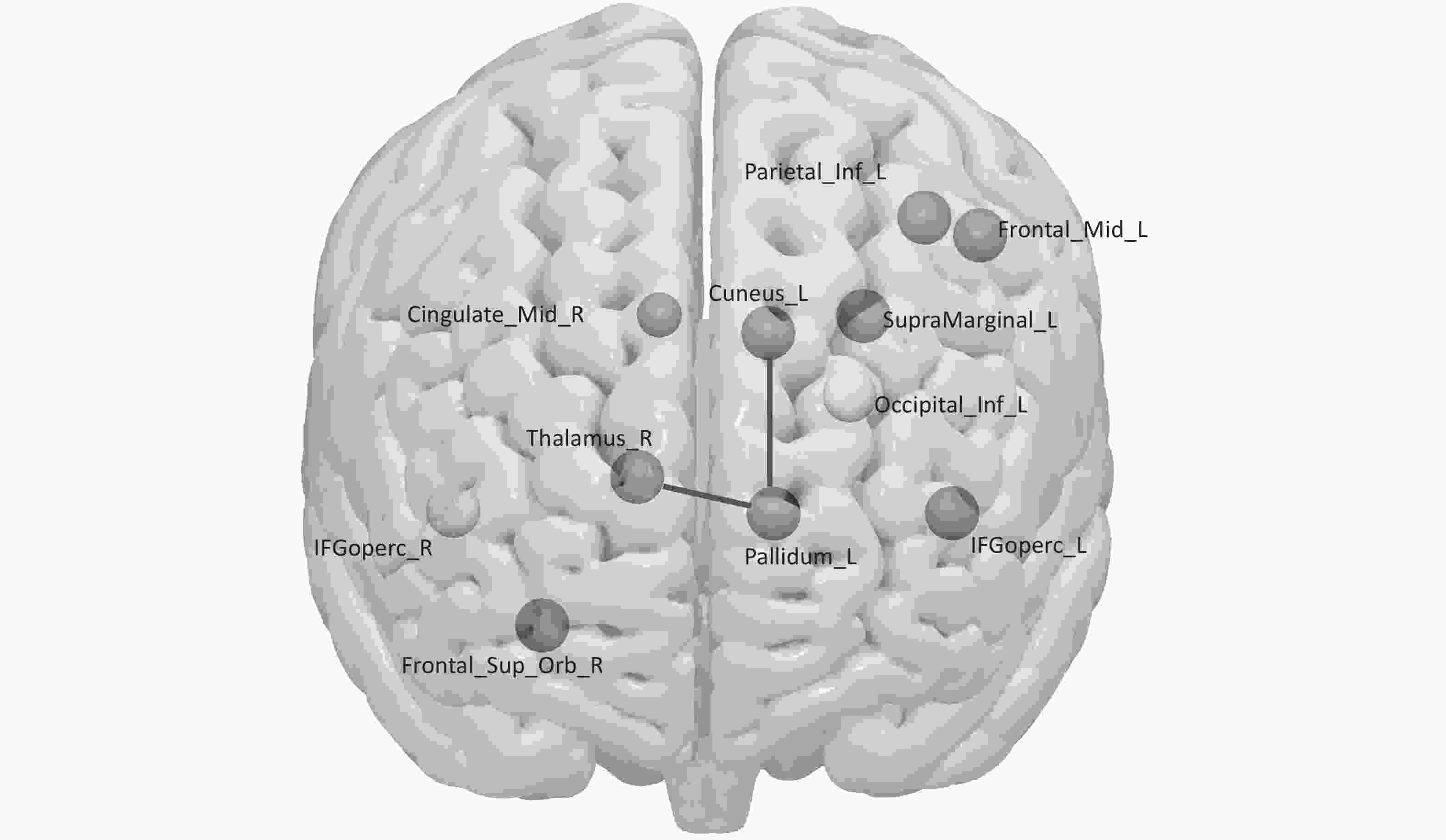
Figure 1. Exploratory brain regions and structural connections in chronic insomnia. Nodes represent regions with nominal group differences (P < 0.05, uncorrected) in functional or structural network analysis. Dark gray: increased metrics; light gray: decreased metrics. Black lines indicate NBS-identified structural connections (P < 0.001, FWE-corrected), linking the left pallidum to the left cuneus and right thalamus. Abbreviations: ORBsup, orbital part of the superior frontal gyrus; IPL, inferior parietal lobule; IFGoperc, opercular part of the inferior frontal gyrus; MFG, middle frontal gyrus; SMG, supramarginal gyrus; MCC, midcingulate cortex; PAL, pallidum; CUN, cuneus; THA, thalamus; IOG, inferior occipital gyrus. L, left; R, right.
Structure-function coupling analysis was performed in 27 participants who underwent both DTI and resting-state fMRI and showed a modest but statistically significant overall correlation (Spearman's r = 0.153, P < 0.001), consistent with previous reports indicating that structure-function relationships are complex and region-dependent[7]. Regional analyses revealed substantial heterogeneity, with the left angular gyrus exhibiting the strongest coupling (r = 0.517), whereas the bilateral supplementary motor areas exhibited negative coupling (r = −0.464 and −0.403), suggesting region-dependent structure-function relationships. The negative coupling observed in the supplementary motor areas may reflect inhibitory or competitive interactions between structural and functional connectivity in these regions (Supplementary Table S5).
Exploratory partial correlation analyses, controlling for age, sex, and education, identified nominal associations between brain region metrics and clinical/cognitive variables, including the right mid-cingulate cortex nodal clustering coefficient with HAMA (r = 0.545, P = 0.004) and right orbitofrontal cortex degree centrality with clock-drawing test performance (r = 0.519, P = 0.009). In the insomnia group, information processing speed showed nominal negative partial correlations with HAMA (r = −0.392, P = 0.043) and PSQI (r = −0.410, P = 0.034). However, none of these associations remained statistically significant after FDR correction (all q > 0.05; Supplementary Tables S4 and S6).
Our findings demonstrate that chronic insomnia is associated with selective cognitive impairment and widespread alterations in both functional and structural brain network topologies. Specifically, patients with insomnia showed significantly lower performance in executive and visuospatial functions as well as impairments in processing speed (DSST), semantic processing, and visuoconstruction (clock drawing). Although global efficiency was preserved at the neural level, suggesting intact whole-brain information transfer, exploratory nodal-level analyses revealed nominal alterations in the frontal, parietal, and limbic regions across both functional and structural networks. More importantly, network-based statistics identified a significant structural connectivity component involving the left pallidum, left cuneus, and right thalamus (P < 0.001, FWE-corrected), indicating preferential disruption of subcortical-cortical pathways in chronic insomnia. Collectively, these findings suggest that chronic insomnia is associated primarily with regional and connectional-level alterations in brain networks rather than with widespread disruption of global network topology.
The lack of significant group differences in global efficiency, together with the failure of the nodal metrics to survive FDR correction, suggest that the neural correlates of chronic insomnia are subtle and spatially distributed, rather than confined to specific regions. This pattern is consistent with a network-level pathophysiological model, in which chronic insomnia affects distributed neural systems rather than selectively targeting a single brain region.
Sex differences in the cognitive effects of chronic insomnia warrant further investigation. Although all analyses were adjusted for sex as a covariate, exploratory sex-by-group interaction analyses revealed significant interactions for episodic memory (P = 0.045) and information processing speed (P < 0.001), suggesting that the cognitive effects of insomnia may differ between men and women. These sex-specific effects may be related to the hormonal influences on sleep architecture and stress reactivity (Supplementary Table S7).
This study has some limitations. First, the cross-sectional design precludes causal inferences; therefore, the conclusions have been interpreted accordingly. Second, the modest sample size (n = 62) may have limited the statistical power to detect small effects, although post hoc power analysis revealed more than 99% power for detecting large effects (Cohen’s d > 0.8). Third, deterministic tractography has well-recognized limitations in resolving crossing fibers, particularly in regions such as the temporo-parietal junction[5,6]. Accordingly, future studies employing probabilistic tractography are required to validate the structural connectivity findings[8]. Fourth, medication use was not systematically recorded. Although our core findings were consistent with those reported in previous studies involving medication-free patients with chronic insomnia[9]. Finally, the inherent challenges associated with reconstructing the human connectome using diffusion tractography have been well documented[10].
Future prospective studies with larger sample sizes, balanced sex distributions, and probabilistic tractography are required to validate these findings. Multimodal integration approaches combining functional and structural data may provide deeper insights into the relationship between brain organization and cognitive function in patients with chronic insomnia.
In conclusion, patients with chronic insomnia exhibited selective cognitive impairment accompanied by widespread functional and structural network alterations. Collectively, these findings enhance our understanding of the neural mechanisms underlying insomnia-related cognitive dysfunction and help identify potential network-based biomarkers.
HTML
Competing Interests All authors declare that they have no competing interests.
Ethics This study was approved by the hospital's Ethics Committee (approval no. 2023ZDSYLL138-P01). All participants provided written informed consent.
Authors' Contributions Study design, experiments, and writing: Jingting Kong. Methodological guidance, study design, and experiments: Zan Wang. Experiments: Mengxue Wang, Xuezi Zhao and Zan Wang. Study Design, Methodological Guidance, and Writing Guidance: Qingguo Ren.
Data Sharing The datasets generated and/or analyzed in the current study are available from the corresponding author upon reasonable request. The supplementary materials will be available in www.besjournal.com.
&These authors contributed equally to this work.
Reference

 Quick Links
 DownLoad: