





-
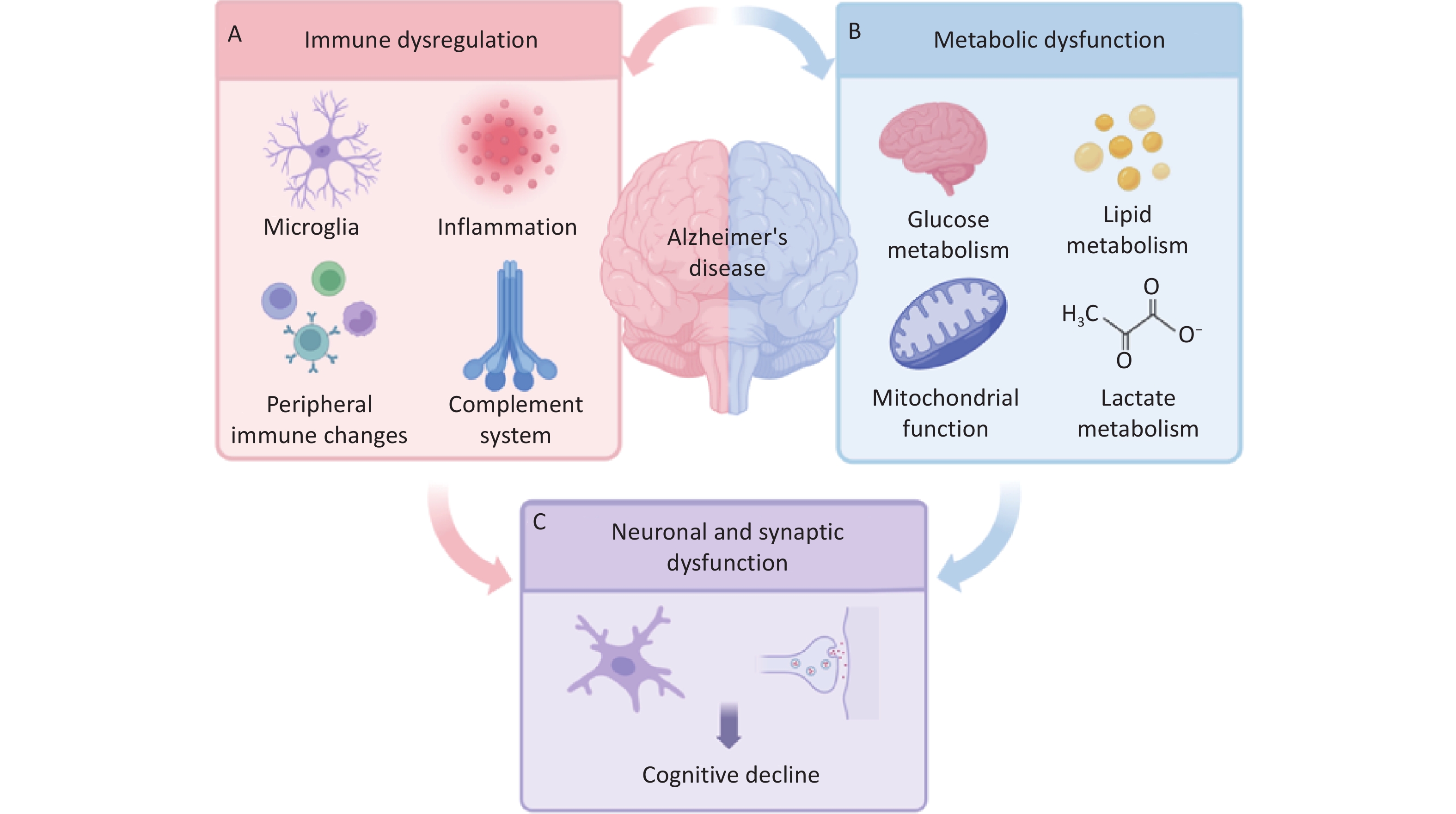
Alzheimer’s disease (AD) is a progressive neurodegenerative disease characterized by cognitive decline. Age is one of the major risk factors for AD, and population aging has made AD an increasing public health challenge. Amyloid-beta (Aβ) deposition and tau pathology are two major features of AD and have guided research and drug development for decades. Anti-Aβ therapies such as lecanemab have demonstrated that reducing amyloid pathology can slow cognitive decline in patients with early AD, although the clinical benefit remains limited and treatment is accompanied by safety and accessibility concerns[1,2]. Besides amyloid and tau pathology, several other mechanisms have been proposed in AD, including neuroinflammation, oxidative stress, synaptic dysfunction, and metabolic abnormalities. Among these, systemic inflammation and impaired energy metabolism are increasingly recognized as important processes involved in cognitive decline in AD progression[3,4] (Figure 1).
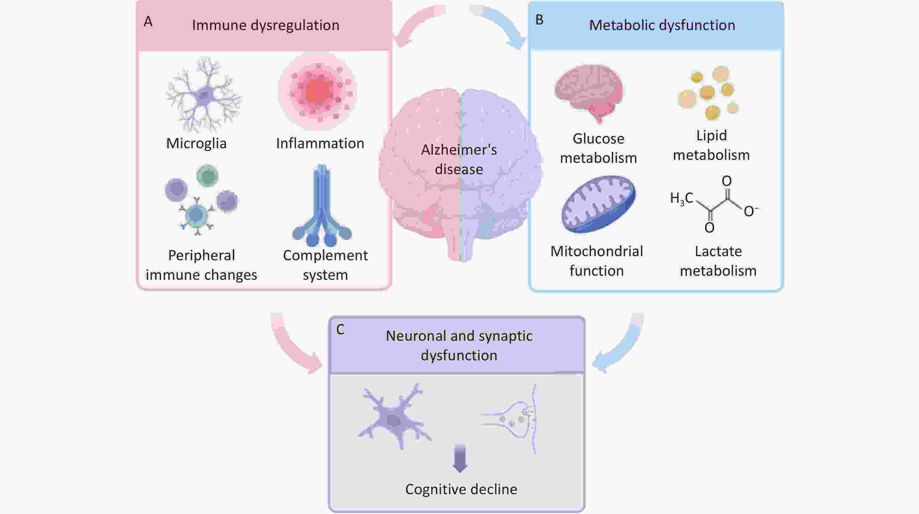
Figure 1. Immune dysregulation and metabolic dysfunction in Alzheimer’s disease. (A) Immune dysregulation in Alzheimer’s disease involves altered microglial activity, inflammatory responses, peripheral immune changes, and complement system activation. (B) Metabolic dysfunction includes abnormalities in glucose and lipid metabolism, mitochondrial function, and lactate metabolism. Immune and metabolic disturbances may interact with each other and contribute to Alzheimer’s disease progression. (C) These alterations are associated with neuronal and synaptic dysfunction, which may ultimately contribute to cognitive decline.
Immune dysregulation is a key feature of AD, involving both the central nervous system and the peripheral immune system. Microglia are the main immune cells in the brain and respond to Aβ deposition, neuronal injury, and other pathological changes. In early disease stages, microglia help clear Aβ and promote tissue repair. However, under persistent stimulation, their inflammatory activity may increase, leading to cytokine release, oxidative stress, and synaptic damage. Abnormal activation of the complement system can enhance synaptic pruning and contribute to the loss of neuronal connections[5]. Adaptive immune responses, particularly T cell activation, can further promote neuroinflammation in AD[6]. These changes in the central nervous system are closely linked to peripheral immune responses[7]. Circulating cytokines, vascular dysfunction, and blood-brain barrier alterations can all influence interactions between the peripheral immune system and the brain[4]. Monocytes are key components of this peripheral response and may contribute to inflammatory regulation and Aβ clearance. Thus, immune abnormalities in AD are not isolated changes in a single cell type or pathway, but rather a complex network involving multiple immune cell types and inflammatory pathways. These immune changes are also reflected in peripheral blood. Monocyte count provides information on systemic immune activity, while serum albumin is influenced by nutritional status, antioxidant capacity, and general physiological condition. The monocyte-to-albumin ratio (MAR) brings these two measures together and may therefore reflect changes in both inflammation and systemic health. In older adults, higher MAR has been associated with poorer memory and lower global cognitive function[8]. These findings suggest that routine blood measurements may offer a practical way to examine the relationship between systemic immune status and cognitive decline.
Metabolic dysfunction is another key aspect of AD. Glucose metabolism dysfunction is one of the most widely studied areas. Diabetes, hyperglycemia, and insulin resistance can affect glucose utilization, vascular function, and inflammatory responses, all of which may influence cognitive function[9]. Systemic lipid metabolism is also closely linked to AD. APOE is the strongest genetic risk factor for late-onset AD. It plays a key role in lipid transport and is closely related to amyloid metabolism, inflammation, and brain energy homeostasis[10]. Other metabolic factors, such as circulating lipids and uric acid, have also been associated with cognitive function and neurodegenerative changes. These findings suggest that systemic metabolic status may shape the biological environment in which AD develops. In the brain, reduced glucose uptake and utilization are common metabolic features of AD and may be present before severe cognitive impairment appears. Glucose metabolism provides substrates for mitochondrial energy production: pyruvate from glycolysis enters the tricarboxylic acid (TCA) cycle, and the resulting reducing equivalents support the mitochondrial electron transport chain. Dysfunction of this process can reduce ATP production and increase reactive oxygen species (ROS) levels[11]. Mitochondrial abnormalities in AD also include mitochondrial DNA damage, altered mitochondrial dynamics, and impaired mitophagy. These changes may disrupt calcium balance, redox homeostasis, neuronal survival, and synaptic function. Lactate metabolism is another component of this metabolic disturbance[12]. Lactate dehydrogenase (LDH) catalyzes the reversible conversion between pyruvate and lactate and influences how these substrates are used for energy production. Recent clinical and transcriptomic analyses show reduced serum LDH activity in patients with AD and altered expression of LDHA and LDHB in specific brain regions, suggesting a link between lactate metabolism and mitochondrial and synaptic function[13].
Immune dysregulation and metabolic dysfunction are closely interconnected in AD and may jointly contribute to neuronal and synaptic injury. Future studies should examine how these immune and metabolic changes evolve and how they interact with AD pathology. Longitudinal studies that combine routine blood tests, cognitive assessments, AD biomarkers, and neuroimaging may help clarify the sequence of these changes and their relationship to AD progression. Multi-omics approaches may further identify links between immune activation and metabolic disturbance across different stages of AD. Mechanistic studies should also investigate how glucose, lipid, and mitochondrial metabolism affect specific immune cell states, and how these metabolic changes in turn regulate inflammatory responses[14]. Clarifying how peripheral inflammation interacts with brain energy metabolism, mitochondrial dysfunction, and synaptic injury would be important for understanding the biological connections between systemic changes and brain pathology. Combining peripheral laboratory markers with brain-specific molecular and metabolic measures may provide a more comprehensive understanding of AD pathogenesis and offer new avenues for early assessment and intervention.
HTML
Reference

 Quick Links
 DownLoad: