


-
Bee stings are common emergency presentations during summer and autumn. Bee venom contains a variety of enzymes, peptides, biogenic amines, and other bioactive substances[1-3]. In addition to causing marked local inflammatory responses, bee venom can trigger systemic allergic reactions[4]. European epidemiological data indicate that systemic allergic reactions occur in 0.3%–7.5% of adults after bee stings, with anaphylactic shock reported in 0.6%–42.8% of those affected. Approximately 70% of the patients develop respiratory and circulatory symptoms[2,3]. Severe bee sting injuries may be complicated by multiple organ dysfunction syndrome (MODS)[1,5]. A large multicenter study conducted in China reported an in-hospital mortality rate of up to 5.1% in patients with severe bee sting injuries[6]. Therefore, early and accurate identification of patients with moderate-to-severe injuries is important for timely assessment of organ function, intensified monitoring, and appropriate emergency management.
The Sequential Organ Failure Assessment (SOFA) score is commonly used to quantify the extent of organ dysfunction and has been applied to severity assessment in patients with bee sting injuries[7]. However, its calculation requires multiple physiological and laboratory parameters, which may limit its practicality for rapid severity stratification in the early phase of emergency care. White blood cell (WBC) count is a conventional marker of systemic inflammatory responses, and changes in WBC count may reflect immune dysregulation and excessive inflammatory activation following bee stings. Lactate dehydrogenase (LDH), which is widely distributed in cells of the myocardium, liver, skeletal muscle, and other tissues, is a sensitive biomarker of tissue and cellular injury[8-10]. Therefore, this retrospective cohort study aimed to investigate the associations of peripheral blood WBC count and LDH levels measured early after admission with severe bee sting injuries and to evaluate their ability to identify moderate-to-severe cases. These readily available laboratory markers may help identify high-risk patients and support early clinical decision-making in the emergency department.
-
This was a single-center, retrospective, exploratory cohort study. Adult patients with bee sting injuries who presented to the Emergency Department of the Wu'an First People's Hospital, Hebei Province, between August 2021 and September 2025 were consecutively enrolled. In this study, “bee sting injury” was used as a broad clinical term encompassing Hymenoptera stings documented in medical records, including stings from bees, wasps, and hornets.
The inclusion criteria were as follows: (1) a definite history of a Hymenoptera sting and fulfillment of the diagnostic criteria specified in the 2018 Chinese Expert Consensus on the Standardized Diagnosis and Treatment of Bee Stings[11]; (2) age ≥ 18 years; (3) presentation within 24 h after the sting; and (4) complete clinical records, including vital signs on admission and laboratory data required for the study.
The exclusion criteria were as follows: (1) inability to exclude bites or stings from other insects; (2) concomitant severe trauma, such as multiple trauma or extensive burns; (3) receipt of treatments before admission that could substantially affect the study variables, such as high-dose glucocorticoid pulse therapy or blood purification therapy; (4) malignancy, end-stage renal disease, chronic liver failure, autoimmune disease, or long-term use of immunosuppressive agents; (5) preexisting chronic organ dysfunction before the bee sting, with a baseline Sequential Organ Failure Assessment (SOFA) score ≥ 2, which prevented differentiation of newly developed or acutely worsened organ injury after the sting; and (6) missing key clinical data that precluded patient classification or analysis of the primary study variables.
-
The bee sting-adjusted SOFA score was calculated for all patients using clinical data obtained early after admission. Only acute organ dysfunction that developed after the bee sting or substantially worsened from baseline was included in the score[12]. Baseline organ function was assessed using medical history, previous laboratory results, long-term medication use, and clinical course after admission for patients with underlying conditions such as coronary artery disease, chronic pulmonary disease, diabetes mellitus, or hypertension. Chronic organ dysfunction attributable to pre-existing diseases was excluded from the score.
Baseline renal function was assessed using previous creatinine and estimated glomerular filtration rate (eGFR) measurements as well as any history of chronic kidney disease or dialysis. Baseline cardiovascular function was assessed based on previous blood pressure measurements, prior use of vasoactive or antihypertensive agents, and post-admission hemodynamic trends. Respiratory, neurological, hepatic, and coagulation functions were individually adjusted with reference to the corresponding SOFA subcomponents.
-
Based on the clinical manifestations and adjusted SOFA scores described above, patients were classified into mild and moderate-to-severe groups. The mild group comprised patients with an adjusted SOFA score of < 2 who had only local or mild systemic reactions and no clear evidence of acute organ involvement related to the bee sting. The moderate-to-severe group comprised patients with an adjusted SOFA score ≥ 2 and at least one newly developed or acutely worsened organ dysfunction attributable to a bee sting[12].
WBC count and LDH levels were not components of the SOFA score and were not used for group classification. Two researchers who were not involved in the patients’ clinical care independently assigned patients to the study groups. Any disagreements were resolved by a third senior researcher after reviewing the medical records. Owing to the retrospective study design, the original database did not retain case-level records of the two researchers’ independent assessments; therefore, inter-rater agreement could not be quantified using the kappa coefficient. Complete blinding of all laboratory results was not feasible during the retrospective review of medical records. The resulting potential for classification bias is discussed under the study limitations.
-
The emergency physicians obtained each patient’s medical history and performed a physical examination upon admission. Baseline data including age, sex, body mass index (BMI), heart rate, respiratory rate, and blood pressure were recorded systematically. For each laboratory variable, the first peripheral venous blood test result obtained within 24 h of presentation to the emergency department was used. The extracted laboratory data included complete blood count parameters, albumin levels, hepatic and renal function indices, electrolyte levels, and LDH levels. The core variables included in the final analytical dataset had no missing values; therefore, no missing data were imputed.
-
Data were analyzed using SPSS Statistics version 26.0 and R statistical software. Given the limited number of positive events (n = 20), measures were taken throughout the analysis to minimize the risk of overfitting.
The normality of continuous variables was assessed using the Shapiro-Wilk test. Normally distributed variables were expressed as the mean ± standard deviation and compared between groups using the independent-samples t-test or Welch’s t-test, as appropriate. Non-normally distributed variables were expressed as median (interquartile range) [M (P25, P75)] and compared using the Mann-Whitney U test. Categorical variables were expressed as frequencies and percentages and compared using the χ2 test. Fisher’s exact test was used when the expected cell count was < 5. All statistical tests were two-sided, and statistical significance was set at P < 0.05.
For multivariable modeling, the events-per-variable (EPV) principle was followed, and the primary logistic regression model was restricted to two core predictors: WBC and LDH. To reduce the potential incorporation bias, blood urea nitrogen (BUN), creatinine (Cr), and mean arterial pressure (MAP) were excluded from the primary model because they overlapped with the organ function components used in the SOFA-based severity classification. Given the skewed distributions, extreme values, and differences in the measurement scales of biomarkers such as WBC and LDH, natural logarithmic transformation followed by z-score standardization was performed to improve the stability of the model estimates and facilitate comparison of the corresponding odds ratios (ORs).
The model performance was evaluated in terms of discrimination, calibration, and clinical utility. Discrimination was quantified using the area under the receiver operating characteristic curve (AUC), and the AUCs were compared between models using the nonparametric DeLong test. To address potential overfitting related to the small sample size, bootstrap resampling was performed 1,000 times, and the optimism-corrected AUC was calculated to assess internal validity. Calibration was performed using the Brier score and a calibration curve. Decision curve analysis (DCA) was performed to quantify the net clinical benefit of the model across threshold probabilities ranging from 0 to 1 and to assess its potential clinical utility.
-
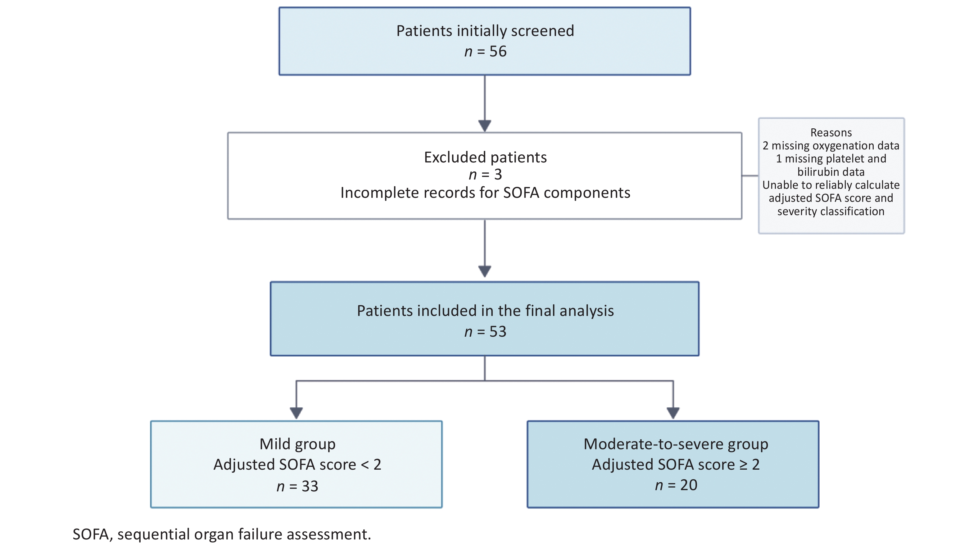
A total of 56 patients with bee sting injuries, who presented to the Emergency Department of the First People’s Hospital of Wu’an between August 2021 and September 2025, were consecutively screened. After three patients with missing data required for SOFA score calculation, 53 patients were included in the final analysis. Based on their clinical manifestations early after admission and bee sting-adjusted SOFA scores, 33 patients were classified into the mild group (adjusted SOFA score < 2) and 20 into the moderate-to-severe group (adjusted SOFA score ≥ 2) (Figure 1).
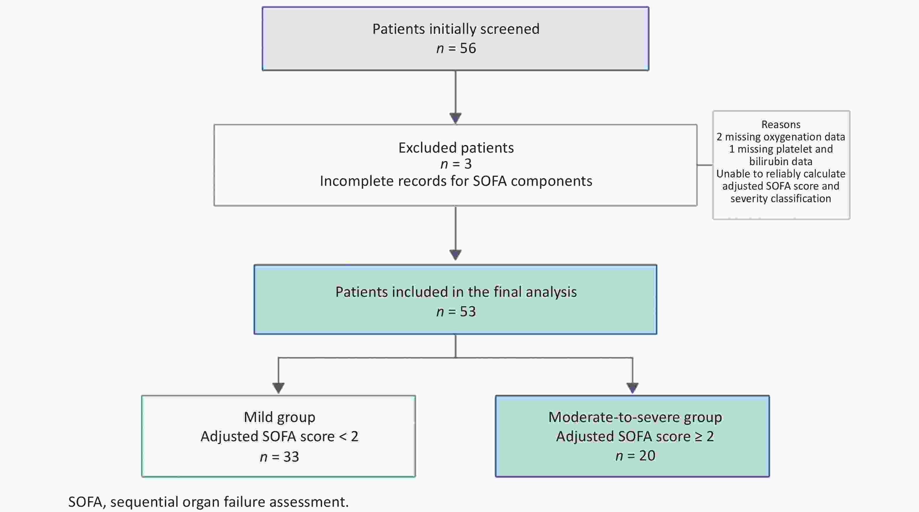
Figure 1. Flowchart of patient screening and group allocation. Three patients were excluded primarily because of incomplete documentation of variables required for SOFA score calculation. Two patients had missing oxygenation parameters, and one patient had missing platelet count and bilirubin data; therefore, reliable calculation of the adjusted SOFA score and severity classification could not be completed. SOFA, Sequential Organ Failure Assessment.
Sex distribution, age, heart rate, respiratory rate, and BMI did not differ significantly between the groups (all P > 0.05). No significant between-group differences were observed in lymphocyte count, hemoglobin level, platelet count, or levels of alanine aminotransferase (ALT), aspartate aminotransferase (AST), serum potassium, sodium, and calcium (all P > 0.05).
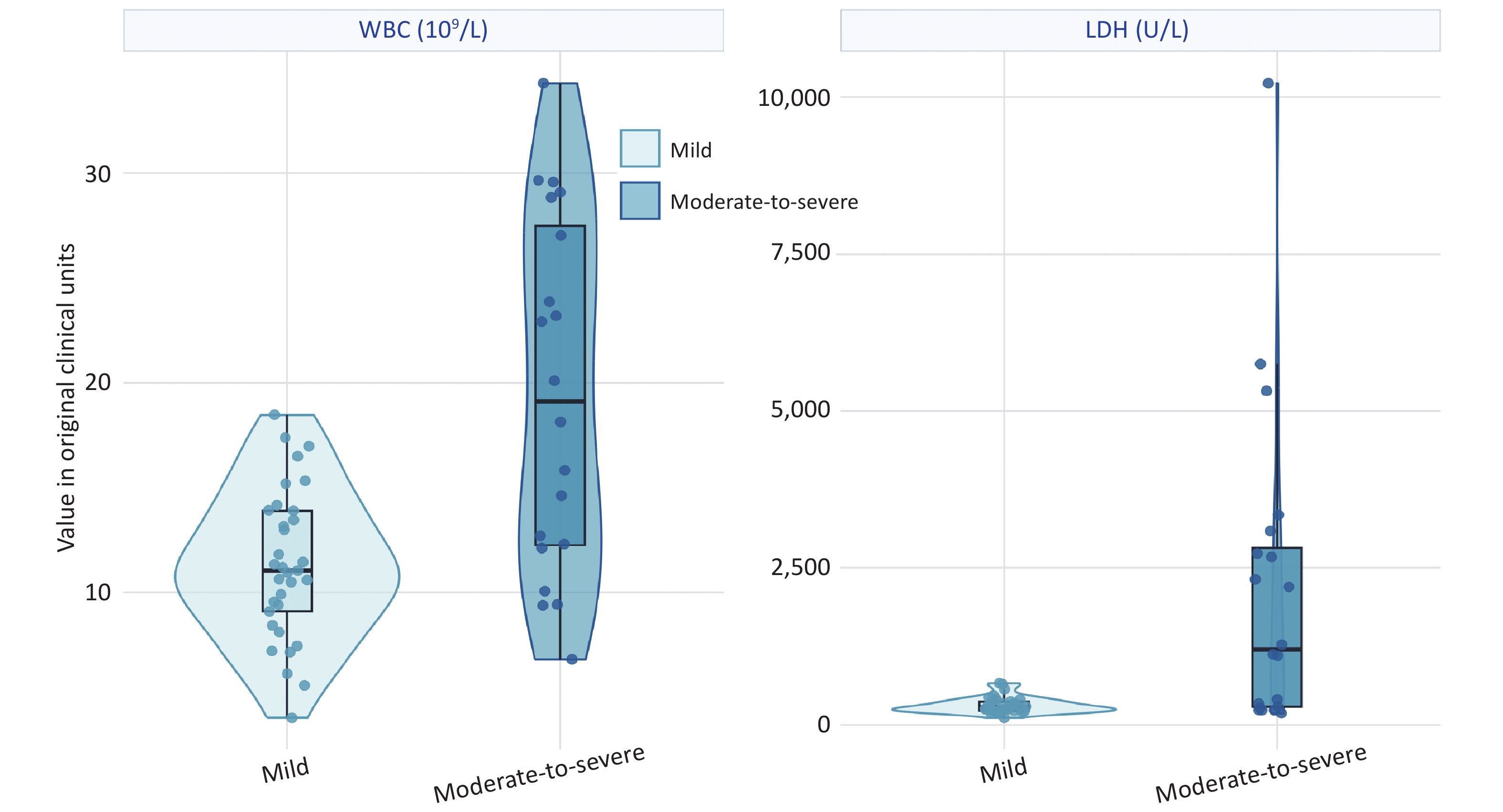
WBC and neutrophil counts were significantly higher in the moderate-to-severe group than in the mild group (WBC: 19.49×10⁹ ± 8.43×10⁹/L vs. 11.30×10⁹ ± 3.57×10⁹/L, P < 0.001; neutrophil count: 15.78×10⁹ ± 7.86×10⁹/L vs. 9.61×10⁹ ± 3.39×10⁹/L, P = 0.003). The moderate-to-severe group also had a significantly lower albumin level (39.49 ± 4.78 g/L vs. 42.66 ± 4.45 g/L, P = 0.021) and significantly higher BUN (8.43 vs. 5.87 mmol/L, P = 0.002), Cr (100.00 vs. 66.00 μmol/L, P = 0.001), and LDH levels (1,199.50 vs. 284.00 U/L, P < 0.001) (Table 1).
Characteristics Mild group
(SOFA < 2, n = 33)Moderate-to-severe group
(SOFA ≥ 2, n = 20)P value Sex (male/female) 11/22 11/9 0.121 Age (years) 65.00 (59.00, 69.00) 66.00 (60.75,73.25) 0.354 Adjusted SOFA score 0.00 (0.00, 1.00) 3.00 (2.00, 5.00) < 0.001 MAP (mmHg) 93.94 ± 16.16 77.68 ± 24.22 0.012 Heart rate (bpm) 83.94 ± 12.98 86.20 ± 12.16 0.526 Respiratory rate (breaths/min) 19.00 (18.00, 20.00) 20.00 (18.00, 21.00) 0.067 BMI (kg/m2) 23.75 ± 2.96 22.82 ± 2.26 0.206 WBC count (× 109/L) 11.30 ± 3.57 19.49 ± 8.43 < 0.001 Neutrophil count (× 109/L) 9.61 ± 3.39 15.78 ± 7.86 0.003 Lymphocyte count (× 109/L) 0.95 (0.72, 1.31) 1.65 (0.74, 3.47) 0.090 Hemoglobin (g/L) 135.85 ± 16.84 138.05 ± 24.50 0.726 Platelet count (× 109/L) 212.79 ± 46.82 206.60 ± 80.54 0.757 Albumin (g/L) 42.66 ± 4.45 39.49 ± 4.78 0.021 ALT (U/L) 26.20 (17.50, 37.30) 45.75 (18.80, 495.95) 0.077 AST (U/L) 35.60 (24.70, 53.00) 116.90 (24.35, 855.90) 0.091 BUN (mmol/L) 5.87 (4.96, 6.80) 8.43 (6.37, 16.86) 0.002 Cr (µmol/L) 66.00 (58.00, 82.00) 100.00 (65.75, 271.00) 0.001 LDH (U/L) 284.00 (224.00, 369.00) 1,199.50 (283.50, 2817.00) < 0.001 K+ (mmol/L) 4.02 (3.80, 4.24) 4.15 (3.70, 4.71) 0.430 Na+ (mmol/L) 142.40 (141.10, 144.60) 140.95 (134.75, 143.53) 0.160 Ca2+ (mmol/L) 2.26 (2.17, 2.38) 2.16 (2.07, 2.30) 0.132 Note. Normally distributed continuous variables are presented as the mean ± standard deviation, non-normally distributed continuous variables as the median (interquartile range) [M (P25, P75)], and categorical variables as the number of patients. Statistical significance was set at P < 0.05. SOFA, Sequential Organ Failure Assessment; MAP, mean arterial pressure; BMI, body mass index; WBC, white blood cell; ALT, alanine aminotransferase; AST, aspartate aminotransferase; BUN, blood urea nitrogen; Cr, creatinine; LDH, lactate dehydrogenase. Table 1. Comparison of baseline clinical and laboratory characteristics between patients with mild and moderate-to-severe bee sting injuries
Three deaths occurred in the moderate-to-severe group. The overall mortality rate was 5.7% (3/53), and the mortality rate in the moderate-to-severe group was 15.0% (3/20). All three deaths were attributed to multiple organ failure secondary to bee sting injuries. The WBC count and LDH levels at admission in the three patients who died were 29.58 × 10⁹/L, 20.09 × 10⁹/L, 34.29 × 10⁹L and 3,087 U/L, 2,675 U/L, 5,750 U/L, respectively.
-
Because WBC count, LDH levels, and other biomarkers showed skewed distributions, extreme values, and differences in the measurement scale, they were natural log-transformed and standardized using z-scores before analysis. Univariable logistic regression analysis showed that respiratory rate, mean arterial pressure (MAP), albumin, Zln(WBC), Zln(neutrophil count), Zln(LDH), Zln(BUN), and Zln(Cr) were significantly associated with the odds of moderate-to-severe bee sting injury (all P < 0.05) (Table 2).
Candidate variable Univariable OR (95% CI) AUC P value Age 0.997 (0.951–1.045) 0.577 0.894 Male sex 2.444 (0.782–7.644) 0.608 0.124 BMI 0.879 (0.711–1.086) 0.599 0.232 Heart rate 1.015 (0.970–1.061) 0.555 0.524 Respiratory rate 1.358 (1.005–1.833) 0.650 0.046 MAP 0.958 (0.928–0.99) 0.722 0.010 Albumin 0.861 (0.756–0.981) 0.692 0.024 Zln(WBC) 4.287 (1.804–10.185) 0.780 < 0.001 Zln(Neutrophil) 2.634 (1.256–5.524) 0.745 0.010 Zln(LDH) 6.286 (2.114–18.690) 0.774 < 0.001 Zln(BUN) 4.464 (1.555–12.818) 0.753 0.005 Zln(Cr) 3.838 (1.450–10.157) 0.764 0.007 Zln(NLR) 0.890 (0.507–1.561) 0.541 0.684 Note. Moderate-to-severe bee sting injury was used as the dependent variable, with the mild group coded as 0 and the moderate-to-severe group coded 1. Age, BMI, heart rate, respiratory rate, MAP, and albumin levels were entered into univariate logistic regression models in their original clinical units. Because WBC, LDH, neutrophil count, BUN, Cr, and the NLR were right-skewed, they were logarithmically transformed and subsequently standardized. OR represents the change in the odds of moderate-to-severe bee sting injury associated with a 1-standard-deviation increase in the corresponding Zln-transformed variable. AUC represents the apparent area under the curve obtained from the univariate ROC analysis. Statistical significance was set at P < 0.05. MAP, mean arterial pressure; BMI, body mass index; WBC, white blood cell; ALT, alanine aminotransferase; AST, aspartate aminotransferase; BUN, blood urea nitrogen; Cr, creatinine; LDH, lactate dehydrogenase; AUC, area under the curve; NLR, neutrophil-to-lymphocyte ratio; CI, confidence interval; OR, odds ratio; ROC, reciever operating characteristic. Table 2. Univariable Logistic regression analysis of the associations between candidate variables and moderate-to-severe bee sting injury
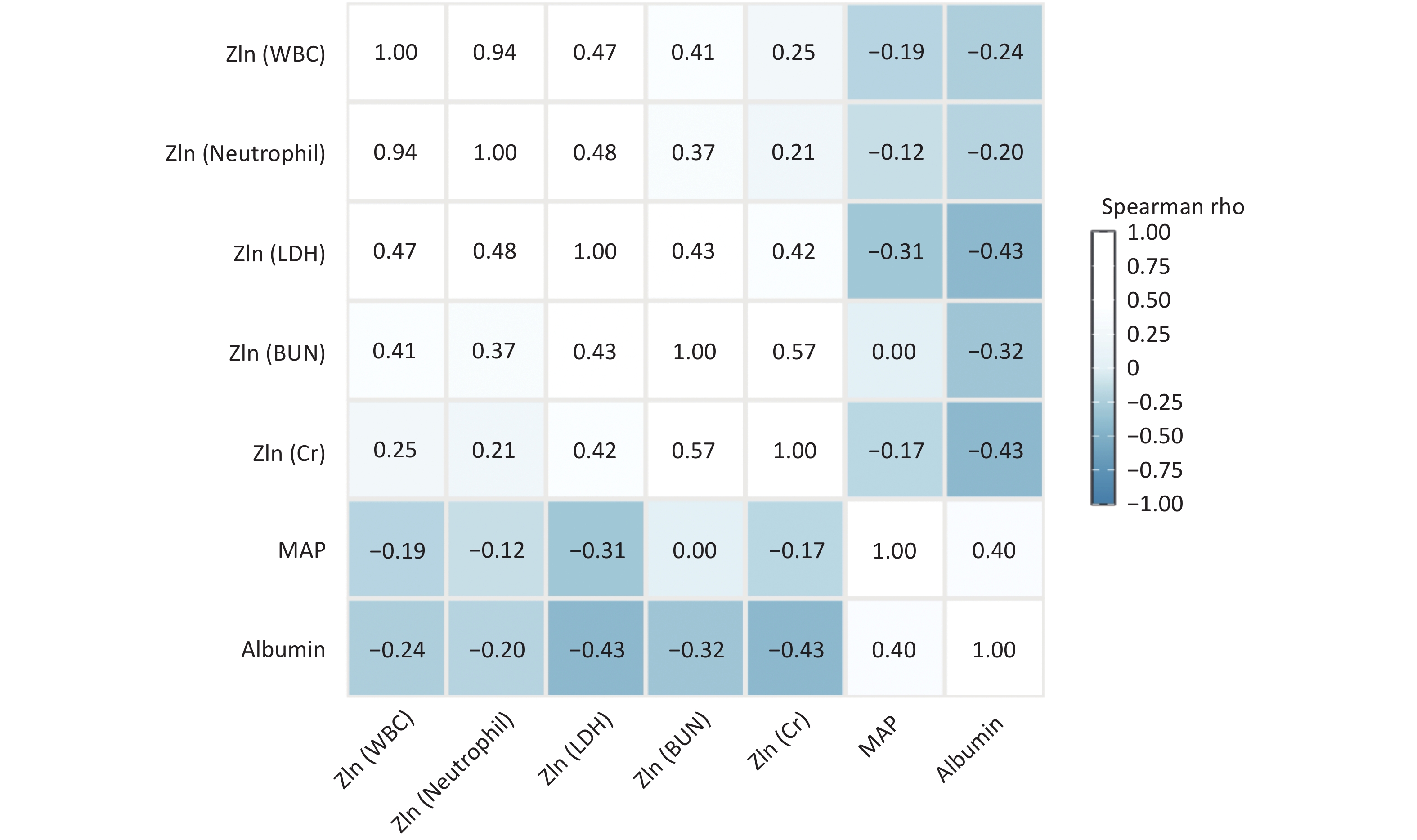
Zln(WBC) and Zln(neutrophil count) were strongly correlated (Spearman’s r = 0.938, P < 0.001). When both variables were included in the same model, the variance inflation factor (VIF) was 5.91 each. Collinearity diagnostics were performed for a candidate variable set comprising the MAP, albumin, Zln(WBC), Zln(neutrophil count), Zln(LDH), Zln(BUN), and Zln(Cr) levels. In this analysis, the VIFs for Zln(WBC) and Zln(neutrophil count) were 7.29 and 7.07, respectively, indicating substantial multicollinearity. The WBC count was selected as the representative marker of systemic inflammatory burden because it is routinely measured and readily available in emergency settings; therefore, the neutrophil count was not included concurrently in the primary model.
BUN, Cr, and MAP reflect renal and circulatory functions and partially overlap with the information used for the SOFA-based severity classification. These variables were excluded from the primary model to reduce the potential incorporation bias. Although albumin level and respiratory rate were statistically significant in the univariable analysis, they were not included in the primary model because the limited number of moderate-to-severe events restricted the available model degrees of freedom. Exploratory sensitivity analyses incorporating relevant clinical variables were performed to assess the robustness of the findings.
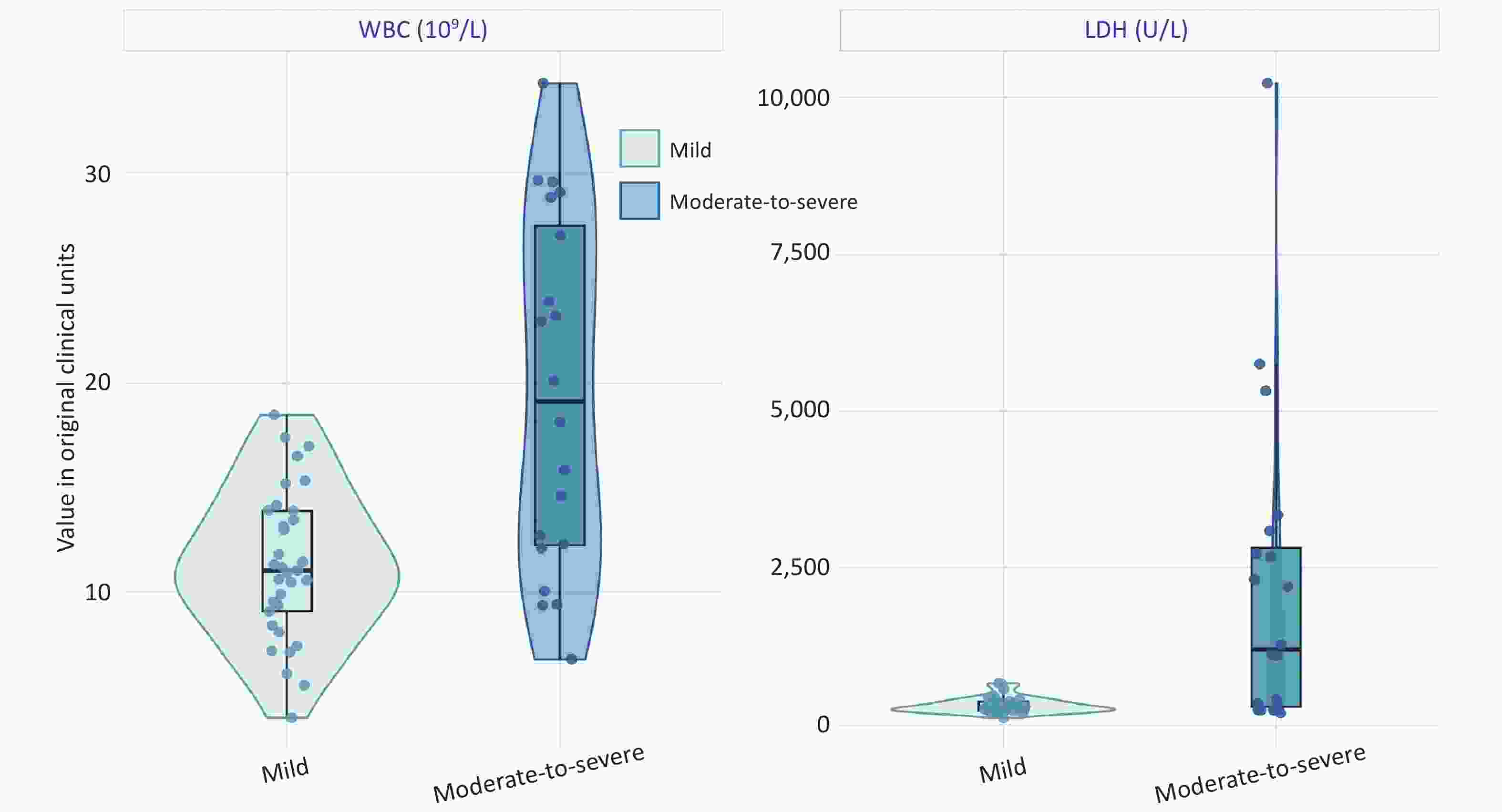
Figure 2. Distribution of WBC and LDH in the mild and moderate-to-severe groups. WBC, white blood cell count; LDH, lactate dehydrogenase.
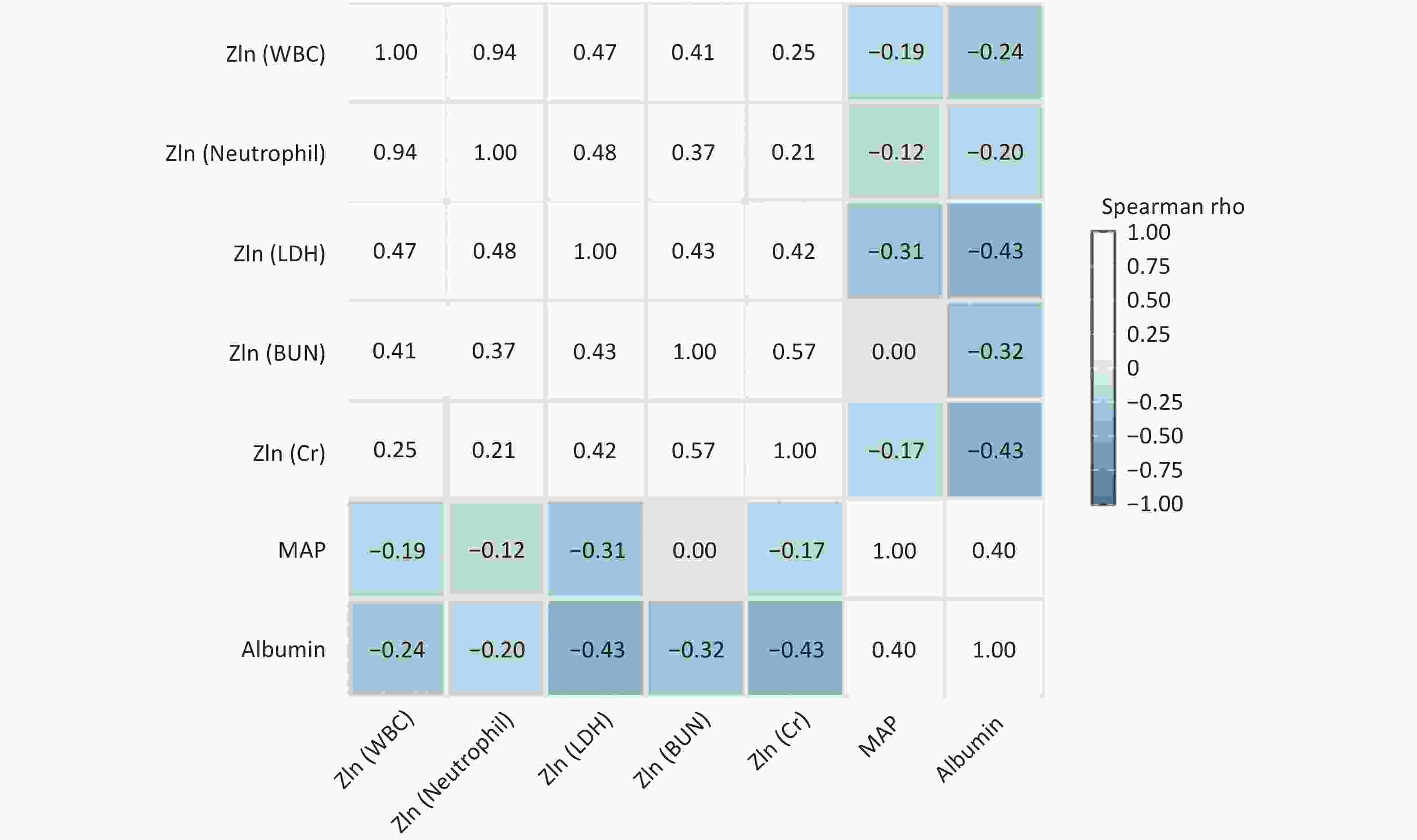
Figure 3. Spearman correlation matrix of the main candidate variables. The correlation matrix presents the Spearman correlation coefficients among the main candidate variables. WBC and neutrophil count were strongly positively correlated, suggesting that their simultaneous inclusion in the logistic regression model could result in multicollinearity. WBC, white blood cell; LDH, lactate dehydrogenase; BUN, blood urea nitrogen; Cr, creatinine; MAP, mean arterial pressure.
-
Given the limited number of outcome events (n = 20), the primary multivariable logistic regression model was restricted to two core predictors, Zln(WBC) and Zln(LDH), in accordance with the EPV principle, to reduce the risk of overfitting. In this exploratory two-predictor model, both Zln(WBC) (OR = 2.778, 95% confidence interval [CI]: 1.057–7.301, P = 0.038) and Zln(LDH) (OR = 4.853, 95% CI: 1.545–15.240, P = 0.007) were associated with moderate-to-severe bee sting injury early after admission (Table 3).
Variable β SE OR (95% CI) P value Zln(WBC), per 1 SD 1.022 0.493 2.778 (1.057–7.301) 0.038 Zln(LDH), per 1 SD 1.580 0.584 4.853 (1.545–15.240) 0.007 Note. WBC and LDH were natural logarithmically transformed and standardized per 1-standard-deviation increase. OR represents the change in the odds of moderate-to-severe bee sting injury associated with a 1-standard-deviation increase in the corresponding variable. The equation for the combined model was: logit(P) = −0.492 + 1.022 × Zln(WBC) + 1.580 × Zln(LDH), Zln(WBC) = [ln(WBC) − 2.558]/0.467, and Zln(LDH) = [ln(LDH) − 6.170]/1.056. The model was developed to evaluate the association between two readily available emergency laboratory indicators and moderate-to-severe injury early after admission. Zln(X) denotes a variable that was natural logarithmically transformed and standardized per 1-standard-deviation increase. WBC, white blood cell; LDH, lactate dehydrogenase; CI, confidence interval; OR, odds ratio; SE, standard error; SD, standard deviation. Table 3. Primary two-predictor Logistic regression model using WBC and LDH to identify moderate-to-severe bee sting injury
In the sensitivity analyses, the effect estimate for Zln(LDH) remained stable and statistically significant across models after adjusting for age and sex, or the addition of one physiological or laboratory variable at a time, including respiratory rate, MAP, albumin, Zln(BUN), or Zln(Cr). The effect estimates for Zln(WBC) remained directionally consistent, although the statistical significance was borderline in some models because of wider CIs. In the model where Zln(neutrophil count) replaced Zln(WBC), the AUC was 0.802, which was lower than that of the primary model, and Zln(neutrophil count) was not statistically significant. Given the limited sample size, these extended models were considered as exploratory sensitivity analyses and could not be substituted for external validation (Table 4).
Model AUC Zln(WBC),
OR (95% CI); PZln(Neutrophil),
OR (95% CI); PZln(LDH),
OR (95% CI); PAdditional covariate
OR (95% CI); PPrimary model
(Zln(WBC)+Zln(LDH))0.827 2.778 (1.057–7.301);
P = 0.038− 4.853 (1.545–15.240);
P = 0.007− + Age + sex 0.879 3.285 (1.000–10.791);
P = 0.050− 7.027 (1.705–28.958);
P = 0.007Age: 0.995 (0.930–1.065);
P = 0.886;
Male sex: 7.74 (1.178–50.871);
P = 0.033+ Respiratory rate 0.897 3.071 (1.101–8.566);
P = 0.032− 7.047 (1.729–28.723);
P = 0.008Respiratory rate: 1.563
(0.983–2.485);
P = 0.059+ MAP 0.848 2.718 (0.986–7.497);
P = 0.053− 3.870 (1.181–12.682);
P = 0.025MAP: 0.974 (0.934–1.016);
P = 0.222+ Albumin 0.827 2.789 (1.057–7.359);
P = 0.038− 4.759 (1.373–16.496);
P = 0.014Albumin: 0.992 (0.811–1.213);
P = 0.938+ Zln(BUN) 0.856 2.551 (0.937–6.946);
P = 0.067− 4.739 (1.366–16.435);
P = 0.014Zln(BUN): 2.823(0.567–14.063);
P = 0.205+ Zln(Cr) 0.858 2.887 (1.024–8.144);
P = 0.045− 4.479 (1.321–15.188);
P = 0.016Zln(Cr): 3.412(0.776–15.012);
P = 0.104Neutrophil
replacing WBC0.802 − 1.425 (0.598–3.395);
P = 0.4245.548 (1.822–16.897);
P = 0.003− Note. Sensitivity analyses were conducted to examine the effects of potential confounders or alternative inflammatory markers on estimates obtained from the primary model. The “Primary model” included only Zln(WBC) and Zln(LDH). “+ Age + sex,” “+ Respiratory rate,” “+ MAP,” “+ Albumin,” “+ Zln(BUN),” and “+ Zln(Cr)” indicate models in which the corresponding covariate or covariates were added to the primary model. “Neutrophil replacing WBC” indicates that Zln(neutrophil count) was entered into the model in place of Zln(WBC). AUC represents the apparent AUC of each model. “-” indicates that the variable was not included in the corresponding model. Zln(X) denotes a variable that was natural logarithmically transformed and standardized per 1-standard-deviation increase. WBC, white blood cell; LDH, lactate dehydrogenase; MAP, mean arterial pressure; BUN, blood urea nitrogen; Cr, creatinine; AUC, area under the curve; CI, confidence interval; OR, odds ratio. Table 4. Exploratory sensitivity analyses of the primary WBC and LDH model
-
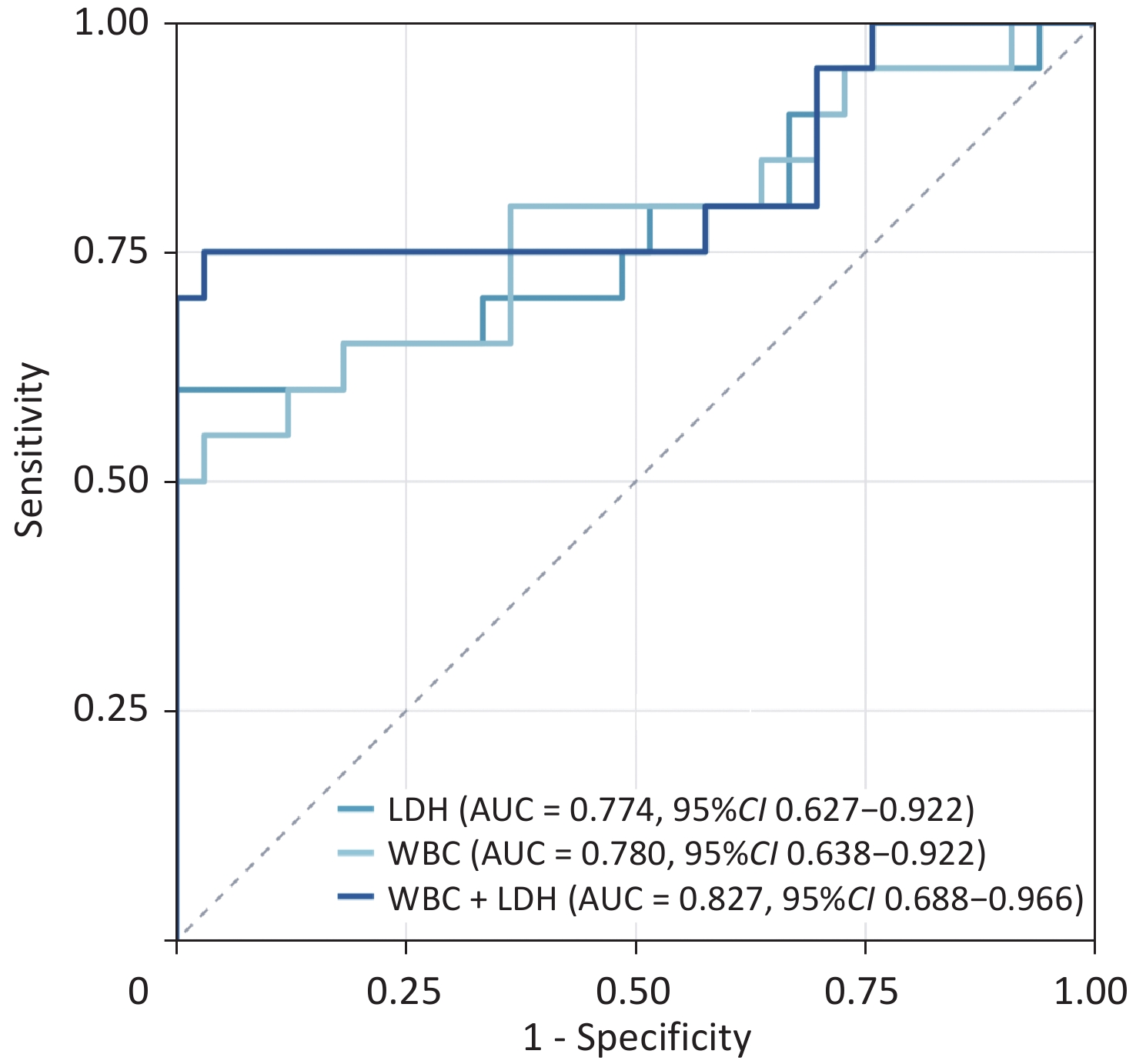
Model discrimination was assessed using the receiver operating characteristic (ROC) curves. In the single-marker analyses, the AUCs for WBC count and LDH levels were 0.780 (95% CI: 0.638–0.922) and 0.774 (95% CI: 0.627–0.922), respectively. LDH had an observed specificity of 100% at an optimal cutoff of >881 U/L. This finding may reflect a small sample size and should not be interpreted as evidence of absolute specificity in external populations. The ROC curve for the combined WBC+LDH model was generated from the predicted probabilities of the logistic regression model and yielded an AUC of 0.827 (95% CI: 0.688–0.966) (Table 5 and Figure 4).
Variable/model AUC (95% CI) Cutoff TP/FP/TN/FN Sensitivity
(%)Specificity
(%)PPV
(%)NPV
(%)DeLong P vs
combinedWBC 0.780 (0.638–0.922) > 17.76×10⁹/L 11/1/32/9 55.0 97.0 91.7 78.0 0.414 LDH 0.774 (0.627–0.922) > 881 U/L 12/0/33/8 60.0 100.0 100.0 80.5 0.178 WBC+LDH 0.827 (0.688–0.966) Predicted
probability ≥ 0.559515/1/32/5 75.0 97.0 93.8 86.5 Reference Note. Moderate-to-severe bee sting injury was defined as a positive outcome, and mild bee sting injury was as a negative outcome. The ROC analyses and optimal cutoff values for WBC and LDH levels alone were based on the original clinical measurements. ROC analysis of the combined WBC + LDH model was based on the predicted probabilities generated by a binary logistic regression model comprising Zln(WBC) and Zln(LDH). For WBC and LDH levels alone, values greater than the respective cutoff values were classified as positive. For the combined model, an observed predicted probability ≥ 0.5595 was classified as positive. All cutoff values were determined using the Youden index. The 95% CIs for the AUCs were calculated using the DeLong method and the AUCs were compared using the DeLong test. LDH showed a specificity of 100% at the optimal cutoff in this cohort because no false-positive cases occurred at this threshold. This finding was strongly influenced by the sample size and case composition, and should not be interpreted as indicating absolute specificity in external populations. WBC, white blood cell; LDH, lactate dehydrogenase; AUC, area under the curve; CI, confidence interval; ROC, receiver operating characteristic; PPV, positive predictive value; NPV, negative predictive value; TP, true positive; FP, false positive; TN, true negative; FN, false negative. Table 5. Discriminatory performance of WBC, LDH, and the combined WBC + LDH model for identifying moderate-to-severe bee sting injury
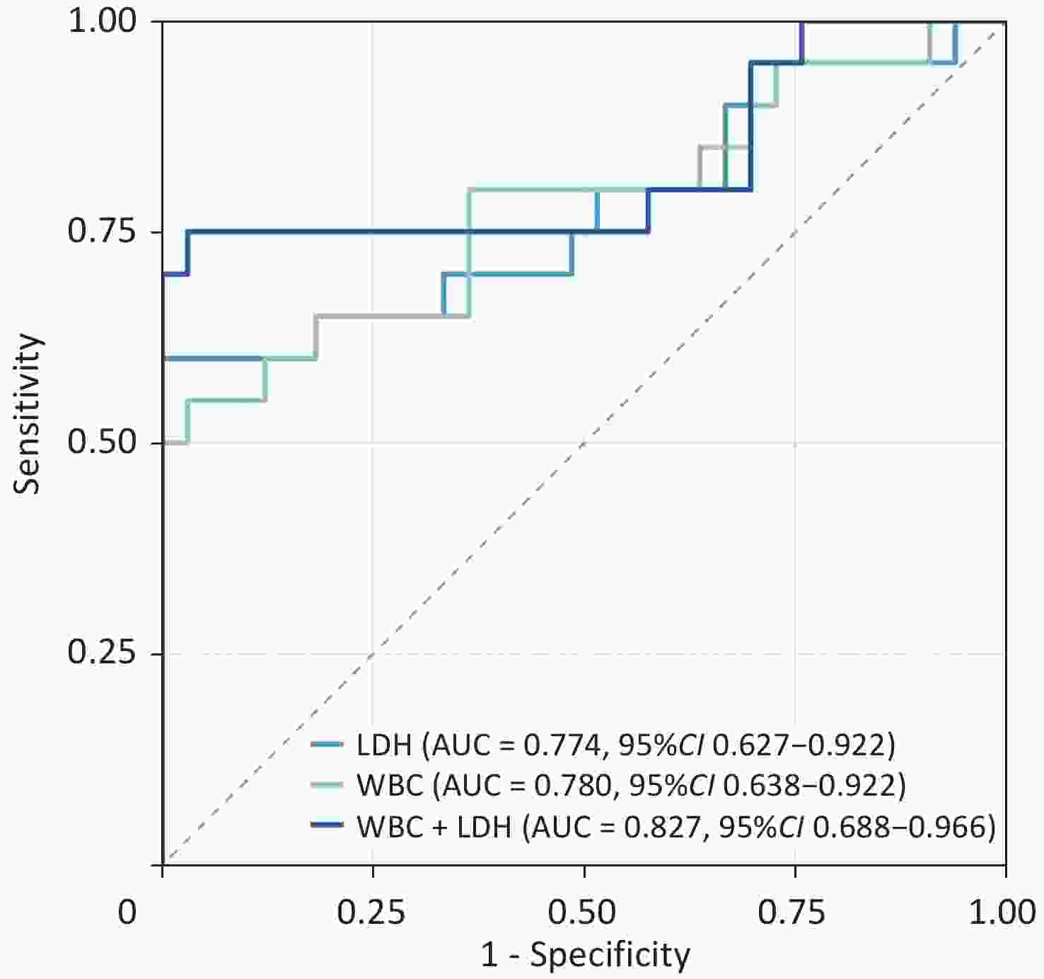
Figure 4. ROC curves of WBC, LDH, and the combined WBC + LDH model for identifying moderate-to-severe bee sting injuries. WBC: AUC = 0.780 (95% CI: 0.638–0.922); LDH: AUC = 0.774 (95% CI: 0.627–0.922); WBC + LDH: AUC = 0.827 (95% CI: 0.688–0.966). The ROC curves were used to evaluate the discriminatory performance of WBC, LDH, and the combined WBC + LDH model for moderate-to-severe bee sting injuries. ROC analyses of WBC and LDH were based on the original measured values, whereas the ROC curve for the combined WBC + LDH model was generated using the predicted probabilities from the logistic regression model. WBC, white blood cell; LDH, lactate dehydrogenase; AUC, area under the curve; CI, confidence interval; ROC, receiver operating characteristic.
Although the AUC of the combined model was numerically higher than that of WBC and LDH alone, the pairwise differences were not statistically significant according to the DeLong test (combined model vs. WBC, P = 0.414; combined model vs. LDH, P = 0.178). Thus, the present analysis did not provide evidence that the combined model was statistically superior to the individual markers. Bootstrap internal validation was performed using a fixed random seed and 1,000 resamples. The apparent AUC of the combined model was 0.827, the mean optimism was 0.013, and the optimism-corrected AUC was approximately 0.814, indicating only modest optimism in the apparent AUC.
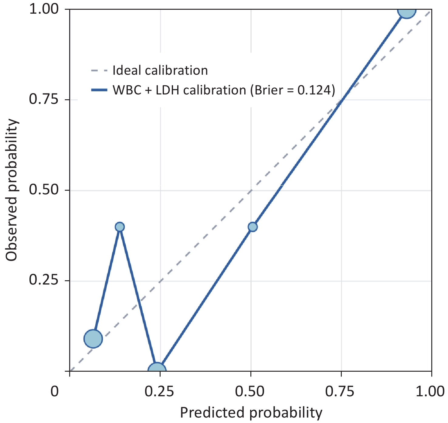
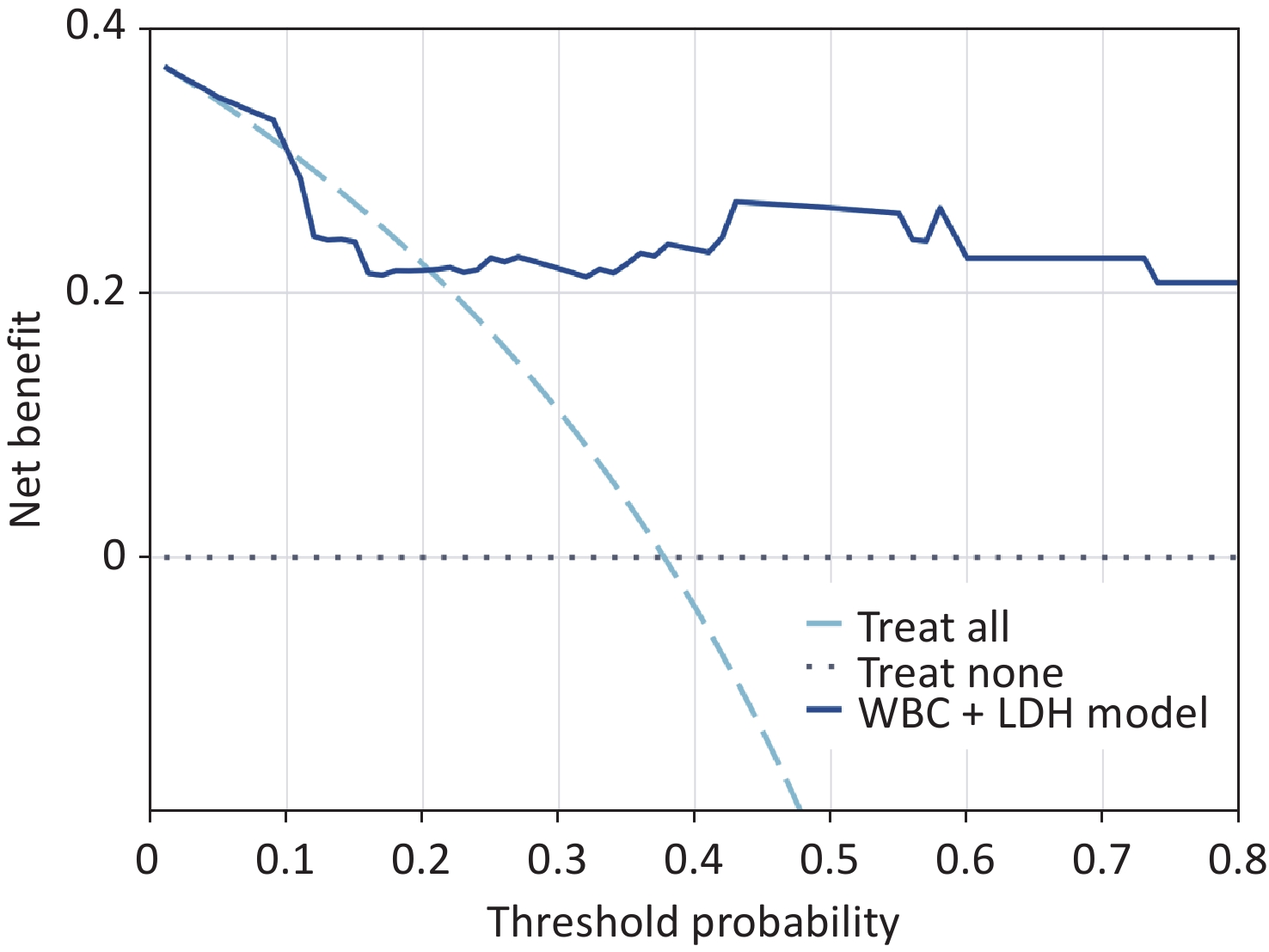
The Brier score was 0.124. The calibration curve showed considerable fluctuation in the low predicted-probability range, likely reflecting the limited sample size; however, no clear systematic deviation was observed (Figure 5). Decision curve analysis (DCA) showed that, across threshold probabilities of 0.20–0.80, the combined model provided greater net benefit than either of the treat-all strategy, in which all patients were classified as having moderate-to-severe injuries, or the treat-none strategy, in which no patients were classified as having moderate-to-severe injuries (Figure 6). These findings were exploratory and based solely on internal validation. The stability and generalizability of the model require further evaluation in an independent cohort.
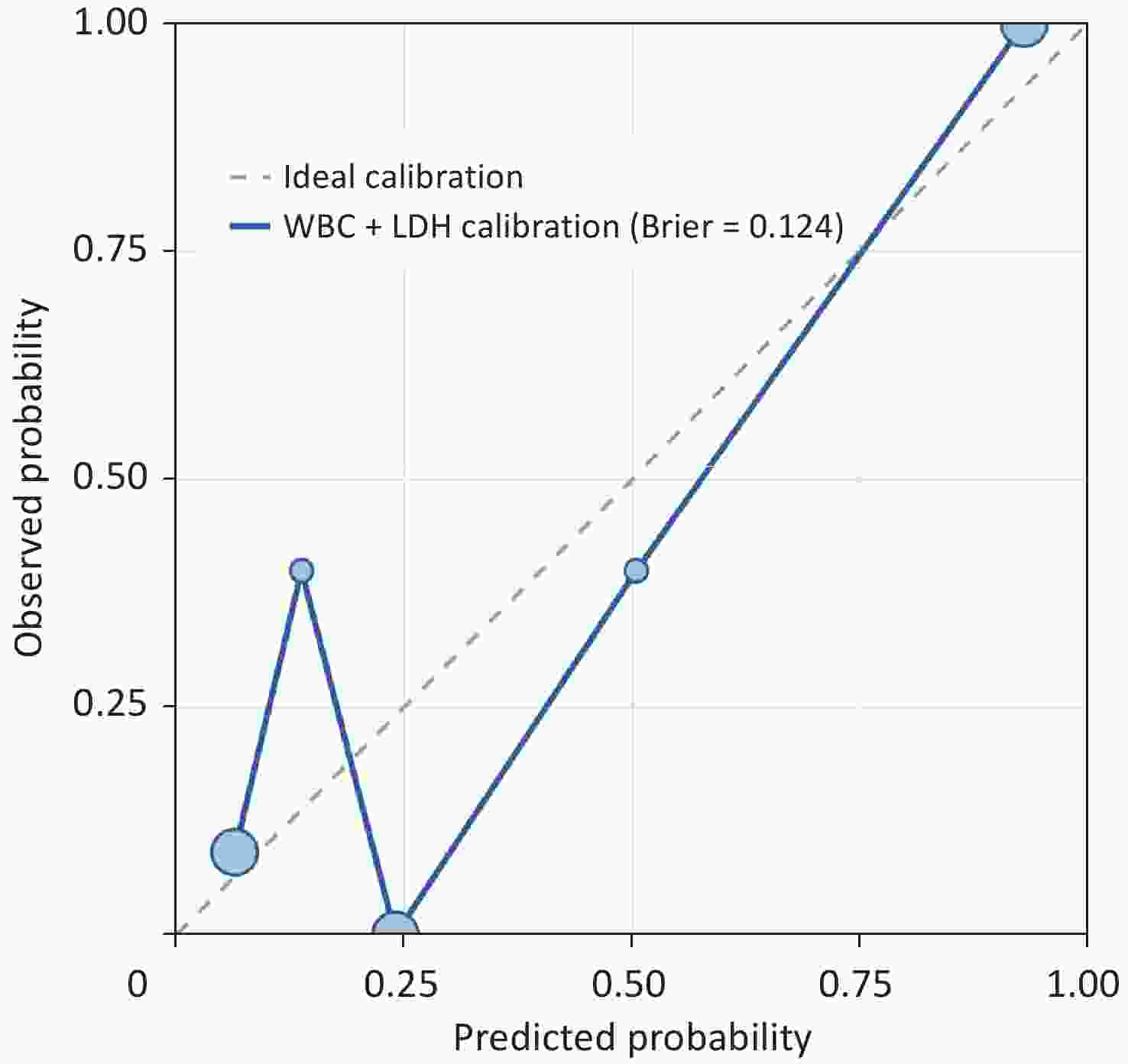
Figure 5. Calibration curve of the combined WBC + LDH model. The calibration curve was used to evaluate agreement between the predicted probabilities generated by the combined WBC + LDH model and the observed outcomes. The dashed line represents perfect calibration, and the solid line represents the apparent calibration curve in the study cohort. The Brier score of the combined model was 0.124, suggesting a degree of agreement between the predicted probabilities and the observed outcomes. WBC, white blood cell; LDH, lactate dehydrogenase.
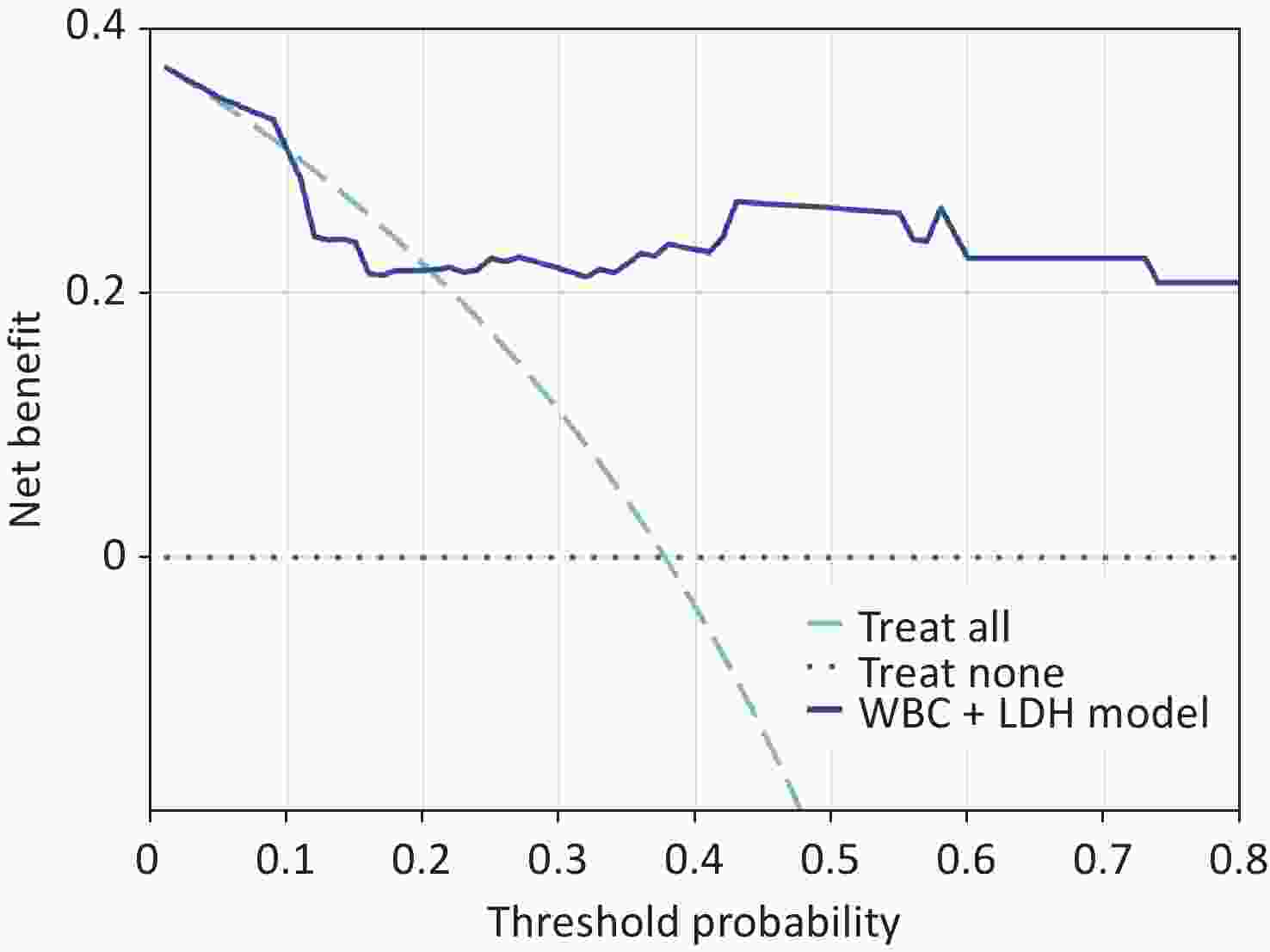
Figure 6. Decision curve analysis of the combined WBC + LDH model. Decision curve analysis was performed to evaluate the potential net clinical benefit of the combined WBC + LDH model across different threshold probabilities. The solid line represents the combined WBC + LDH model, the dashed line represents the treat-all strategy, in which all patients were managed as having moderate-to-severe injuries, and the dotted line represents the treat-none strategy, in which no patients were managed as having moderate-to-severe injuries. The apparent analysis suggested that, across threshold probabilities of approximately 0.20–0.80, the combined model might provide a greater net benefit than either of the treat-all or treat-none strategy. WBC, white blood cell; LDH, lactate dehydrogenase; AUC, area under the curve; CI, confidence interval; ROC, receiver operating characteristic.
-
This study included 53 patients with bee sting injuries who were stratified according to their clinical manifestations and bee sting-adjusted SOFA scores. The WBC count and LDH levels measured early after admission showed potential value in assessing injury severity. Compared to those of the mild group, the moderate-to-severe group had significantly lower MAP and higher levels of systemic inflammatory markers, including WBC and neutrophil counts, as well as markers of organ dysfunction and tissue injury, including LDH, Cr, and BUN. These findings are consistent with greater systemic inflammatory activation, hemodynamic compromise, and multiorgan involvement in patients with more severe injuries. The combined WBC and LDH model had an AUC of 0.827 and greater sensitivity than those of either marker alone. However, the AUC differences were not statistically significant according to the DeLong test, and bootstrap internal validation indicated modest optimism in model performance. Therefore, WBC count and LDH levels may serve as adjunctive markers for the early identification of moderate-to-severe bee sting injuries in the emergency department. The exploratory cutoffs identified in this study require external validation and should not replace SOFA-based assessment or comprehensive clinical judgment[13].
Bee stings are common medical emergencies caused by venom injection from Hymenoptera. Venom delivery is rapid and involves a relatively high load. A single sting may inject approximately 140–150 μg of venom, and nearly 90% of the venom sac contents may be delivered within 20 s. Because much of the venom may be injected within a short period, removal of the stinger after this initial phase may have limited effects on the amount of venom already delivered. The inflammatory and toxic responses to bee venom are influenced by several factors, including the number of stings, individual susceptibility, and baseline physiological status[2,4-6].
The pathophysiology of bee sting injury involves complex interactions between the biochemical properties of the venom and host immune response. Elevated WBC and LDH levels are not the direct causes of multiple organ dysfunction; rather, they reflect systemic inflammatory activation and the burden of tissue and cellular injury, respectively. Bee venom is a complex mixture of both biologically active and toxic compounds. Melittin, phospholipase A2, and other constituents may activate mast cells through both immunoglobulin E (IgE)-dependent and IgE-independent pathways[14,15], leading to the release of histamine, chemokines, and other inflammatory mediators[5,16-18]. These mediators can recruit and activate neutrophils and amplify local and systemic inflammatory responses[19-21], providing a biological rationale for using WBC as a marker of inflammatory burden[22].
Heparin released from mast cells may partially counteract the effects of melittin; however, the pore-forming activity of melittin can directly disrupt the membranes of erythrocytes, skeletal muscle cells, and hepatocytes, resulting in the release of intracellular LDH into circulation[17,18]. In this study, the median LDH level was 1,199.50 U/L in the moderate-to-severe group, compared with 284.00 U/L in the mild group. This difference may be attributable, at least in part, to the direct cytotoxic effects of bee venom and may also reflect metabolic disturbances and tissue hypoxia associated with systemic inflammatory activation[8,9,23,24]. The interactions between venom-induced inflammation and coagulation may further aggravate tissue injury. Previous studies suggested that melittin and phospholipase A2 activate the kinin cascade and promote bradykinin release, thus contributing to vasodilation and coagulation abnormalities. These mechanisms may partly explain the lower MAP observed in the moderate-to-severe group[2]. Myoglobin and other intracellular components released during rhabdomyolysis and hemolysis may exacerbate organ dysfunction through oxidative stress and contribute to persistent LDH elevation[19,21].
In our study, all three deaths occurred in the moderate-to-severe group, and all three patients had markedly elevated WBC and LDH levels at admission. One illustrative case involved a 71-year-old woman who presented within 24 h after a bee sting. Her admission electrocardiogram showed ST-segment elevation in leads V2–V6, accompanied by a marked elevation in troponin levels, raising the initial suspicion of acute myocardial infarction (Supplementary Figure S1A). Subsequent coronary angiography showed no corresponding coronary abnormalities; however, her condition rapidly progressed to MODS. Laboratory testing showed a WBC count of 34.29 × 10⁹/L and a LDH level of 5,750 U/L, accompanied by marked elevations in creatine kinase (CK; 8,287 U/L) and AST (3,570.7 U/L) and the development of coagulation dysfunction. The patient died within 24 h after admission (Supplementary Figure S1B). This case suggests that extreme elevations in WBC and LDH levels may be associated with extensive tissue and cellular injury and multiple organ failure. The underlying pathophysiology may involve the combined effects of direct venom cytotoxicity and an excessive immunometabolic response, although confirmation of the exact mechanism requires pathological or autopsy evidence. All patients who died had underlying diseases, including asthma, hypertension, or diabetes mellitus. Larger studies are needed to evaluate the relationships of comorbidities and physiological reserve with severe outcomes after bee stings. When marked elevations in WBC count and LDH levels are accompanied by hemodynamic instability, renal dysfunction, myocardial injury, or evidence of rhabdomyolysis early after admission, emergency physicians should consider the possibility of extensive tissue injury and multiorgan involvement and promptly undertake comprehensive organ function assessment and intensified monitoring.
Previous studies have reported associations between WBC count and LDH levels, bee sting-related organ injury, and adverse outcomes[6]. However, the proposed LDH thresholds varied according to the evaluated clinical outcomes. Wang et al. reported that an admission LDH level > 463.5 U/L was associated with an increased risk of acute kidney injury[25], whereas Zhang et al. used LDH ≥ 2,200 U/L as a cutoff for a high risk of death[26]. Accordingly, the WBC > 17.76 × 10⁹/L and LDH > 881 U/L thresholds identified in this study should be regarded as exploratory cutoffs derived from the present cohort. However, their clinical applicability requires validation in independent cohorts. LDH had an observed specificity of 100% at the exploratory optimal cutoff because no false-positive cases occurred above this threshold in the present cohort. This estimate was strongly influenced by the small sample size and case composition and may be lower in larger external populations. However, this finding should not be interpreted as evidence of stability or absolute specificity.
WBC and LDH testing are rapid, inexpensive, and widely available, making these markers potentially useful in emergency clinical assessments. Marked elevations in WBC count and LDH levels may serve as adjunctive warning signs of moderate-to-severe bee injury and warrant intensified monitoring. Decisions regarding management in the resuscitation area, multidisciplinary consultation, or urgent transfer to a higher-level hospital should be based on the patient’s hemodynamic status, the severity of the allergic reaction, and the extent of organ involvement. This study assessed injury severity early after admission, rather than the risk of subsequent delayed deterioration. Therefore, the proposed cutoffs should not be used as the sole criteria for automatic admission to the intensive care unit or initiation of a specific treatment.
-
The principal limitations of this exploratory study were its single-center retrospective design and modest sample size, which may have introduced selection bias and limited the generalizability of the findings. Some potentially relevant sting-related factors and prehospital interventions were not consistently documented, and only laboratory measurements obtained early after admission were analyzed, precluding assessment of biomarker trajectories. Although bootstrap internal validation showed only modest optimism in model performance, external validation was not performed. Therefore, the proposed cutoffs should be interpreted as preliminary and confirmed in larger prospective multicenter cohorts.
-
Bee venom may cause multi-organ injury through direct cytotoxic effects and excessive inflammatory responses. WBC and LDH are readily available and inexpensive biomarkers of these pathophysiological processes and may serve as adjunctive markers for early severity stratification in patients with bee sting injuries. Their use in primary care settings and emergency departments may support the further assessment of organ function, intensified monitoring, and timely referral.
HTML
Study Design and Participants
Bee Sting-adjusted SOFA Score
Patient Classification
Data Collection and Study Variables
Statistical Analysis
Clinical Characteristics of the Two Groups
Univariable Analysis, Variable Selection, and Collinearity Diagnostics
Primary Logistic Regression Model and Sensitivity Analyses
Model Performance: Discrimination, Calibration, and Clinical Utility
Competing Interests The authors declare that they have no competing interests.
Ethics This retrospective study was approved by the Ethics Review Committee of the Wu'an First People's Hospital. Given the retrospective design and anonymized data analysis, the requirement for informed consent was waived by the Ethics Review Committee. This study was conducted in accordance with the Declaration of Helsinki and the relevant institutional regulations.
Authors’ Contributions Data analysis, interpretation, and manuscript drafting: Yiyang Liu and Xuebin Pei. Conceptualization and supervision: Yanqiang Jiang, Zhidong Fang, and Xinhua He. Laboratory analysis and quantification: Kun Zhang and Bianfang Liu. Drafting, reviewing, and editing: Junli Chen. All authors read and approved the final manuscript.
Data Sharing The supplementary materials will be available in www.besjournal.com.
&These authors contributed equally to this work.

 Quick Links
 DownLoad: