


-
Human adenoviruses (HAdVs) constitute a diverse family of double-stranded DNA viruses within the Adenoviridae family[1]. Currently, the Human Adenovirus Working Group officially categorizes them as species A to G, spanning more than 110 genotypes, based on their genomic, immunological, and structural properties[2]. Among them, HAdV-D exhibits the highest degree of genomic diversity[3]. HAdV-D, particularly HAdV-D8, HAdV-D37, and HAdV-D19, is primarily associated with ocular infections, particularly severe and highly contagious epidemic conjunctivitis (EKC)[3]. However, HAdV-D is increasingly implicated in acute respiratory infections (ARIs), broadening its clinical and public health significance[4]. This continuous intraspecies variation is driven primarily by homologous recombination[2-4]. Genomic surveillance indicates that recombination events frequently occur within the hypervariable regions of the major capsid genes, including the penton base, hexon, and fiber, as well as the immune-modulating E1 and E3 transcription units. These genetic exchanges can generate novel chimeric strains with altered pathogenic profiles and extended tissue tropism[4]. Recent epidemiological data strongly indicate that newly emergent recombinant HAdV-D strains are increasingly crossing historical tissue boundaries, occasionally triggering severe pediatric pneumonia[2]. The recently designated HAdV-D115 is an example of this evolutionary dynamic[2]. A retrospective review of pediatric ARIs in Beijing from 2015 to 2021 by Wang et al[2], identified HAdV-D115 as newly emergent in a child presenting with severe respiratory symptoms. As the precise correlation between specific recombination events and disease severity requires further elucidation, detailed molecular characterization of emerging strains is essential. This study reports a detailed clinical, genomic, and virological characterization of a severe HAdV-D115 infection in a pediatric patient, using a systematic bioinformatics approach to investigate the molecular basis of the genomic dynamics and clinical implications of this virus.
Diagnostic and molecular protocols were initiated after the admission of a child with atypical clinical manifestations, detected during routine virological surveillance of ARIs. A throat swab was obtained from a 4-year-old boy who had been urgently admitted to Beijing Children’s Hospital with severe respiratory disease. Initial diagnostic screening using a multiplex quantitative real-time polymerase chain reaction (qPCR) assay designed for common respiratory viruses confirmed the presence of HAdV nucleic acid. Following established surveillance laboratory protocols for HAdV epidemiology, initial attempts were made to genotype the virus directly from the clinical specimen using targeted PCR amplification and subsequent Sanger sequencing of the three major adenovirus capsid genes, namely the hexon, penton base, and fiber genes[5]. Standard laboratory diagnostic algorithms specify that when preliminary trigene genotyping reveals phylogenetic discordance among these major capsid genes, the isolate should be classified as a highly suspicious recombinant. To definitively ascertain the evolutionary standing of such variants, rule out transient coinfections, and isolate viable pathogen, viral isolation and full-length genome sequencing is performed on all highly suspicious recombinant isolates. The PCR-positive clinical specimen was inoculated onto confluent monolayers of A549 cells, a human alveolar basal epithelial cell line known to be highly permissive to a broad spectrum of human respiratory adenoviruses. Once the characteristic cytopathic effect (CPE) emerged, the culture was harvested and subjected to three consecutive rounds of rigorous plaque purification under an agarose overlay to ensure that the isolate represented a single, homogeneous, and truly clonal viral population. Viral genomic DNA was then extracted directly from the clarified cell lysate using the QIAamp MinElute Virus Kit (QIAGEN, Germany). A high-fidelity sequencing library was constructed using the NEBNext Ultra DNA Library Prep Kit (NEB, USA), and whole-genome sequencing was performed using the Illumina NovaSeq PE150 platform. High-quality clean data were assembled using SOAPdenovo (v2.04), SPAdes, and ABySS, and integrated using Contig Integrator for Sequence Assembly (CISA). Sequence alignment was performed using MAFFT v7.526. For comprehensive phylogenetic mapping, a comparative dataset was constructed using verified reference sequences encompassing all currently recognized HAdV genotypes, from type 1 through 116 (Supplementary Table S1), and whole-genome neighbor-joining (NJ) phylogenetic trees were generated in MEGA 12 using the Kimura 2-parameter model with 1,000 bootstrap replicates. To accurately delineate homologous recombination breakpoints without the analytical interference of highly chimeric circulating strains, SimPlot v3.5.1 analysis was restricted to the genetically stable prototype strains from HAdV serotypes 1 through 52, utilizing a standard 1,000 bp sliding window and a 200 bp step size (Supplementary Figure S1). In vitro biological replication kinetics were evaluated by infecting A549 cell monolayers (MOI 0.1) and quantifying the replication efficiency over 120 h using a 50% tissue culture infectious dose (TCID50) assay.
The patient's clinical presentation exhibited atypical features that differed from those of usual pediatric respiratory adenovirus infections. Prior to admission, the child had a persistent dry cough for 2 months and had developed a acute fever 5 days prior to admission, which had peaked at 40.0 °C. Physical examination revealed pharyngeal congestion, tonsillar hypertrophy, and cervical lymphadenopathy. Chest auscultation suggested extensive lower respiratory tract involvement, which was confirmed by a radiograph showing bilateral bronchopneumonia. Notably, pronounced extrapulmonary manifestations were observed. Abdominal imaging revealed hepatosplenomegaly with minimal ascites. Laboratory profiles showed mild anemia (hemoglobin 110 g/L) and leukocytosis (8.32 × 10⁹/L). A significant divergence in acute-phase reactants was observed: the C-reactive protein (CRP) level was within the normal physiological limit (< 8.0 mg/L), whereas the serum amyloid A (SAA) level was markedly elevated (66.4 mg/L). SAA is an acute-phase protein synthesized predominantly by the liver. In the context of viral infections, the finding of highly elevated SAA levels without CRP level elevation is frequently associated with systemic inflammatory responses and hepatic macrophage activation, distinguishing it from typical localized bacterial infections. The patient was treated with intravenous azithromycin, budesonide nebulization, and empirical ceftriaxone. Although the fever resolved and acute respiratory distress eased by Day 4 of admission, the hepatosplenomegaly and ascites persisted, delaying discharge. Endemic respiratory adenoviruses typically lack the tropism required to induce pronounced hepatosplenomegaly in immunocompetent children. This clinical presentation provided a rationale for investigating whether the isolated viral strain possessed specific genomic alterations that facilitated systemic dissemination.
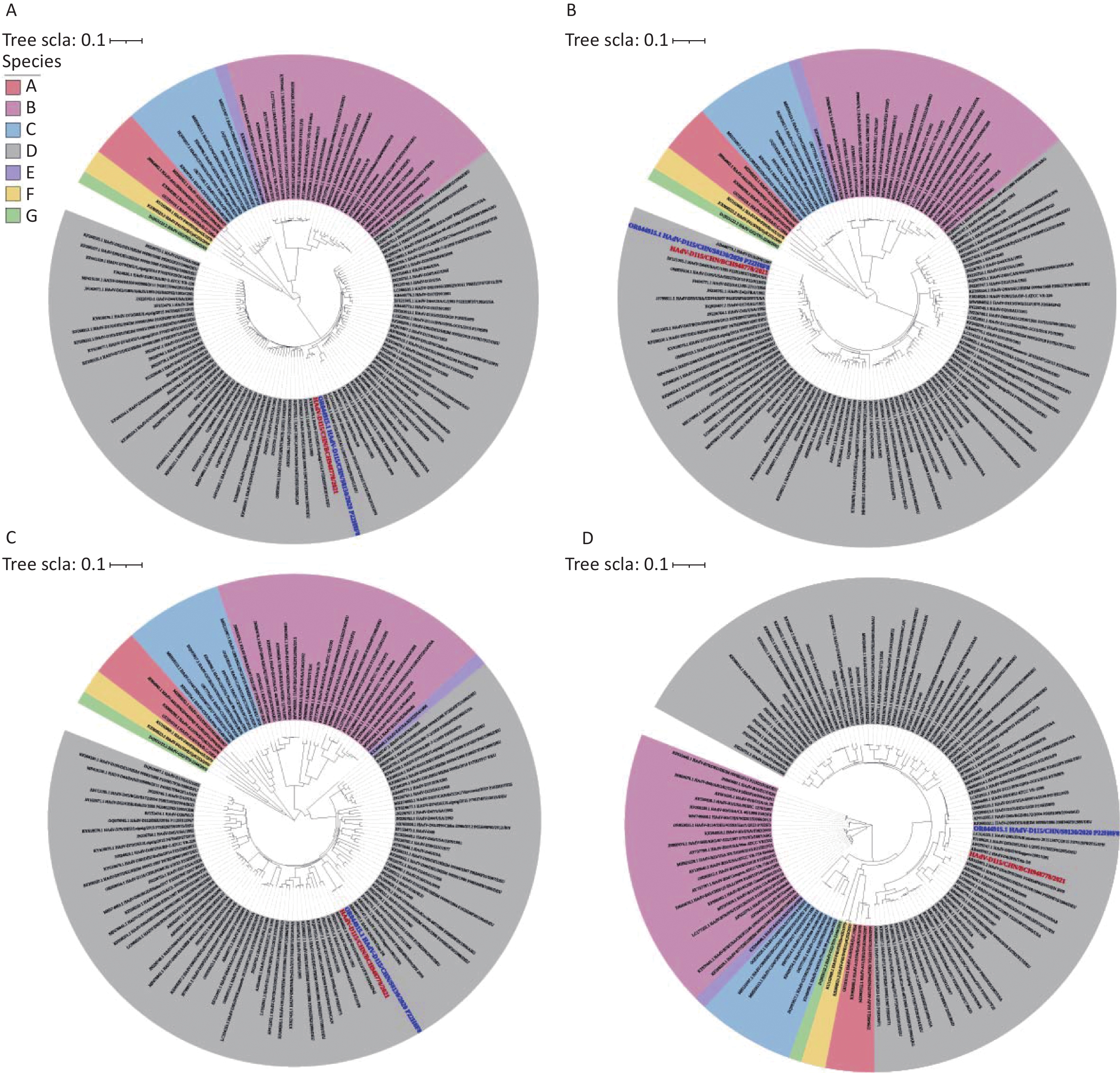
The initial trigene screening provided the initial molecular evidence of the atypical nature of the pathogen. Phylogenetic analysis demonstrated that the hexon (Figure 1A), penton base (Figure 1B), and fiber (Figure 1C) genes each clustered into distinct HAdV-D genotypic lineages, indicating multi-parental recombination. Subsequent whole-genome assembly and characterization of the plaque-purified isolate, designated CHN/BCH948778/2021, confirmed a genome length of 35,141 bp and a GC content of 56.14%. A whole-genome BLASTn query confirmed its identity as HAdV-D115, demonstrating 99.93% sequence identity with CHN/BJ-S8130/2020 (GenBank Accession: OR044915), which was officially designated as a strain by the HAdV Working Group in 2024[2].
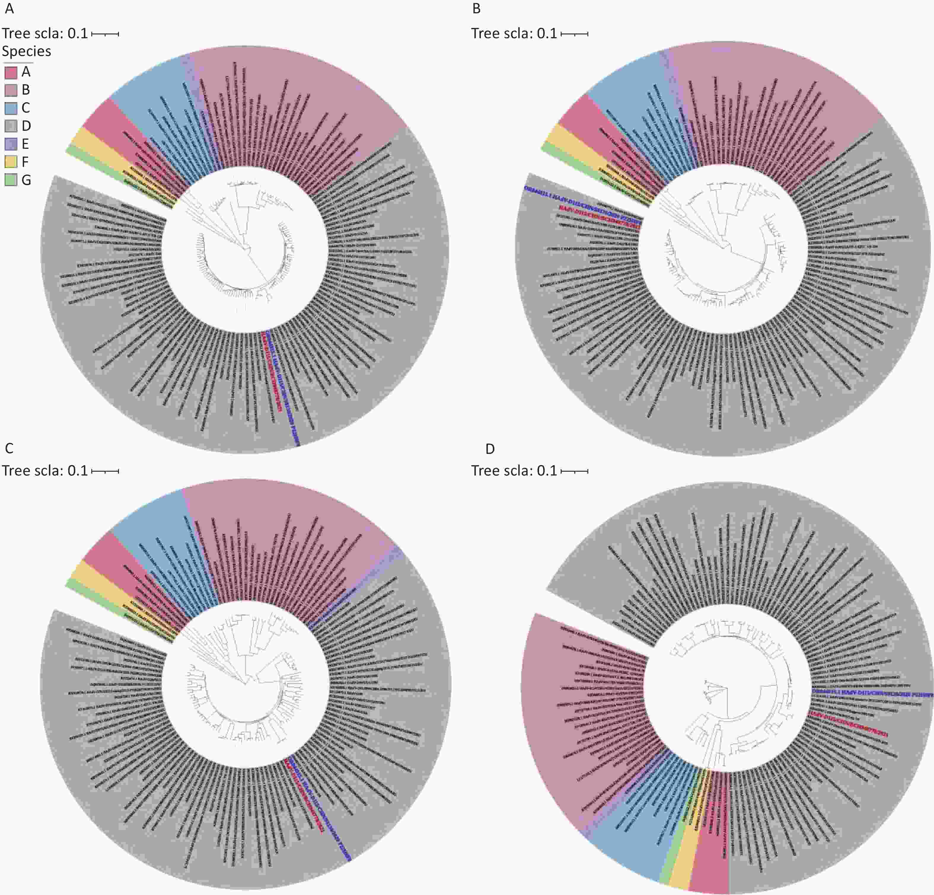
Figure 1. Neighbor-joining (NJ) phylogenetic trees based on the nucleotide sequences of the hexon (A), penton base (B) fiber (C), and whole genome sequence (D) of the isolate CHN/BCH948778/2021 belonging to species D and those from GenBank using the Neighbor-Joining method of MEGA 12 with 1,000 bootstrap replicates. The sequence from the study is shown in red, and the reference strain of HAdV-D115 is shown in blue. HadV, human adenovirus.
Despite the high nucleotide sequence homology, sequence alignment revealed a specific functional divergence within the early transcription unit 3 (E3)[6]. The E3 region encodes the membrane proteins involved in modulating the host immune response[6]. A primary component is the Receptor Internalization and Degradation (RID) complex, a heterodimer composed of the RIDα (E3 10.4 kDa) and RIDβ proteins[6]. The RID complex operates as a multitarget sorting machinery that clears pro-apoptotic death receptors, most notably Fas (CD95) and TRAIL receptors, to prevent extrinsic apoptosis induced by cytotoxic T lymphocytes and natural killer cells, while concurrently downregulating the tyrosine kinase epidermal growth factor receptor (EGFR) to suppress pro-inflammatory signaling[6]. The previously sequenced 2020 reference strain (CHN/BJ-S8130/2020) contains a 9-nucleotide deletion within the E3 10.4 kDa gene that results in a truncated RIDα protein. Although complete knockouts of the RID genes prevent the complex from internalizing death receptors[6,7], the specific functional and structural impact of this particular 3-amino acid truncation has not been experimentally characterized. Because RIDα is a compact, 91-amino acid integral membrane protein, its function depends on precise alignment between the transmembrane domain (required for RIDβ heterodimerization) and the C-terminal sorting motif (for clathrin adaptor recruitment)[7]. Therefore, a 3-amino acid deletion could disrupt its topology and impair complex assembly. In contrast, the wild-type sequence in CHN/BCH948778/2021 suggests a fully functional RID complex. By delaying host cell apoptosis, this mechanism extends the primary replication window, and the resulting high local viral burden provides a biological basis for the systemic dissemination observed clinically.
The structural architecture and putative tissue tropism of the isolate were further delineated using comprehensive phylogenetic and recombination analyses. The whole-genome NJ tree (Figure 1D) revealed that CHN/BCH948778/2021 clusters within the HAdV-D115 lineage. SimPlot analysis confirmed a P22H8F8 recombination pattern (Supplementary Figure S1). The genomic segment encompassing the penton base originated from an HAdV-D22 lineage, the hexon gene originated from an HAdV-D8 lineage, and the fiber gene originated from an HAdV-D8 lineage, with the remaining backbone aligning with HAdV-D53. Integration of the HAdV-D8 hexon affects viral entry mechanics. Although many adenovirus species depend primarily on the fiber knob for cellular attachment, certain HAdV-D strains use a noncanonical entry mechanism[8]. Structural analyses, including high-resolution cryo-electron microscopy, have demonstrated that HAdV-D hexon proteins interact directlywith CD46, a complement regulatory receptor expressed almost universally across nucleated human cells, via their solvent-exposed hypervariable regions[8]. By incorporating the HAdV-D8 hexon, HAdV-D115 acquires an established binding affinity for CD46. Given the ubiquitous expression of CD46 on human nucleated cells, this interaction may facilitate viral dissemination from the primary site of infection[8]. This receptor profile explains the extrapulmonary involvement and hepatosplenomegaly observed clinically.
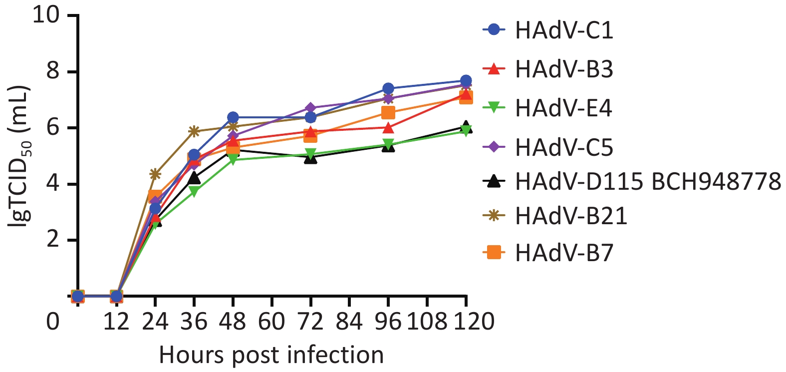
This CD46-mediated interaction likely functions in parallel with the receptor specificity of the chimeric fiber protein[9]. Unlike coxsackievirus and adenovirus receptor (CAR) which is used by species C respiratory adenoviruses, species D adenovirus fibers, such as those derived from HAdV-D8 and D37, exhibit an affinity for binding to sialic acid-containing glycans. Sialic acids are terminal carbohydrates abundantly expressed in various non-pulmonary tissues, including hepatocytes and mucosal endothelium[9]. Although sialic acid engagement by the parental HAdV-D8 fiber is canonically associated with strictly localized epidemic keratoconjunctivitis (EKC)[1], its novel integration with a CD46-interacting hexon creates a synergistic, dual-receptor entry mechanism. This unique receptor profile may circumvent traditional tissue restrictions, differentiating this recombinant from typical localized pathogens and contributing to its atypical systemic spread. This altered receptor usage also provides context for the in vitro viral growth kinetics. At 120 hours post-infection, the D115 isolate reached a maximum titer of 106.05 TCID50/mL, performing marginally better than HAdV-E4 but significantly lower than the respiratory prototype HAdV-C1 (107.69 TCID50/mL) (Figure 2). A549 is an immortalized human alveolar adenocarcinoma cell line. Although A549 cells express both CAR and CD46, this specific tumor line is intrinsically highly permissive to CAR-dependent species C adenoviruses[8], which typically exhibit rapid and highly efficient replication cycles in these cells. Species D adenoviruses generally exhibit slower replication kinetics and lower yields in standard A549 cultures than those of species C strains, regardless of their clinical virulence[10]. Therefore, the reduced in vitro growth of HAdV-D115 in A549 cells does not necessarily indicate diminished clinical virulence or a replication defect in vivo. Rather, it demonstrates the limitations of relying on a single immortalized cell line to model the in vivo replicative fitness of recombinant adenoviruses with altered, systemic receptor specificities. Further studies using primary human tissue cultures or in vivo models that accurately reflect human receptor distribution and immune dynamics, are required to quantify the true biological fitness of HAdV-D115.
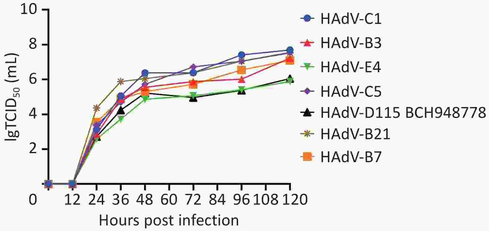
Figure 2. Growth curve of CHN/BCH948778/2021 (HAdV-D115) in A549 cells. The growth kinetics of HAdV strain CHN/BCH948778/2021 were assessed in A549 human lung epithelial cells. Cells were infected at a multiplicity of infection (MOI) of 1, and viral titers were measured at 0, 12, 24, 36, 48, 72, 96 and 120 h post-infection (hpi) using a 50% tissue culture infection dose (TCID50). The values shown are the mean viral titers (TCID50/mL) from three independent experiments. HadV, human adenovirus.
In summary, the clinical and virological characterization of this HAdV-D115 isolate highlights the impact of homologous recombination on the evolutionary trajectory of HAdVs. The severe bronchopneumonia and accompanying hepatosplenomegaly documented in this case were associated with a virus possessing an intact E3 RID complex, a CD46-binding hexon, and a sialic acid-targeting fiber. As the epidemiology of HAdV-D expands to include systemic pediatric infections, the potential for genetic recombination to alter viral tissue tropism must be recognized. Continued genomic surveillance, integrated with structural and functional laboratorystudies, is essential for tracking emergent chimeric strains, evaluating their clinical implications, and guiding future treatment strategies.
HTML
Competing Interests The authors declare that they have no competing interests.
Ethics This study was conducted in accordance with the Declaration of Helsinki. The collection and use of clinical samples were approved by the Ethics Committee of Beijing Children's Hospital (approval no. [2021]-E-131-Y). Written informed consent was obtained from the patient’s legal guardians for the use of clinical data and samples for research purposes and for the publication of the patient’s case details.
Authors’ Contributions Investigation: Ling Jing, Ying Lin, Xiaolei Guan, Yue Cui, Qiuping Li, Xiangpeng Chen, Ju Yin, Yun Zhu. Data analysis, visualization, interpretation, and drafting of the manuscript: Ling Jing, Ying Lin, Xiaolei Guan, and Yun Zhu. Conceptualization, Design of the study, Funding acquisition, Supervision: Baoping Xu, Yun Zhu and Zhengde Xie. Writing – review & editing: Ling Jing, Yun Zhu and Zhengde Xie.
Data Sharing All relevant data are included in the manuscript and the supplementary materials. The supplementary materials will be available in www.besjournal.com.
&These authors contributed equally to this work.
Reference

 Quick Links

 DownLoad: