


-
Hand, foot and mouth disease (HFMD) is a major public health concern, particularly in infants and young children[1]. HFMD imposes a serious burden worldwide, particularly in East and Southeast Asian countries[2]. A key pathogen responsible for HFMD is Coxsackievirus A16 (CVA16), a member of the genus Enterovirus in the family Picornaviridae[3]. CVA16 infection may result in mild and self-limiting symptoms as well as severe clinical outcomes such as encephalitis, myocarditis, pneumonitis, and even death[3]. Based on complete VP1 sequencing, CVA16 has three genotypes (A, B, D)[1]. Genotype B is divided into subgenotypes B1–B4, with B1 comprising three clades: B1a, B1b, and B1c[4]. Epidemiological data from 2000 to 2023 indicate that clades B1a and B1b alternated as the dominant strains in Fujian Province and mainland China[5,6]. Since 2017, the B1c clade has been detected in Liaoning[1], Beijing[4], and Guangdong[2], and Fujian. Surveillance data for the full year 2023 in Fujian Province showed that B1c has become the predominant clade[5].
The biological determinants underlying the fitness and potential epidemiological impacts of these clades, especially the newly dominant B1c clade, remain largely unknown. In particular, whether the B1c clade has gained a transmission advantage owing to its unique phenotypic traits, thereby driving the shift in epidemic genotypes, remains to be elucidated. To address this knowledge gap, the present study undertook a comparative analysis of the biological characteristics of clades B1a, B1b, and B1c and investigated their potential associations with HFMD trends in Fujian Province. Elucidating these relationships is essential as the findings may provide a valuable scientific foundation for the rational design of vaccines and therapeutics, with significant implications for enhancing HFMD prevention and control measures.
Based on phylogenetic analyses of VP1 and full-length genomic sequences of CVA16 strains isolated from Fujian Province from 2010 to 2024, along with the prototype strain G-10 and representative GenBank strains, three representative strains (B1a-2019FJNP228, B1b-2014FJLY198, and B1c-2024FJNDJC012) were selected and identified as spherical particles approximately 30 nm in diameter by transmission electron microscopy after negative staining (Supplementary Figures S1–S3).
All three subgenotype B1 clades of CVA16 induced a cytopathic effect (CPE) in RD cells at all three multiplicities of infections (MOIs) tested. In contrast, none of the three clades induced CPE in HEp-2 cells at any MOIs, even after 144 h. Only one representative strain of the B1c clade produced CPE in Vero cells (Table 1). Viral titers (50% cell culture infectious dose [CCID50]) were determined on RD cells using supernatants collected from groups that exhibited ≥ 75% CPE and from groups showing no CPE at 144 hours post-infection (hpi). Viral titration confirmed that RD cells efficiently supported replication of all three clades, whereas HEp-2 cells did not support substantial replication of any clade. Vero cells supported replication only for the B1c clade, albeit with lower efficiency than in RD cells (Supplementary Figure S4). CVA16 interacts with host receptors to enter susceptible cells. Specifically, CVA16 utilizes cell-surface heparan sulfate as its attachment receptor and subsequently binds to the key functional receptor human scavenger receptor class B member 2 to accomplish viral attachment, endocytosis, and uncoating, thereby initiating viral proliferation and replication[3]. Our findings on the cellular susceptibility to CVA16 align with previous reports[7]. Importantly, although the predominant epidemic strain of CVA16 in Fujian Province has shifted to the B1c clade, RD cells remain the most sensitive and reliable cell line for CVA16 isolation. Therefore, in routine clinical laboratory surveillance, RD cells are still recommended as the preferred cell line for CVA16 isolation to avoid clade omission due to cell selectivity, and to improve the reliability and standardization of pathogen surveillance data.
MOI/h RD cells Vero cells B1a clade B1b clade B1c clade B1a/B1b clade B1c clade 0.5 0.1 0.01 0.5 0.1 0.01 0.5 0.1 0.01 0.5 0.1 0.01 0.5 0.1 0.01 0 - - - - - - - - - - - - - - - 24 - - - + + - - - - - - - - - - 48 + + - +++ ++ + + + - - - - + - - 72 +++ ++ + +++ +++ +++ +++ ++ - - - ++ + - 96 +++ +++ +++ - - - +++ ++ + 120 - - - ++ + 144 - - - +++ ++ Note. “-” indicates no CPE (< 5%); “+” indicates mild CPE (25%–50%); “++” indicates moderate CPE (50%–75%); “+++” indicates severe CPE (> 75%). CPE, cytopathic effect; CVA16, coxsackievirus A16; MOI, multiplicity of infection. Table 1. CPE induction by CVA16 subgenotype B1 in RD and Vero cells at 0–144 hpi under different MOIs
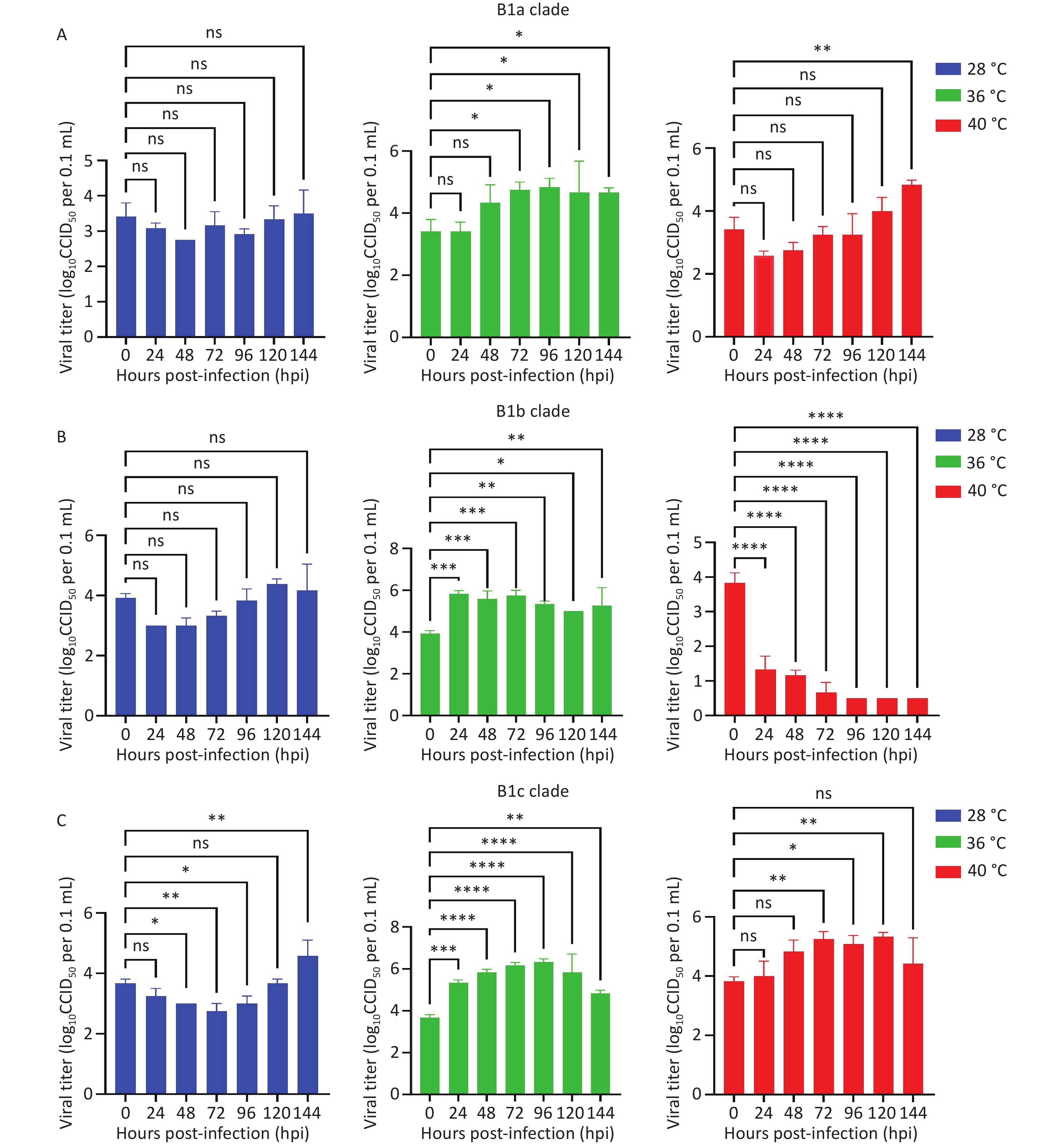
Temperature is an important modulator of enterovirus population-level transmission, with warmer conditions linked to enhanced outbreak risk of EV-A71, independent of viral genetic adaptation[8]. Our investigation of the CVA16 subgenotype B1 revealed distinct temperature sensitivity profiles among the three clades in RD cells. At 28°C, none of the three clades induced significant CPE or viral replication, with a log increase in CCID50 titer of < 1. At 36 °C, all three clades replicated efficiently, with peak titers ranking B1c > B1b > B1a, and the B1b clade showed the fastest CPE progression, appearing at 24 hpi and exceeding 50% by 48 hpi. At 40 °C, both B1a and B1c remained temperature-insensitive: B1a exhibited late-onset CPE starting at 120 hpi, whereas B1c induced rapid CPE with cell rounding at 72 hpi and over 75% cytopathic effect by 96 hpi (Figure 1, Supplementary Table S1, and Supplementary Figures S5 and S6). In contrast, the B1b clade was temperature-sensitive, showing no obvious CPE and a progressive decrease in titer. Peak titers for all clades occurred at 36 °C, and temperatures deviating from 36°C, namely 28°C and 40 °C, limited efficient CVA16 replication. Epidemiological data show that B1b was undetectable in Fujian Province in 2022 and 2024, whereas B1c emerged in October 2023 and rapidly displaced B1a as the predominant strain[5]. The B1b clade exhibited lower in vitro replication efficiency at 40°C compared to B1a and B1c, whereas B1c demonstrated the highest replication capacity and thermotolerance across all tested temperatures. This difference in thermotolerance may affect the stability of viral replication under febrile host conditions or environmental thermal stress, thereby influencing transmission dynamics. Given that CVA16 transmission is positively correlated with temperature, with outbreak risk increasing significantly during warmer seasons[8], and that Fujian Province has a subtropical monsoon climate characterized by long, hot, and humid summers that facilitate year-round HFMD transmission, these factors may collectively explain the gradual replacement of B1b by B1a and B1c, with B1c emerging as the predominant circulating clade. However, this inference is based solely on in vitro experimental data, and because the host body temperature is tightly regulated by homeostatic mechanisms, further animal experiments are required to validate these findings.
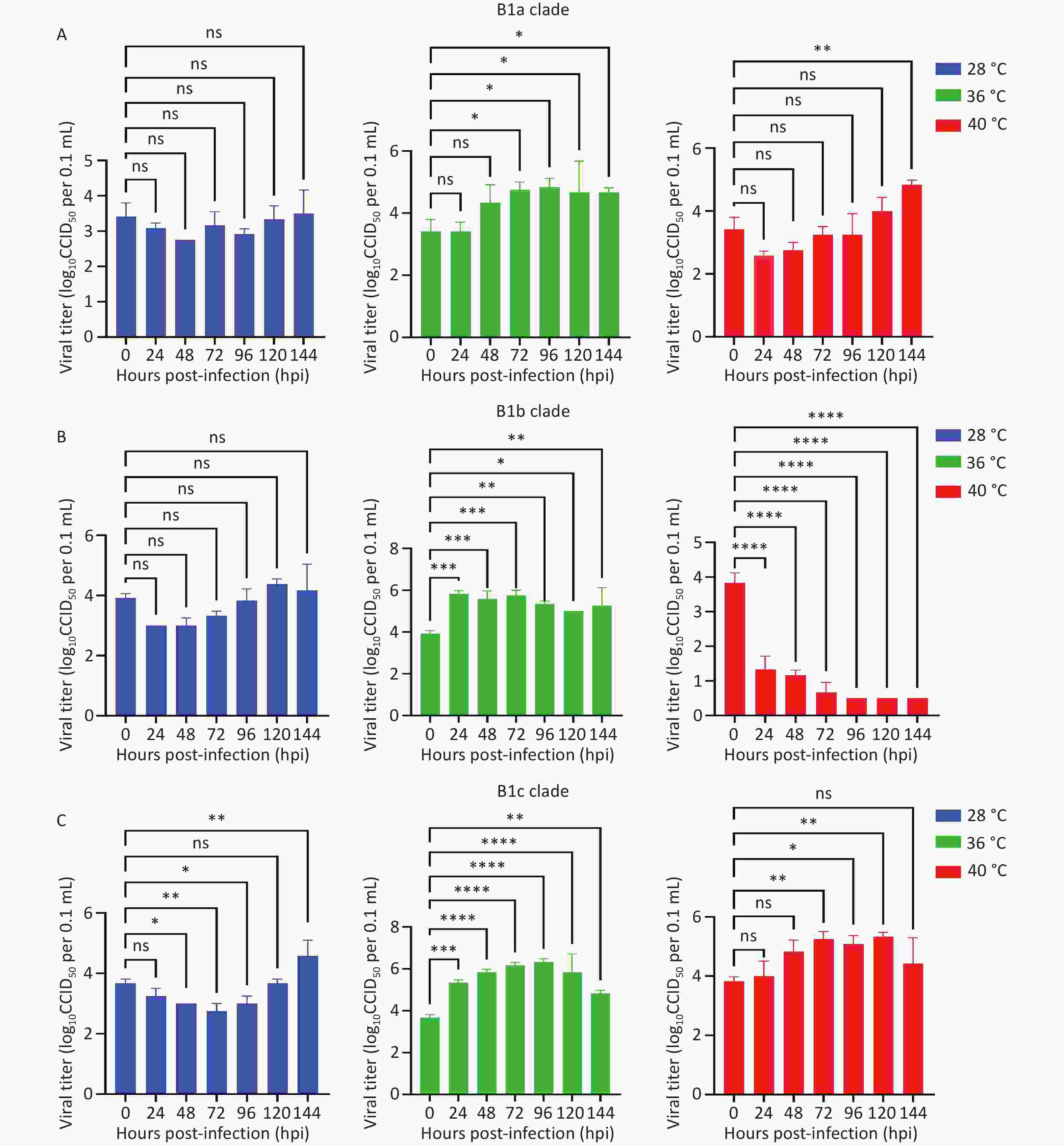
Figure 1. Replication kinetics of CVA16 B1a, B1b, and B1c clades at three different cultivation temperatures. (A) B1a clade representative strain. (B) B1b clade representative strain. (C) B1c clade representative strain. Data are presented as mean ± SD of three biological replicates. Statistical significance was determined by one-way ANOVA; ns, not significant (P > 0.05); *P < 0.05; **P < 0.01; ***P < 0.001; ****P < 0.0001. CVA16, Coxsackievirus A16; CCID50, 50% cell culture infectious dose.
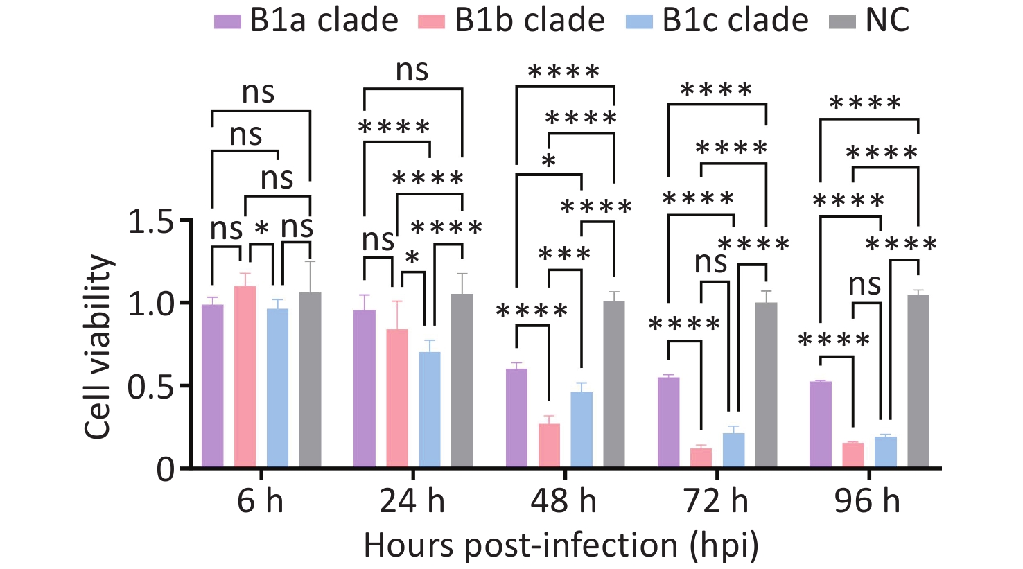
RD cells were infected with the three CVA16 clades at an MOI of 0.1, and cell viability was assessed using the CCK-8 assay. RD cell proliferation was significantly inhibited by all three clades. At 6 hpi, no statistically significant differences were observed among the clades. By 24 hpi, the inhibitory effect was ranked as follows: B1c > B1b > B1a, although inhibition by the B1a clade alone did not reach statistical significance. Significant differences persisted from 48 to 96 hpi, with cytotoxicity consistently ranked in the following order: B1b > B1c > B1a. The induction of apoptosis in infected host cells represents a critical virus-host interaction, balancing the restriction of viral replication with the facilitation of viral spread[9]. Evaluation of the cytotoxicity of the three CVA16 clades on RD cells revealed that all clades inhibited cell growth, with a cytotoxicity hierarchy of B1b > B1c > B1a (Figure 2). These in vitro phenotypic differences identified distinct biological profiles among the clades, with implications for viral fitness that warrant further investigation. CVA16 is a major HFMD pathogen in Fujian Province. Epidemiological surveillance has indicated that the currently circulating dominant clades B1a and B1c (with B1c being primary) have become predominant as the B1b clade has declined. These epidemiological observations are consistent with our in vitro findings that B1c and B1a exhibit lower cytotoxicity compared to B1b. HFMD cases associated with CVA16 in Fujian Province were predominantly mild, with only a few severe cases. This epidemiological pattern may be attributed to the generally lower virulence of CVA16 than that of EV-A71, whereas the lower in vitro cytotoxicity of the currently circulating B1c and B1a clades may also partially explain their emergence as dominant strains.
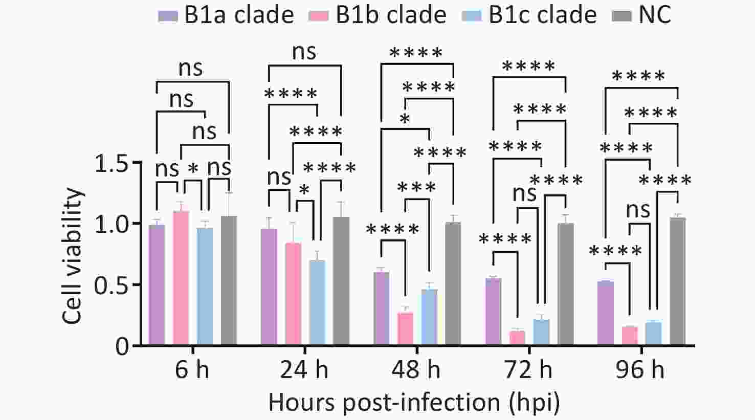
Figure 2. Inhibition of RD cell proliferation by CVA16 B1a, B1b, and B1c clades at different time points. Data are presented as mean percentage of inhibition ± SD from three independent replicate samples. Statistical significance was determined by two-way ANOVA (ns, not significant, P > 0.05; *P < 0.05, **P < 0.01, ***P < 0.001, ****P < 0.0001). NC, normal control.
In summary, this study provides a comparative analysis of key biological characteristics, including cell tropism, thermostability, replication kinetics, and cytotoxicity, among the three evolutionary clades (B1a, B1b, and B1c) of the CVA16 subgenotype B1 at the cellular level. Our findings indicate that RD cells are more suitable than Vero and HEp-2 cells for culturing and isolating the three CVA16 virus lineages. Representative strains of the B1a and B1c clades are heat insensitive, whereas the B1b representative strain is heat sensitive. Furthermore, the B1c representative strain demonstrated a relatively superior in vitro replication capacity compared to B1a and B1b, without greater cytotoxicity than the B1b representative strain. Notably, despite the stronger in vitro replication capacity of B1c, epidemiological data from Fujian Province have revealed that B1c and B1a are the predominant clades, with B1c being dominant, whereas B1b is gradually declining. This phenomenon aligns with the "virulence-transmission trade-off hypothesis"[10] in evolutionary biology, which posits that pathogen virulence evolves in concert with transmission fitness rather than independently. In support of this, our identified phenotypic differences among the clades of CVA16 subgenotype B1 likely reflect variations in viral fitness that warrant deeper molecular characterization. In conclusion, the emergence of B1c as a new dominant CVA16 clade may increase the epidemic intensity of CVA16-associated HFMD.
This study has two main limitations. First, only one representative strain per clade was selected for the phenotypic experiments, which limits the generalizability of the conclusions. Second, the findings were based solely on in vitro analyses and lacked in vivo investigations. Therefore, future studies integrating animal experiments, continuous molecular epidemiological surveillance, and larger sample sizes are warranted to validate our findings and monitor the evolution of CVA16.
HTML
Author Contributions Shujing Li: Conceptualization, Investigation, Formal Analysis, Writing – Original Draft.
Yuwei Weng: Supervision, Project Administration.
Wei Chen: Conceptualization, Supervision, Writing – Review & Editing.
Wenxiang He and Shaoyong Xu: Investigation, Data Curation, Formal Analysis.
All authors have read and approved the final version of the manuscript.
Ying Zhu: Visualization, Investigation, Funding Acquisition, Data Curation, Formal Analysis.
Data Sharing The supplementary materials will be available at www.besjournal.com.
&These authors contributed equally to this work.
Reference

 Quick Links

 DownLoad: