


HTML
-
Experimental studies on animals and cultured human cell lines support a role for phenolic compounds in the prevention of cardiovascular diseases, cancers, neurodegenerative diseases, diabetes, and osteoporosis. However, it is uncertain if these results of polyphenol intake affect disease prevention in humans[1]. Ethnomedicinal plants have made a tremendous contribution to the discovery of new drugs against different diseases, including cancer[2]. Investigation of medicinal plants to discover potent anticancer compounds with fewer side effects is warranted[3].
Cancer is a hyperproliferative disorder in which invasion and angiogenesis lead to tumor metastasis[4]. In fact, many novel natural compounds, such as favonoids (honokiol, magnolol, jaceosidin, and casticin), sesquiterpenes (parthenolide, costunolide, isoalantolactone, and alantolactone), alkaloids (evodiamine), diterpenoids (oridonin and pseudolaric acid B), and polyphenolics (wedelolactone) with potential utility as anticancer agents have been discovered[5]. The phenolic groups in polyphenols can accept an electron to form relatively stable phenoxyl radicals, thereby disrupting chain oxidation reactions in cellular components[6]. Numerous lines of evidence suggest that the whole plant may be better than its active component[7]. In addition, herbal antioxidants are reported to modify cell survival and DNA repair efficacy by downregulating activation of receptor tyrosine kinases and NF-κB[8]. The antioxidant activities of medicinal plants are attributed to their phenolic contents due to the redox properties of phenolics, which allow them to act as reducing agents, hydrogen donors, and singlet oxygen quenchers[9].
Species of the genera Thymus, Teucrium, Hydrophyllum, and Clematis are widespread on all continents of the world, and many such species are present in the Mediterranean[10, 11]. The aerial parts of the plants used in this report have an aromatic taste and are used as favoring and anti-infammatory agents[12, 13]. Clematis species were formerly used as a remedy for venereal disease (syphilis). Externally, it is used for chronic skin conditions. It is also used to treat gout, rheumatism, and bone disorders; as an antimalarial agent; as an antidote for snake bites; in the treatment of dysentery; and as a diuretic[14, 15]. In traditional medicine, herbal medicines belonging to the genus Thymus are employed for internal use in the oral treatment of coughs, upper respiratory infections, acute and chronic bronchitis, whooping cough, and catarrh[16]. To the best of our knowledge, this is the first report on the cytotoxic effects of extracts obtained from T. alopecurus.
This report focuses on evaluation of the biological effects of four plant species from the same location and grown under the same geographic and climactic conditions. Crude methanolic extracts of the tested plants displayed antioxidant and antibacterial activity. At high doses, the methanolic extracts induced cell viability arrest in a multiple myeloma cell line and exhibited high cytotoxic potential in cancer cell lines. These effects may be related to the total phenolic contents of the extracts as potential sources of natural antioxidants.
-
Stock solutions of different fractions of methanolic solutions (1 mg/mL) were prepared in methanol, stored at -20 ℃, and then diluted as needed in cell culture medium and for other experiments. MTT was purchased from Sigma-Aldrich (St. Louis, MO, USA). Penicillin, streptomycin, Roswell Park Memorial Institute (RPMI)-640 medium, and fetal bovine serum (FBS) were obtained from MediaTek, Inc. (Herndon, VA, USA). Butyl-hydroxytoluene (BHT), ascorbic acid, ferric chloride (FeCl3) (0.1%), potassium ferricyanide [K3Fe(CN)6] (1%), 2, 2-diphenyl-1-picrylhydrazyl (DPPH), ferric reducing antioxidant power (FRAP), trichloroacetic acid (TCA) (10%), ascorbic acid (1%), and phosphate buffer (0.1 mol/L; pH 6.6) were obtained from Sigma-Aldrich Chemical Co. (St. Louis, MO, USA). Phenolic standards quercetin, rutin, gallic acid, catechin, caffeic acid, coumarin, coumaric acid, epicatechin, and vanillic acid were obtained from Sigma-Aldrich.
-
The human multiple myeloma cell line U266 was obtained from the American Type Culture Collection (ATCC, Manassas, VA, USA).
-
The tested bacteria used in this report including Staphylococcus aureus (S. aureus), Escherichia coli (E. coli), Klebsiella pneumoniae (K. pneumoniae), and Bacillus subtilis (B. subtilis) were purchased from the ATCC.
-
Chloramphenicol (CFM) (10 μg/μL); Amoxicillin-clavulanic acid (AMC) (30 μg/μL); Ticarcillin (TIC) (75 μg/μL); Cefixim (cystites) (CFM) (10 μg/μL); Cefoxitin (FOX) (10 μg/μL); Cefotaxim (CTX) (10 μg/μL); Ceftazidim (CAZ) (10 μg/μL); Aztreonam (ATM) (5 μg/μL); Ertapenem (ETP) (10 μg/μL); Imipenem (IPM) (10 μg/μL); Ciprofloxacin (CIP) (5 μg/μL); Gentamicin (CN) (10 μg/μL); Tobramycin (TOB) (10 μg/μL); Cefoxitime (FOX) (30 μg/μL); Fosfomycin (FF) (200 μg/μL); Cefotaxime (CTX) (30 μg/μL); and Ceftazidime (CAZ) (10 μg/μL) were obtained from Sigma-Aldrich Chemical Co. (St. Louis, MO, USA).
-
Extract Preparation Medicinal plants used in this report were extracted by maceration with methanol (MeOH) for 24 h. The extracts were kept in vials at -80 ℃ until the corresponding analysis. Methanolic extracts were evaporated to dryness at 40 ℃ under vacuum conditions in an evaporator system. The extract was then filtered and concentrated under vacuum at 4 ℃, yielding a waxy material.
Analysis of Methanolic Extracts with High Performance Liquid Chromatography High performance liquid chromatography (HPLC) analyses were carried out at 30 ℃ using a C18 column (4.6 mm × 150 mm) × 5 mm in a Thermo Fisher apparatus, and the injected volume was 20 μL. The mobile phase consisted of acetonitrile (A) and 2% glacial acetic acid (pH = 2.6) (B). The flow rate was 0.5 mL/min. The elution gradient was 30%-95% (B) for a total run time of 45 min, starting from 5% solvent A and 95% solvent B, decreasing to 65% solvent B over 30 min, and to 30% over 10 min. Pure compounds used in this study, namely gallic acid, catechin, vanillic acid, coumarin, epicatechin, coumaric acid, cinnamic acid, rutin, and quercetin were used as external standards. All standards were dissolved in methanol and injected under the same chromatographic conditions as the extracts. Phenolic compounds of plant extracts were identified by comparing their retention times with those of pure standards. The results are expressed as micrograms of each compound per gram dry weight (μg/g DW).
Total Phenolic Content Total phenolic content was assessed by the method of Singleton and Rossi[17] using Folin-Ciocalteu reagent. Methanolic extracts were mixed with 10% Folin-Ciocalteu reagent and distilled water. After 5 min at room temperature, a 7.5% Na2CO3 solution was added to the mixture. Optical density was measured at 760 nm versus a blank prepared without extract with a Shimadzu (UV-2401PC, Kyoto, Japan) spectrophotometer. Phenolic content was calculated by comparison with a Gallic acid standard curve (0-500 μg/mL) (y = 0.004x, R2 = 0.9977) and expressed as micrograms gallic acid equivalents (GAE) per milligram dry weight extract (μg GAE/mg DW). The experiments were carried out in triplicate.
The evaluation of flavonoid content was carried out according to Dewanto et al.[18]. Methanolic extracts was mixed with sodium nitrite solution (5%, 75 μL). After incubation for 6 min, aluminium chloride solution (10%, 150 μL) and sodium hydroxide (1 mol/L, 500 μL) were added to the mixture. The absorbance was determined at 510 nm with a Shimadzu (UV-2401PC, Kyoto, Japan). Flavonoid content was calculated by comparison with a quercetin standard curve (0-100 μg/mL) (y = 0.0093x, R2 = 0.9672) and expressed as micrograms quercetin equivalents (QE) per milligram dry weight of extract (μG QE/mg DW).
Condensed tannin was assayed by the method of Makkar and Becker[19] using vanillin reagent. Methanolic extracts were mixed with vanillin. Afterwards, concentrated HCl was added to the mixture. The absorbance was read at 500 nm with a Shimadzu (UV-2401PC, Kyoto, Japan). Condensed tannin was calculated by comparison with a catechin standard curve (0-250 μg/mL) (y = 0.001x + 0.0049, R2 = 0.9792) and expressed as micrograms catechin equivalents (CE) per milligram dry weight extract (μg CE/mg DW).
-
DPPH Assay For the assessement of the capacity of methanolic extracts to scavenge DPPH, the modified DPPH method was used as described by Brand-Williams et al.[20], using ascorbic acid as a standard solution. To the methanolic fraction (1, 10, 100, 200 μg/mL) of four species, methanolic solution of DPPH was added. Loss of DPPH color was measured spectrometricaly at 515 nm using a UV/visible spectrophotometer (Spectronic Genesys 8, Rochester, NY, USA).
Ferric Reducing Antioxidant Power Assessment of the FRAP of methanolic extracts was carried out by the modified method of potassium ferricyanide-ferric chloride as described by Oyaizu[21]. Potassium ferricyanide and phosphate buffer were added to the extract diluted in distilled water (100, 200, 300, 400, or 500 μg/mL) or various BHT standard solutions (100, 200, 300, 400, or 500 μg/mL). Then, TCA was added. FeCl3 and distilled water were added to the upper layer solution after centrifugation (3, 000 rpm/10 min). Absorbance was measured at 700 nm using a spectrophotometer (Spectronic Genesys 8, Rochester, NY, USA).
Cell Culture U266 cell lines were grown in RPMI 1640 with 10% FBS. Culture media were supplemented with 100 U/mL penicillin and 100 μg/mL streptomycin. Cells were split twice a week and cultures were maintained in a humidified incubator (5% CO2 at 37 ℃).
Cytotoxicity Assays The cytotoxic effect of methanol extracts of different species used in this report on cell viability was determined using the modified MTT method as previously described[22]. Briefly, U266 cells (5 × 10² cells/well), seeded in triplicate in a 96-well plate, were incubated with increasing concentrations of ME-TA (0, 50, 60, 70, 80, or 100 μg/mL), ME-TAl (0, 50, 60, 70, 80, or 100 μg/mL), ME-CF (0, 50, 60, 70, 80, or 100 μg/mL), and ME-HT (0, 50, 60, 70, 80, or 100 μg/mL) until the indicated time points (24, 48, and 72 h) at 37 ℃. Control wells were incubated with vehicle. Then, the cells were incubated with MTT solution for 2 h, the extraction buffer was added, and the cells were incubated at 37 ℃ overnight. Optical densities were measured at 570 nm by a 96-well multiscanner (Dynex Technologies; MRX Revelation) using the extraction buffer as a blank.
-
All data are presented as mean ± standard deviation (SD), n = 3 for all experiments. Extraction, phenolic content and other experimental assays were carried out in triplicate. For cytotoxicity assays, three independent assays were performed. Statistical analysis of methanolic extract activity on cell viability was performed using two-way analysis of variance (ANOVA), followed by Tukey's multiple comparisons test (P < 0.05).
Chemicals
Cell Lines
Bacteria
Antibiotics
Plant Material
Free Radical Scavenging Activity
Statistical Analysis
-
In this report, methanol was selected as the solvent to extract the bioactive compounds of four medicinal plants (Figure 1, Table 1). The mass yields of methanol extracts of the aerial parts of these species were found to be from 7.3% to 37% (w/w), respectively, with ME-CF being predominant (Figure 2A); the lowest yield was detected in ME-TAl.
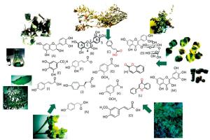
Figure 1. Chemical structures of catechin (A), epicatechin (B), cinnamic acid (C), rutin (D), gallic acid (E), caffeic acid (F), vanillic acid (G), coumarin (H), coumaric acid (I), tyrosine (J), vanillin (K), flavone (L), catechin hydrate (M), hydroxyphenylacetic acid (N), ferulic acid (O), phenolic content present in collected plants with cytotoxic and antibacterial effects.
N° Code Scientific Name Family Voucher Specimen Local Name Localisation Part Used; Extraction and Solvent Used Traditional Therapeutic Indications References 1 ME-TAI Teucruim olopecurus Lablaceae n.1122 H'chichit Ben Salem Jbel Orbata-Gafsa-Tunisia Upper parts; Methanol Anti inflammatory drug (Guesmi et al., 2017)[12] 2 ME-TA Thymus hirtus sp.olgeriensis Lamiaceae n.1188 Moujecha Jbel Orbata-Gafsa-Tunisia Upper parts: Methanol Ulcers and testis toxicity (Guesmi etal., 2014; Guesmi et al., 2016)[13, 30] 3 ME-CF Clematis flommula Ranuncuiaceae Nar Berda Jbel Orbata-Gafsa-Tunisia Leaves; Methanol Rheumatism and anti inflammation (Guesmi et al., 2018; Reivera et al., 2008)[25] 4 ME-HT Hydrophyllum tuberculatum Rutaceae Mnitna ZannouchGafsa- Tunisia Upper parts; Methanol Anticancer, antioxydant, antibacterial, anti-HIV, uterus-relaxing activity (Raissi 2016; Adnan ct al., 2001)[37] Table 1. The Collected Plant Parts, Collection Sites, Ethnobotanical Indications, and Percentage Yields of Methanolic Extracts

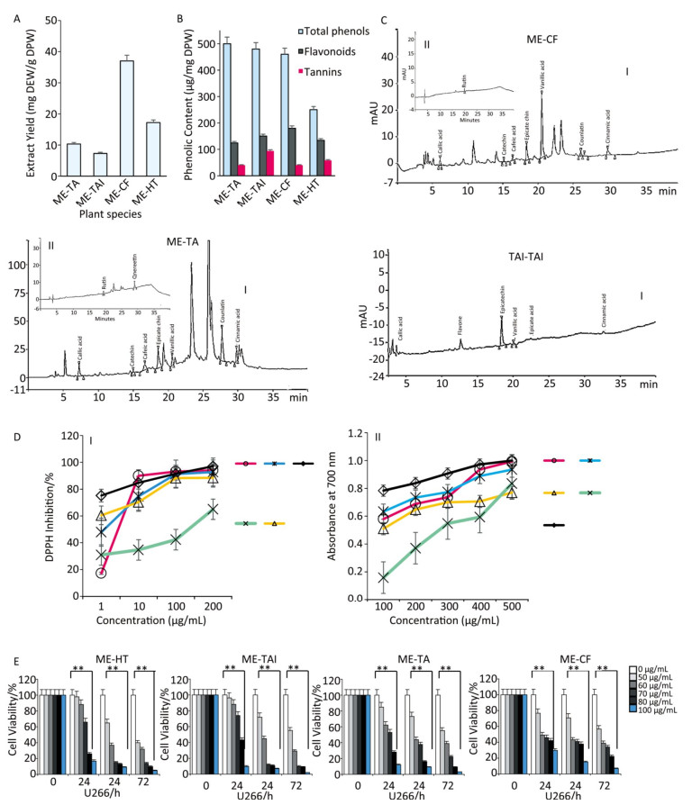
Figure 2. (A) Yield of the methanolic extracts of species used in this study. TAME = Thymus algeriensis methanolic extracts, CFME = Clematis flammula methanolic extracts, TAlEM = Teucrium alopecurus methanolic extracts, HTEM = Hydrophyllum tuberculatum methanolic extracts. (B, C) HPLC chromatogram of phenolic compounds detected at 280 nm (Ⅰ) and flavonoids (Ⅱ), of methanolic extracts. (D) antiradical potential of methanolic extracts using DPPH (Ⅰ) and FRAP (Ⅱ) assay; (E) Cytotoxicity of different extracts obtained from TAEM, TAlEM, CFEM, and HTEM toward U266 cancer cell lines. Tumor cells were pre-treated with various concentrations of extracts and incubated for 72 h. Cell viability was analyzed with the MTT assay.
As shown in Figure 2B, the total phenolic contents of different methanolic extracts were determined from a gallic acid standard curve and expressed as μg GAE/mg DW. In this report, the phenolic content ranged from 250 ± 18 μg GAE/mg DW (C. flammula) to 500 ± 11 μg GAE/mg DW (T. algeriensis) for phenolic acids, 124 ± 4 to 180 ± 12 μg QE/mg DW for flavonoids, and 40 ± 7 to 94 ± 8 μg CE/mg DW for tannin content; the lowest mean values were obtained for ME-HT. In contrast, ME-TAl showed the highest total phenolic content. In turn, the latter showed a lower content of flavonoids and tannins in comparison with other plants. The obtained values of the flavonoid compounds in leaf extracts were found to range between 5.09 and 19.81 mg QE/g extract. Clematis flammula leaves revealed higher levels of tannin content (4.94-7280.52 mg tannic acid equivalents/g extract).
Based on the above results, the methanolic extracts of the four plants constitute a promising natural product, being particularly rich in phenolic acid derivatives, as well as rutin (Figure 2C). As shown in Table 2, we detected the presence of a high quantity of phenolic acids in ME-TAl and in ME-CF, including vanillic acids, coumarin, and epicatechin.
Compounds Methanolic Extracts ME-TA ME-TAI ME-CF Approximate RT (min) Content (μg/g dw) Area (counts) Width 1/2 (s) Approximate RT (min) Content (μg/g dw) Area (counts) Width 1/2 (s) Approximate RT (min) Content (Mg/g dw) Area (counts) Width 1/2 (s) Gallic acid 7.23 745 112 339229 11.7 6.00 266 ±23 289764 12.54 7.23 311±5 636005 10.9 Catechin 14.98 16±5 147892 22.7 15.08 18±3 159830 13.06 14.98 19 ±5 55709 12.8 Caffeic acid 16.64 26± 14 78109 9.0 - - - - 16.64 89 ±26 540775 30.9 Ferulic acid 17.83 42 ±6 115097 10.9 18.37 25 ±3 610987 19.45 - - - - Epicatechin 18.55 136 ±11 59027 11.2 18.50 162 ±47 524696 11.50 18.55 125 ±32 1479481 14.9 Rutin 20.01 89 ±3 256110 12.5 - - - - 20.01 45 ±15 89619 12.5 Vanillic acid 20.57 615±41 131110 11.4 15.96 792 ± 2 36126 14.90 20, 57 7182 ± 68 419659 13.0 Coumarin 27.70 234 ± 17 677045 12.2 - - - - 27.70 38 ±7 2211453 13.3 Coumaric acid 30.00 124 ±11 628065 11.8 30.00 22 ±15 233541 11.04 - - - - Flavone - - - - 9.16 66 ±10 679210 10.60 - - - - Quercetin 17.56 126 ±16 628017 11.3 - - - - - - - Cinnamic add 29.78 - 203634 11.5 - - — - 29.78 348 ± 56 500006 11.5 Table 2. Phenolic Compounds Identified in Methanolic Extracts by HPLC
-
In the DPPH assay, the MeOH extract of Thymus algeriensis exhibited the highest reducing activity followed by ME-CF, ascorbic acid, ME-TAl, and ME-HT fractions at 100 and 200 μg/mL (Figure 2DⅠ). In the FRAP assay, ME-TA and ME-TAl displayed more potent radical scavenging activity than ME-CF and ME-HT (Figure 2DⅡ). BHT exhibited the highest radical scavenging activity among all samples.
-
Our findings suggested that methanolic extracts of Thymus and Teucrium might act as chemopreventive agents with antioxidant properties, offering effective protection against in vitro U266 proliferation in a dose-dependent manner. Of the four methanolic extracts, two showed the potential to inhibit U266 cell viability (Figure 2E). Induction of apoptosis was increased significantly in tumor cells. Our results clearly demonstrated that the ME-TAl and ME-TA were the most active, whereas the ME-CF and ME-HT were the least active. These findings suggest that treatment with methanolic extracts rich in phenolic acids may provide an enhanced therapeutic response in human multiple myeloma cells. However, 50 μg/mL appeared to have no significant effect on cell proliferation in tumor cells. Moreover, the induction of apoptosis in tumor cells using the MTT assay was apparently associated with the antitumor activity of the respective natural preventive agents. Similar results were observed by several workers in different tumor cells exposed to natural products. Moreover, our results clearly show that Hydrophyllum tuberculatum extract significantly induced U266 cytotoxicity in a dose-dependent manner.
-
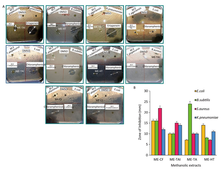
Our findings, validated through bacterial inhibition, showed that methanolic extracts have potential novel applications as nutraceuticals and microbial inhibitors. Taken together, our results suggest that methanolic fractions (1 mg/mL) acted as bacterial inhibitors and suppressed the growth of bacterial strains when compared with antibiotics used in this report (Table 3), with a large inhibition zone detected in B. subtilis (24 mm), to the same extent as 0.10 μg of chloramphenicol, when bacteria were treated with ME-TA and in S. aueus (22 mm) after treatment with ME-TAl (Figure 3A). Furthermore, our data revealed that ME-HT showed a moderate effect on B. subtilis, S. aureus, and K. pneumoniae (Figure 3).
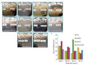
Figure 3. Antibacterial activity of the hydrophobic fraction of Teucrium alopecurus. Plant essential oils (1 μL) were applied to a filter disc (Whatman No 5 mm) onto the seeded top layer of the agar plates containing tested bacteria (E. coli, S. aureus, B. subtilis, and K. pneumoniae). Essential oil was tested with chloramphenicol, and ertapenem discs as positive controls. DMSO discs were used as negative controls (A). The plates were incubated at 37 ℃ for 24 h, and the zone of inhibition diameter was determined (B).
Methanolic Extracts and Antibiotics Inhibition Zones (mma) of Bacterial Strains in Presence of Me-OH Extracts and Antibiotics Gram + Gram - Plant name région S. aureus B. subtilis E. coli K. pneumoniae K. oxycota Control: DMSO * * * * - ME-CF Jbel Orbata-Gafsa 22 16 16 12 - ME-TAl Jbel Orbata-Gafsa 15 10 10 14 - ME-TA Jbel Orbata-Gafsa 10 24 7 10 - ME-HT Zannouch-Gafsa 7 8 14 11 - Antibiotics (μg/μL) ETP (10 μg/μL) * * * 32 24 CFM (10 μg/μL) 26 23 30 - * ATP10K (10 μg/μL) * * 18 * 21 CTX (30 μg/μL) * * 35 * 35 CIP (5 μg/μL) * * 37 * 40 CT (10 μg/μL) * * 24 * 19 CFM (5 μg/μL) * * 30 * 33 IPM (10 μg/μL) * * 33 * 30 CAZ (10 μg/μL) * * * * 24 TIC (75 μg/μL) * * * * * FOX (30 μg/μL) * * 25 * 37 CN (10 μg/μL) * * 19 * 22 TOB (10 μg/μL) * * 18 * 22 FF (200 μg/μL) * * 21 * 18 UA (30 μg/μL) * * 29 * 24 AMC (30 μg/μL) * * 21 * 25 AX (10 μg/μL) * * * * * Note. Me-OH: Methanol; S. aureus: Staphylococcus.auerus; E.coli: Eshershia.coli; K.pneumonie: Klebsiellla pneumoniae; B.subtilis: Bacillus subtilus; a: inhibition zone diameters; *: no activity detected; (-): not tested; AMC: Amoxicilline-acid clavulanic, PRL: Piperacillin; TIC: Ticarcillin; CFM: Cefixim (cystites); FOX: Cefoxitin; CTX: Cefotaxim, CAZ: Ceftazidim; ATM: Aztreonam; ETP: Ertapenem; IPM: Imipenem, NA: Nalidixic acid; NOR: Norfloxacin; CIP: Ciprofloxacin; AK: Amikacin; CN: Gentamicin; TOB: Tobramycin; TGC: Tigecyclin (E.coli); FF: Fosfomycin; CL: Cefalexin; TZP: Piperacillin-Tazobactam 1/2. Table 3. Antibacterial Activity of Methanolic Extracts and Antibiotics Against four Bacterial Strains
Extraction Yield and Identification of Phenolic Compounds
Evaluation of Antioxidant Activity
Cytotoxicity Assay
Antibacterial Activity
-
As are the vast majority of Lamiaceae plants, Thymus are recognized as being strongly aromatic and are widely used as spices to enhance sensory attributes such as the taste and aroma of foods[23]. It is important to note that methanolic extracts contain higher phenolic contents than those found in other reports in distinct species used in this study. Results obtained by determination of the phenolic content of Clematis (C. gouriana) extracts revealed promising bioactive contents with phenolic values of 18.19 ± 0.10 to 22.17 ± 0.10 mg GAE/g extract, respectively[24], while flavonoid values ranged between 2.83 ± 0.01 and 6.52 ± 0.08 mg QE/g extract. In another study, C. flammula showed only weak to moderate capacity[25] and its phenolic compounds were reported to account for 11.54-55.08 mg CE/g extract. Following analysis of the aerial parts of Haplophyllum tuberculatum, the known lignan diphyllin and two new alkaloids were isolated: haplotubinone and haplotubine[26].
Radical oxygen species (ROS) are products of normal cellular metabolism and play vital roles in the stimulation of signaling pathways in plant and animal cells in response to changes in intra- and extracellular environmental conditions[27]. Recently, interest has increased considerably in finding naturally occurring antioxidants for use in food or medicinal materials to replace synthetic antioxidants, which are being restricted due to their carcinogenicity[28]. Phenolic compounds are very reactive in neutralizing free radicals by donating a hydrogen atom or an electron, chelating metal ions in aqueous solutions[29]. The antiradical potential of different species used in this study was investigated by using DPPH and FRAP assays. The results obtained concur with other researchers who reported that aqueous acetone extract of Clematis gouriana showed higher antioxidant activity with an IC50 value of 129.11 and 25.35 μg/mL against DPPH and ABTS free radicals, respectively[24]. Moreover, Guesmi et al.[30] showed that the DPPH values of T. algeriensis were ranged from 81.00% ± 0.26% to 93.00% ± 0.06%. Moreover, researchers have reported that various Thymus and Teucrium species have potential radical scavenging activities[31]. Furthermore, Atmani et al.[25] found that the organic (chloroform) fraction of Clematis flammula showed high activity against linoleic acid peroxidation with an IC50 value of 4.6 μg/mL, whereas the aqueous phase of a chloroform extraction showed moderate scavenging activity against DPPH (IC50 = 25.02 μg/mL). In another study, Rana et al.[24] reported that the antioxidant yield of C. gouriana ranged from 16.87 ± 0.27 to 24.48 ± 0.13 mg/g of Trolox equivalents in different extracts as measured by the FRAP assay.
Natural bioproducts could be used to enrich our arsenal against diseases, including cancer. Our results clearly demonstrate that the ME-TAl and ME-TA were the most active, whereas the ME-CF and ME-HT were the least active. In our previous study, we reported that Teucrium alopecurus essential oil had cytotoxic effects and potentiated the cytotoxicity induced by TNF, capecitabine, 5-fuorouracil, and thalidomide in KBM5 cell lines[12]. The higher contents of total flavonoids and ortho-diphenols expected in Thymus pulegioides essential oil are responsible for its anti-proliferative effect against Caco-2 cells[32]. These findings suggest that treatment with methanolic extracts rich in phenolic acids may provide an enhanced therapeutic response in human multiple myeloma cells.
Extracts from some Teucrium species potentiate the cytotoxic and proapoptotic effects of the anticancer drugs vincristine, vinblastine, and doxorubicin against a panel of cancer cell lines[33]. Stankovic et al.[34] showed that methanolic extracts of Teucrium scordium subsp. Scordioides, which has the highest phenol content, induced the highest levels of superoxide anion radical after 72 h of treatment and that phenolics may be, at least in part, the source of its cytotoxic activity against the HCT-116 human colon cancer cell line.
In addition, a study on human melanoma cells reported that treatment with EOs induces DNA damage in cancer cells, which is an indicator of apoptosis[35]. Studies from many authors and those from our research group have investigated the preventive and therapeutic properties of bioactive compounds (terpenes, flavonoids, phenolic acids, and others) for their antioxidant and antitumor properties. Indeed, it has been reported that a combination of botanicals and/or their active principle components might be more efficacious than any one botanical alone[7]. Previous studies revealed that phenolic acids, flavonoids, and terpenes isolated from Thymus and Teucrium species exhibited cytotoxic activities against some human cancer cell lines[12]. Among the flavonoids, quercetin has shown great potential to exhibit anti-inflammatory and antioxidant properties. In fact, quercetin has been reported to possess anticancer activity against benzo(a)pyrene-induced lung carcinogenesis in mice[36]. According to our literature review, our data provide the first evidence that ME-TA and ME-TAl functions as a broad-range anticancer agent by effectively triggering apoptosis in cancer cells. Our findings are also consistent with recent publications indicating that Haplophyllum tuberculatum extract caused apoptosis in CCRF-CEM cells via alteration of the mitochondrial membrane potential[37]. The main mechanisms that mediate the cytotoxic effects of essential oils[38] and other bioactive compounds include the induction of cell death by activation of apoptosis and/or necrosis processes, cell cycle arrest, and loss of function of essential organelles. This report provides evidence for the potential effect of phytochemicals obtained from the tested plants to be used as therapeutic agents for the treatment of cancer. Further studies in humans and animals will be required to investigate the potent activities of different botanical parts of these species as anticancer bioproducts under downregulation of protein expression levels.
In recent years, the use of herbal medicines and food supplements containing botanical ingredients, as alternative therapy for infectious diseases, has been intensified due to their high content of antimicrobial agents such as polyphenols, i.e., favonoids, tannins, and alkaloids[16]. Results of antibacterial activity are consistent with data obtained by Guesmi et al.[13], who showed that weak activity of water and acetonitrile/water fractions of species cultured in Tunisia, including L. multifida, Pituranthos tortuosus, Teucrium alopecurus, Teucrium polium, and T. algeriensis, was observed for Salmonella typhi and Bacillus subtilis at 3 mg/disc, while Bacillus cereus was sensitive to all samples (3 mg/disc). Ilic et al.[39] reported the synergistic effect of the combination of the essential oil from Thymus glabrescens Willd. and chloramphenicol, which leads to a significant reduction in the MIC value of the synthetic drug, thus, minimizing its side effects. These data were confirmed by many researchers, who reported the antibacterial effect against these strains with essential oil, methanol, and other solvent extracts obtained from Thymus algeriensis[30, 40]. It is extremely important to point out that the antibacterial effects of essential oils in Thymus hirtus sp. algeriensis are strictly related to the presence of 1, 8-cineole (14.12%-19.96%)[30]. It has already been documented that thymol[41], borneol[42], and phenolic compounds inhibit growth inhibition of gram-negative and gram-positive bacteria (Streptococcus pyogenes, Helicobacter pylori, E. coli).
-
Based on traditional use and epidemiological investigations, these four methanolic extracts, obtained from plants adapted to the Mediterranean climate and growing in the mountains of Tunisia, exhibit antioxidant, cytotoxic, and antibacterial effects, which might be related to their total phenolic contents. Further studies are needed to elucidate the mechanism of the in vivo and in vitro pharmacological effects of these compounds.

 Quick Links
 DownLoad: