


-
With the wide application of nuclear energy and nuclear medicine technology, the influence of radiation on the environment and human health has garnered increasing attention by researchers[1-3]. In order to provide accurate references for the diagnosis and treatment of post radiation damage in clinical and public health, it is necessary to use precise instruments to measure the sensitive changes in biomarkers like cytokines, and to develop new methods or techniques to accurately evaluate the required radiation dose.
Radiation can induce damage to human health, including cell death, inflammation, organ damage, and tumor generation. Radiation-induced immune response usually occurs at the early stage, and then the mentioned cell death, inflammation, etc., events come up in exposed cells, animals or humans. Many studies have shown that radiation affects the immune system in varying degrees[4-6]. Measurement of the level of cytokine alteration in plasma is one of the most commonly used methods to evaluate the level of irradiation and risk of disease[7-10]. Enzyme-linked immunosorbent assay (ELISA) or western blotting have also been used for this purpose, but their results have been unsatisfactory. Based on our preliminary tests and other reports, the levels of interleukin (IL)-6 and IL-8 by ELISA in irradiated mice were relatively measured, but most of the other cytokines levels could not be obtained by ELISA[11,12].
The meso-scale discovery (MSD) assay is a bioanalysis platform that utilizes electrochemiluminescence (ECL), as a signal detection technique, unlike the colorimetric or chemiluminescent reaction in ELISA. The MSD assay consists of similar experimental procedures as in traditional ELISA, but is superior in many aspects[13, 14]. The MSD Quick Plex 120 instrument applies robust and sensitive ECL technology to quantify single and multiple target analytes. Interestingly, this assay accurately determines the quantity of analytes in complex biological matrices with improved throughput in a cost-effective and timely manner. For example, the MSD assay can be used to measure many biomarkers with clinical implications, and perform immunogenicity testing in a broad range of samples such as blood, plasma and tissue[8,15]. More recently, the MSD assay was used to assess the immunological effect of severe acute respiratory syndrome coronavirus 2 (SARS-CoV-2) vaccinations[16]. MSD ECL allows simultaneous multiplexing of up to ten different analytes in a well. Thus, the MSD platform requires almost 50-fold samples less than that in an ELISA experiment. MSD technology also has other advantages such as absolute quantitation, short processing time, low sample requirement (≥ 25 µL), higher sensitivity, better dynamic range, reduced signal-to-noise ratio, ready-made single analyte, and multiplex kits with excellent performance and lot-to-lot consistency
(https://www.nebiolab.com/meso-scale-discovery-msd-electrochemiluminescence-ecl/ ).In order to estimate the risk induced by radiation as well as understand the difference between low and high linear energy transfer (LET) ionizing irradiation-induced risks, we used the MSD technique to quantify cytokine levels in mouse plasma collected at different time points (1 h, 24 h and 7 d) after carbon-ion or X-ray irradiation. Approximately 29 cytokines (including inflammatory cytokines, pro-inflammatory cytokines and immune factors) were detected using this assay.
-
Adult female Kunming mice (20–24 g, purchased from Laboratory Animal Center, The Institute of Lanzhou Veterinary Research, Lanzhou, China) were housed and cared to comply with the regulations of the Institute of Modern Physics, Chinese Academy of Sciences (Lanzhou, China). The Committee for Animal Use at the Institute of Modern Physics approved all experimental procedures. We carried out animal experiments by trying to minimize the number of animals used in this study. Water and food were available ad libitum in plastic cages. All mice were acclimated from shipping for one week before treatment.
-
Carbon-ions with an LET of 30 keV/μm were generated at the Heavy Ion Research Facility in Lanzhou (Institute of Modern Physics, Chinese Academy of Sciences, Lanzhou, China) at mean dose rates of 5 Gy/min. X-rays were generated by XRad225 (PRECISION X-RAY) at a dose rate of 2 Gy/min.
Mice were randomly divided into four groups (15 mice in each group) according to the following radiation dose: 0, 0.5, 2.0 and 4.0 Gy of the two aforementioned irradiation types. After irradiation, five mice were maintained per cage and supplied with standard laboratory chow and water ad libitum. At 1 h, 24 h and 7 d after irradiation, the mice were sacrificed for plasma harvesting.
-
Blood was collected from the eyeballs of mice at the designated time points after irradiation, and stored inanticoagulated collection tubes. After 15 min, blood samples were centrifuged at 825 ×g for 10 min at 4 °C, and then the supernatants were collected. The plasma samples were stored at −80 °C.
-
MSD assay was performed according to the U-Plex kit (Meso Scale Discovery, Rockville, MD, USA) manufacturer’s instructions using 25 μL of sample (plasma) or standard. Samples were measured using an MSD instrument with the ability to detect ten kinds of cytokines in each well. The following 29 types of cytokines, including inflammatory cytokines, pro-inflammatory cytokines and immune factors were measured: IFN-γ, IL-1β, IL-2, IL-4, IL-5, IL-6, IL-9, IL-10, IL-12p70, IL-15, IL-16, IL-17A, IL-17A/F, IL-17C, IL-17E/IL-25, IL-17F, IL-21, IL-22, IL-23, IL-27p28/IL-30, IL-31, IL-33, IP-10, KC/GRO, MCP-1, MIP-1α, MIP-2, MIP-3α and TNF-α.
-
Cytokine profiling: data are presented as mean ± SE of three independent experiments. All statistical analyses were performed using SPSS version 22.0. The statistical significance (P value) was determined using unpaired two-tailed Student’s t-test. P < 0.05 was considered statistically significant between two-sample comparisons.
Mathematical modeling: a multiple linear regression model was used to develop a combination of multiple cytokines to predict the exposure degree (radiation dose). This analysis was performed using SPSS version 22.0 software. The fitting graphics were generated using Origin 9.1.
-
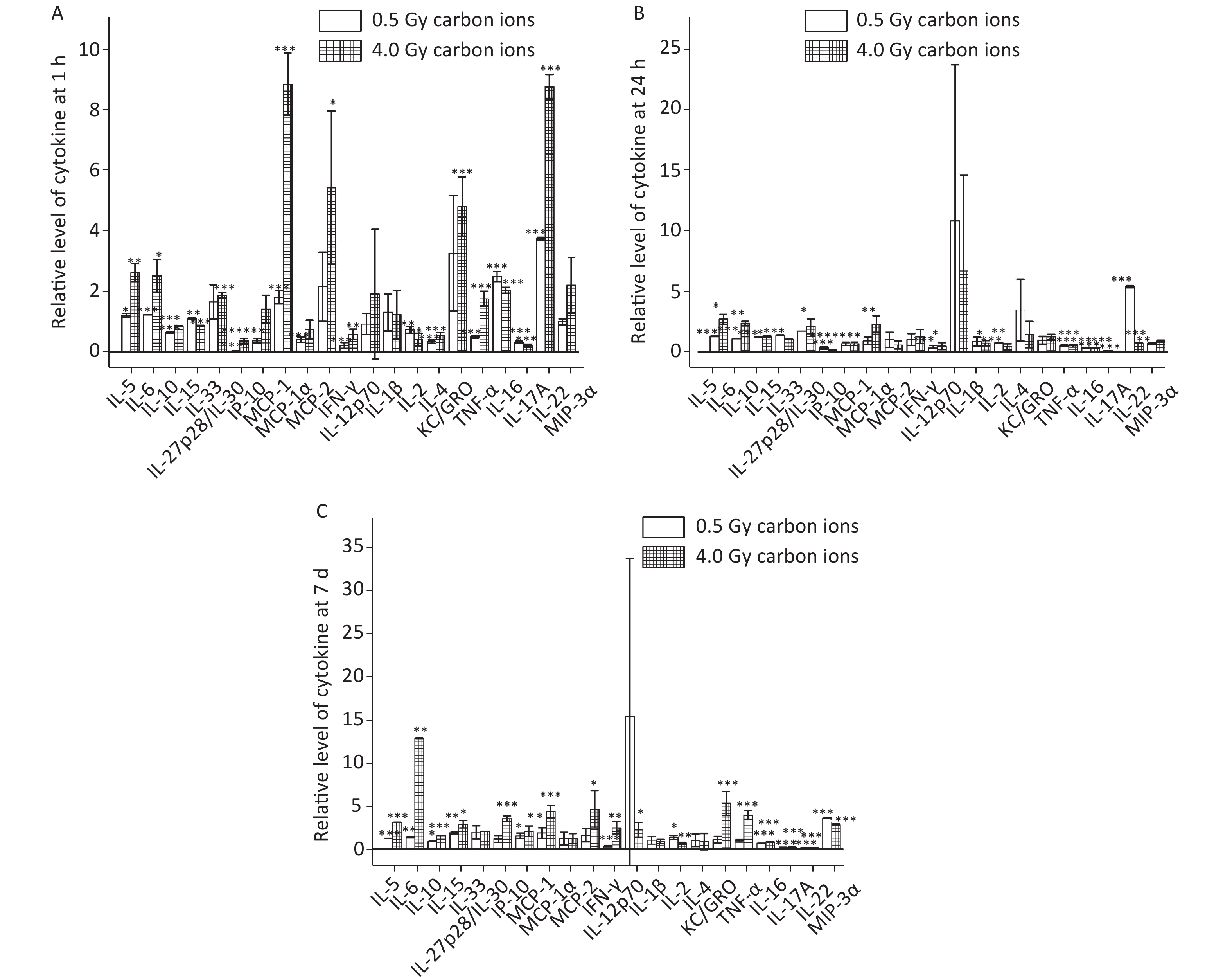
To determine the alterations of cytokines and identify the specific cytokine signatures in response to carbon-ion irradiation, the 29 plasma cytokines listed in the Materials and Methods section (subsection 4) were quantified using the MSD assay. However, only some of the cytokines (IL-5, IL-6, IL-10, IL-15, IL-33, IL-27p28/IL-30, IP-10, MCP-1, MIP-1α, MIP-2, IFN-γ, IL-12p70, IL-1β, IL-2, IL-4, KC/GRO, TNF-α, IL-16, IL-17A, IL-22 and MIP-3α) could be detected. We hypothesized that the expression of IL-9, IL-17A/F, IL-17C, IL-17E/IL-25, IL-17F, IL-21, IL-23 and IL-31 might be too low to detect. Figure 1 shows the relative levels of the detectable cytokines in the plasma of mice after 0.5 Gy and 4.0 Gy carbon-ion irradiation. The relative levels of 12 cytokines (IL-5, IL-6, IL-10, IL-15, MCP-1, IFN-γ, IL-2, IL-4, TNF-α, IL-16, IL-17A and IL-22) at 1 h (Figure 1A), 11 cytokines (IL-5, IL-6, IL-10, IL-15, IP-10, IFN-γ, IL-2, TNF-α, IL-16, IL-17A and IL-22) at 24 h (Figure 1B), and 12 cytokines (IL-5, IL-6, IL-10, IL-15, IP-10, MCP-1, IFN-γ, IL-2, IL-16, IL-17A, IL-22 and MIP-3α) at 7 d post carbon-ion radiation (Figure 1C), significantly difference compared to those in the control groups, a P value of < 0.05 using a Student’s t-test (compared to the control group).

Figure 1. Relative levels of plasma cytokines after total body irradiation (TBI) of mice with 0.5 Gy and 4 .0 Gy carbon-ion radiation. (A) Relative levels of cytokines 1 h after radiation. (B) Relative levels of cytokines 24 h after radiation. (C) Relative levels of cytokines 7 d after radiation.*P < 0.05, **P < 0.01, and ***P < 0.001. Unpaired two-tailed Student’s t-test
-
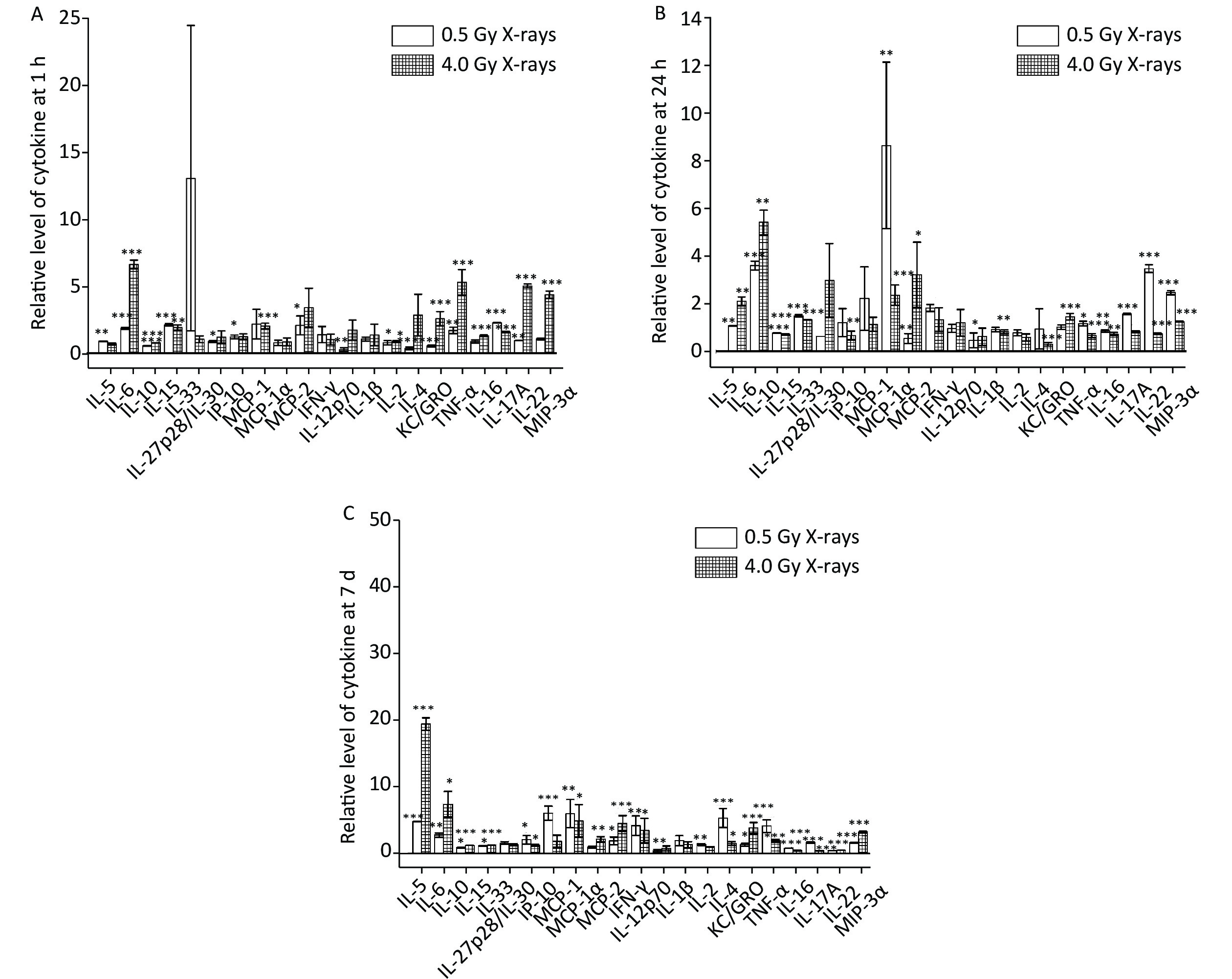
To further determine whether cytokines responded differently depending on radiation type, we measured the levels of the same 29 cytokines in mice after X-ray irradiation. The detectable cytokines after X-ray irradiation were similar to those in the case of carbon-ion irradiation. Figure 2 shows that 10 cytokines (IL-5, IL-6, IL-10, IL-15, MIP-2, IL-2, KC/GRO, TNF-α, IL-17A and IL-22) at 1 h (Figure 2A), 11 cytokines (IL-5, IL-6, IL-10, IL-15, MCP-1, MIP-1α, TNF-α, IL-16, IL-17A, IL-22 and MIP-3α) at 24 h (Figure 2B), and 14 cytokines (IL-5, IL-6, IL-10, IL-15, MCP-1, MIP-2, IFN-γ, IL-4, KC/GRO, TNF-α, IL-16, IL-17A, IL-22 and MIP-3α) at 7 d (Figure 2C), responded significantly to 0.5 Gy and 4.0 Gy of X-ray irradiation, a P value of < 0.05 using a Student’s t-test (compared to the control group).

Figure 2. Relative levels of plasma cytokines in total body irradiation (TBI) in mice after 0.5 Gy and 4.0 Gy X-ray radiation. (A) Relative levels of cytokines 1 h after radiation. (B) Relative levels of cytokines 24 h after radiation. (C) Relative levels of cytokines 7 d after radiation. *P < 0.05, **P < 0.01, and ***P < 0.001. Unpaired two-tailed Student’s t-test
-
As mentioned above (Figures 1 and 2), different kinds of cytokines responded notably to carbon-ion or X-ray irradiation at different time points. In particular, 8 cytokines (IL-2, IL-5, IL-6, IL-10, IL-15, IL-17A, IL-22 and TNF-α) at 1 h, 8 cytokines (IL-5, IL-6, IL-10, IL-15, IL-16, IL-17A, IL-22 and TNF-α) at 24 h, and 10 cytokines (IFN-γ, IL-5, IL-6, IL-10, IL-15, IL-16, IL-17A, IL-22, MCP-1 and MIP-3α) at day 7 d after radiation were sensitive to both the types of radiation. In addition, we found that the levels of some cytokines showed a dose-dependent effect and differed for low and high LET ionizing irradiation at different time points.
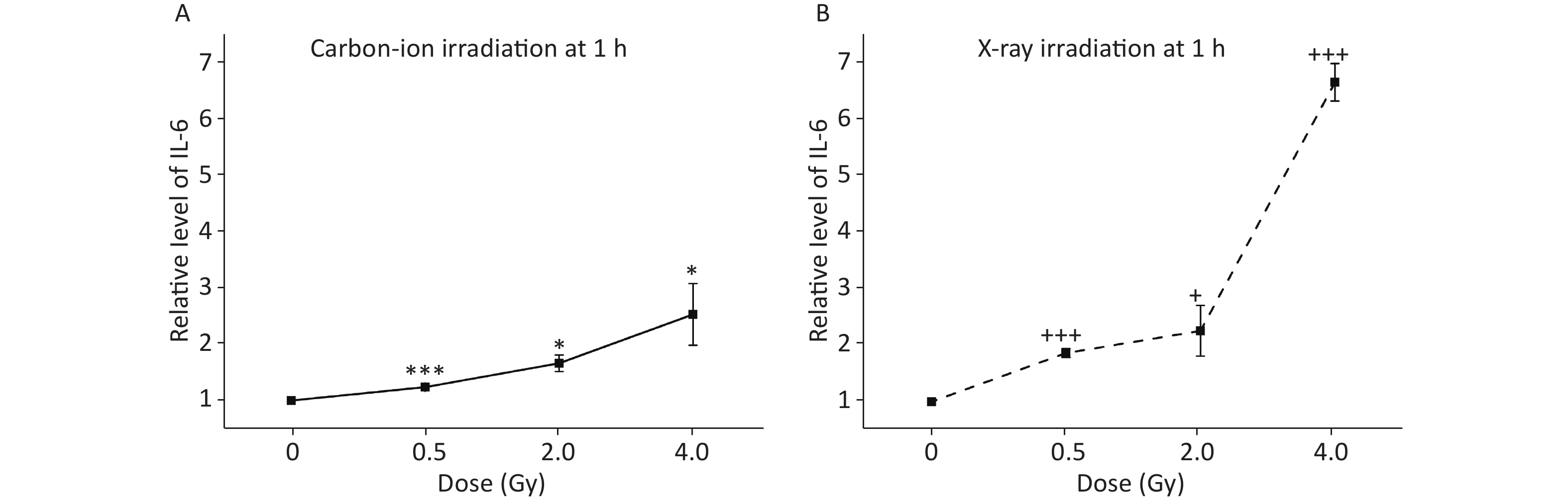
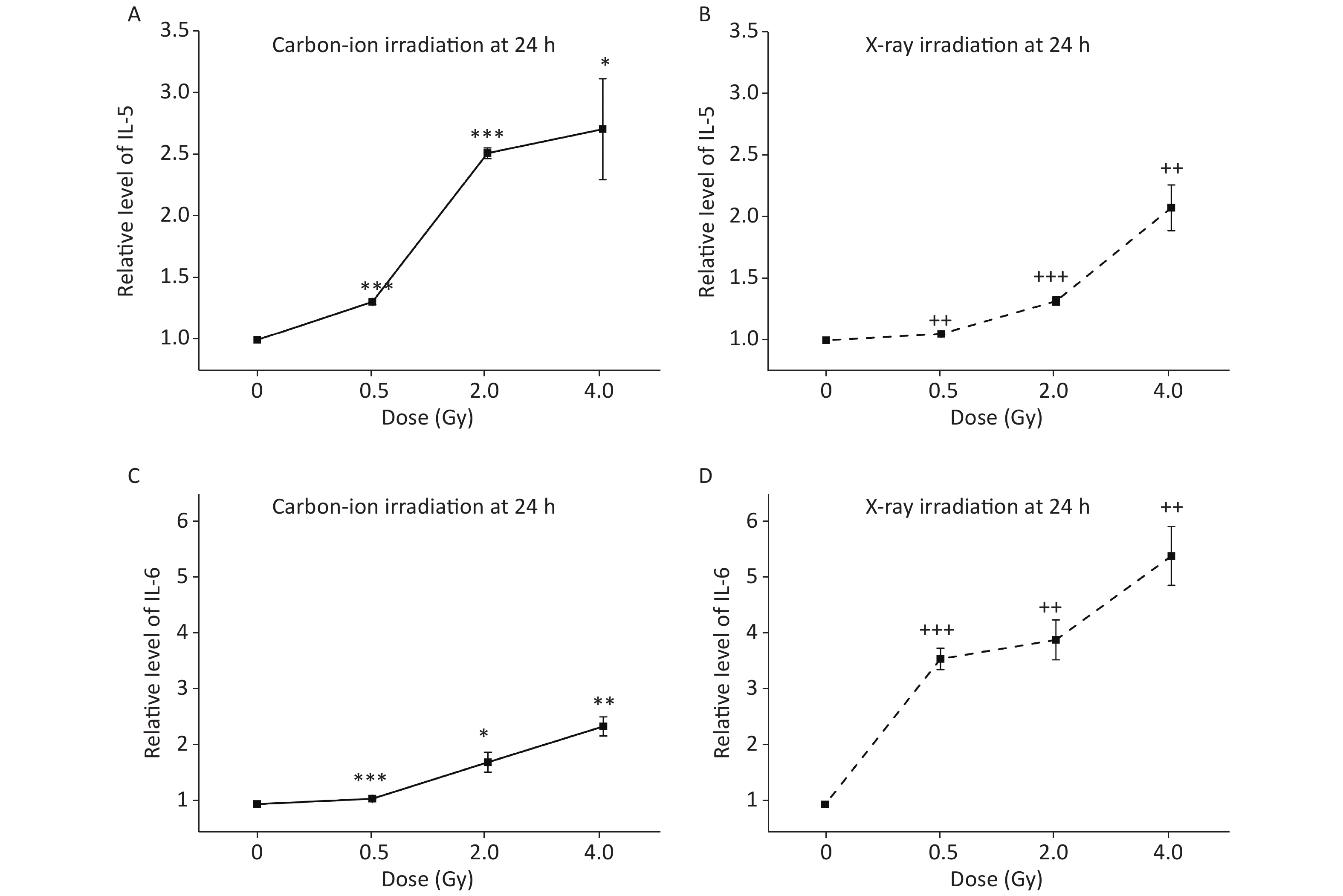
As shown in Figure 3, the relative levels of IL-6 increased significantly 1 h with after a higher dose of carbon-ion (Figure 3A) and X-ray (Figure 3B) irradiation. Figure 4 shows that the relative levels of IL-5 (Figure 4A and B) and IL-6 (Figure 4C and D) 24 h after carbon-ion or X-ray irradiation increased significantly in a dose-dependent manner. At the time point of 7 d after radiation, we found that the relative levels of 3 cytokines, IL-5 (Figure 5A and B), IL-6 (Figure 5C and D) and IL-15 (Figure 5E and F), increased significantly with a higher dose of carbon-ion or X-ray irradiation.
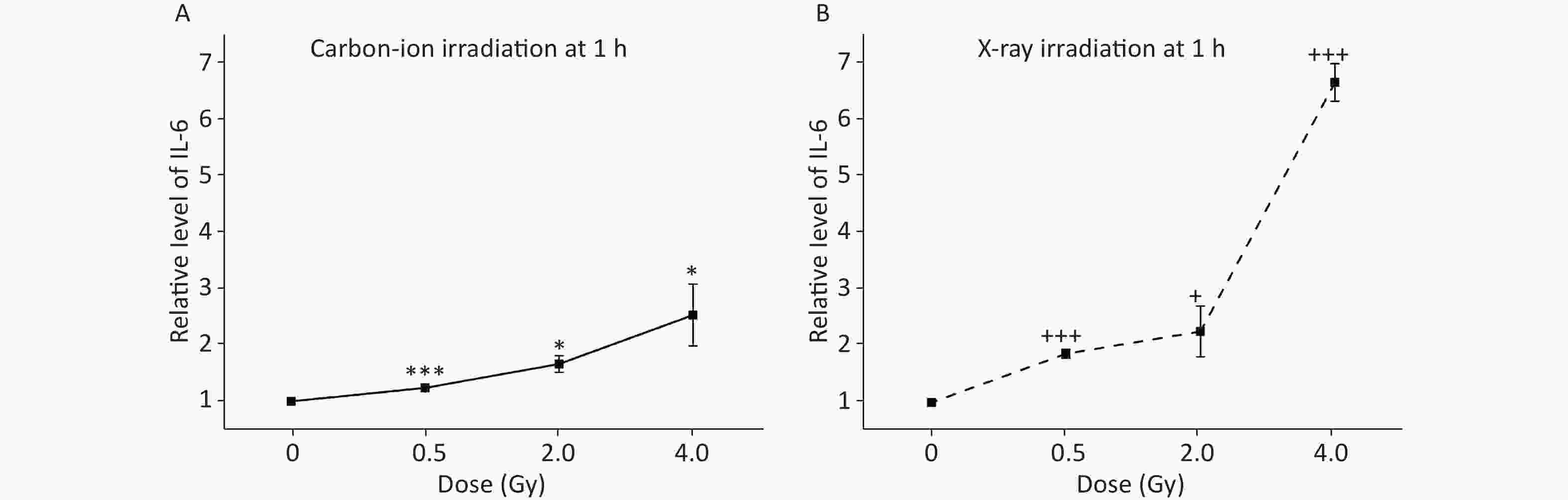
Figure 3. Relative levels of IL-6 in the plasma of total body irradiation (TBI) in mice after 1 h exposure to carbon-ion (A) and X-ray (B) radiation. *P < 0.05 and ***P < 0.001, carbon-ion irradiated groups vs. unirradiated group; +P < 0.05 and +++P < 0.001, X-ray irradiated groups vs. unirradiated group. Unpaired two-tailed Student’s t-test

Figure 4. Relative levels of IL-5 and IL-6 in the plasma of total body irradiation (TBI) in mice after 24 h exposure to carbon-ion (A and C repectively) and X-ray (B and D respectively) radiation. *P < 0.05, **P < 0.01 and ***P < 0.001, carbon-ion irradiated groups vs. unirradiated group; ++P < 0.01 and +++P < 0.001, X-ray irradiated groups vs. unirradiated group. Unpaired two-tailed Student’s t-test
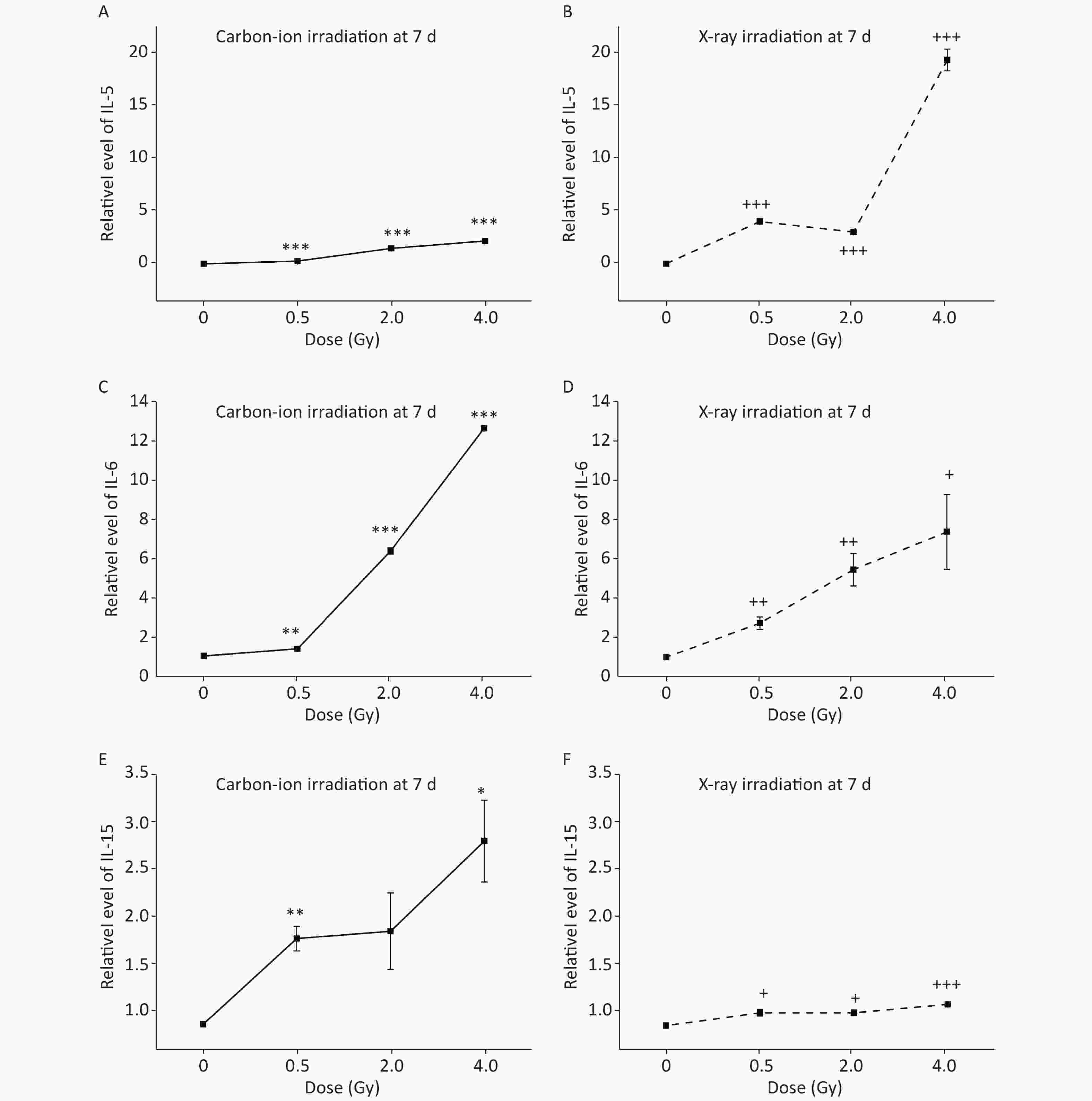
Figure 5. Relative levels of IL-5, IL-6 and IL-15 in the plasma of total body irradiation (TBI) mice after 7 d exposure to carbon-ion (A, C, E) and X-ray (B, D, F) radiation. *P < 0.05, **P < 0.01, and ***P < 0.001, carbon-ion irradiated groups vs. unirradiated group; +P < 0.05, ++P < 0.01, and +++P < 0.001, X-ray irradiated groups vs. unirradiated group. Unpaired two-tailed Student’s t-test
-
Estimating the ED of the patient who has been exposed to radiation is the first task of a radiobiologist, in order to provide reference for clinical treatment and estimate radiation risk. Thus, using a precise instrument to measure the relevant biomarkers, and then determining the potential dose using a suitable mathematical model is important. In this study, by using the precise and high-throughput MSD assay according to the data of standard samples provided by the manufacturer, we found that the minimum expression values of IL-5, IL-6 and IL-15 were measured up to 4.0076 pg/mL, 16.4538 pg/mL and 0.4150 pg/mL, respectively. In addition, we found that the relative levels of IL-6 at 1 h, IL-5 and IL-6 at 24 h, and IL-5, IL-6 and IL-15 at 7 d after radiation increased in a dose-dependent manner with both carbon-ion and X-ray radiation in mice. Thus, we assumed that these cytokines measured using the MSD assay met the essential characteristic criteria of radiosensitive biomarkers, and could be used as radiation indicators. Therefore, we tried to develop mathematical models by combining the relative levels of cytokines at different time points using multiple linear regression analysis.
To perform multiple linear regression analysis, the irradiation doses were used as the dependent variable, and the relative levels of cytokines at 1 h, 24 h or 7 d after carbon-ion or X-ray irradiation were used as independent variables. We obtained the exclusive models of multiple linear regression Equations (1)–(6) that could predict the ED for high LET or low LET radiation at different time points. X in each equation represents the relative level of the corresponding cytokine, which was normalized with the unexposed control based on MSD measurements. Here, the value of r2 in each equation represents the determination coefficient, which is close to 1, indicating that this equation is highly linearly dependent. The exclusive equations of the model for estimating the ED of carbon-ion or X-ray radiation are shown below:
1 h after irradiation
ED(carbon ions) = 1.107 + 0.385(XIL-6-1h) r2 = 0.742 (1)
ED(X-rays) = 0.468 + 0.807(XIL-6-1h) r2 = 0.85 (2)
24 h after irradiation
ED(carbon ions) = −4.163 + 0.748(XIL-5-24h) +3.123(XIL-6-24h) r2 = 0.822 (3)
ED(X-rays) = −2.78 + 2.189(XIL-5-24h) + 0.412(XIL-6-24h) r2 = 0.989 (4)
7 d after irradiation
ED(carbon ions) = −2.016 + 1.465(XIL-5-7d) −0.001(XIL-6-7d) + 0.436(XIL-15-7d) r2 = 0.99 (5)
ED(X-rays) = −5.194 + 0.152(XIL-5-7d) − 0.014(XIL-6-7d) + 5.171(XIL-15-7d) r2 = 0.964 (6)
Similarly, we attempted to establish universal models to estimate the ED for an unknown quality of incident radiation. To perform multiple linear regression analysis, the irradiation doses were used as the dependent variable, and data on the relative levels of cytokines derived from both types of radiation at 1 h, 24 h or 7 d were used as independent variables. We obtained the universal models of multiple linear regression Equations (7)–(9) that could predict the ED for an unknown quality of incident radiation at different time points. X in each equation represents the relative level of the corresponding cytokine, which was normalized with the unexposed control based on MSD measurements. The value of r2 in each equation represents the determination coefficient. The universal equations of the model for estimating the ED of unknown quality of radiation are shown below:
1 h after irradiation
ED(all data) = 0.819 + 0.471(XIL-6-1h) r2 = 0.703 (7)
24 h after irradiation
ED(all data) = −3.08 + 2.485(XIL-5-24h) + 0.387(XIL-6-24h) r2 = 0.85 (8)
7 d after irradiation
ED(all data) = −1.938 + 0.219(XIL-5-7d) − 0.001(XIL-6-7d) + 1.803(XIL-15-7d) r2 = 0.882 (9)
-
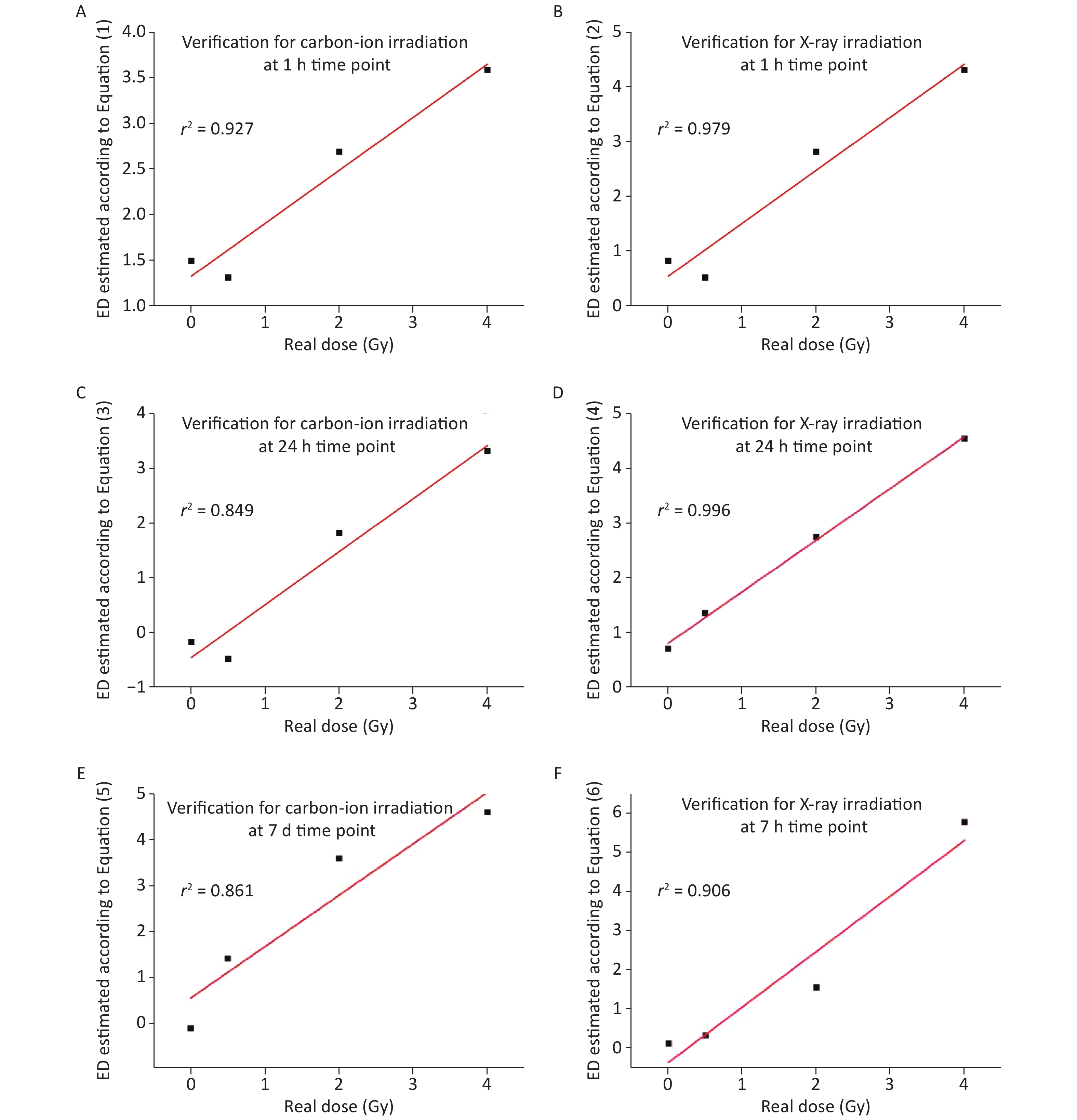
To verify the accuracy of our models, cross-validation was performed using the data from a separate experiment. The measured relative values of the cytokine levels after carbon-ion or X-ray irradiation were substituted into the exclusive Equations (1)–(6), and then a set of ED values were obtained from the equations. Furthermore, regression analysis showed that ED values calculated from the exclusive model had a positive linear correlation to practical radiation doses, and the value of r2 in each correlation analysis was greater than 0.84 (Figure 6A–F). This suggested a good degree of fitting, and that every equation was appropriate to predict the ED after the incident radiation. Our results showed that the equation of estimation for X-ray exposure is better than that for carbon-ion exposure.
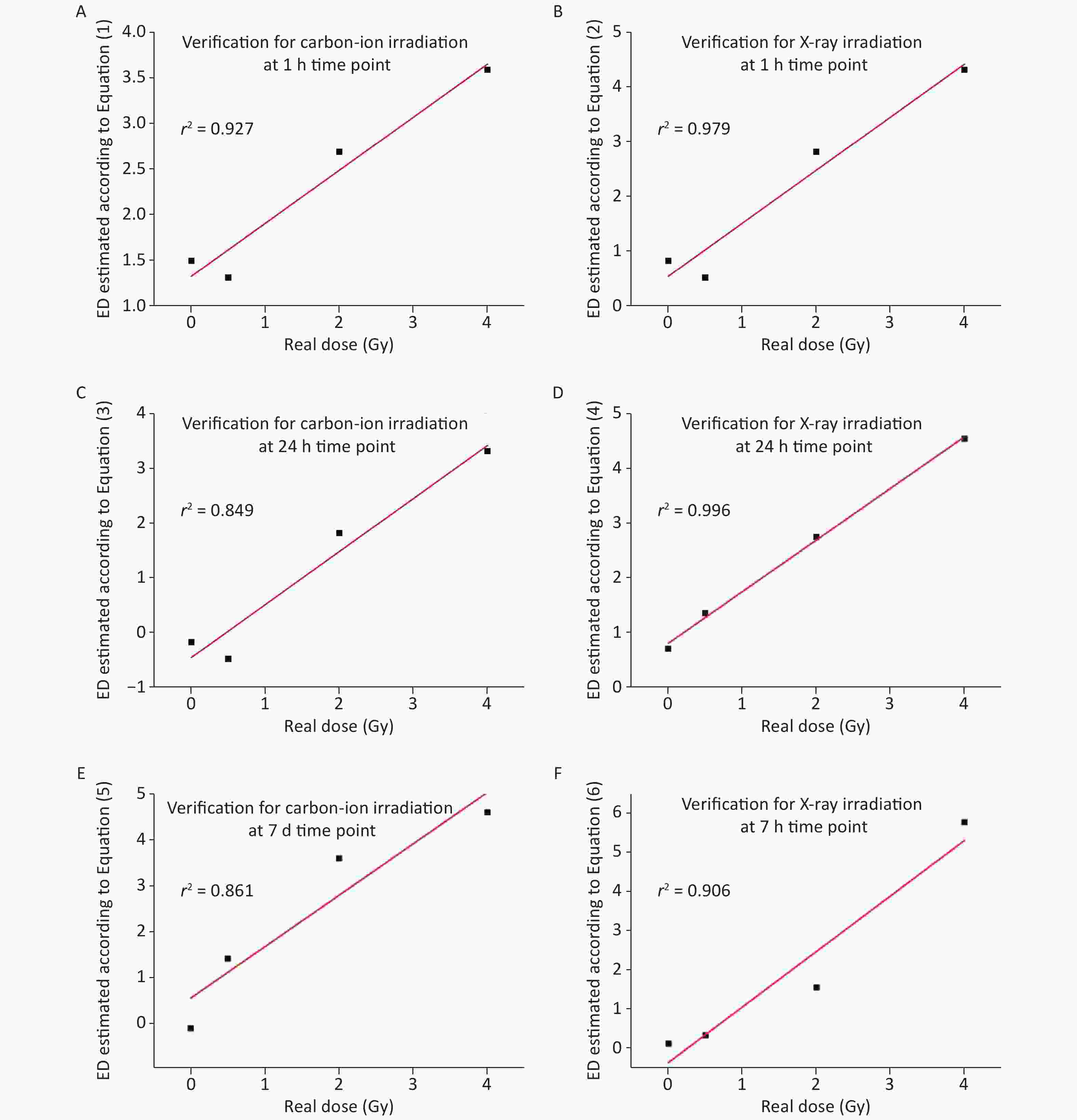
Figure 6. Verification of exclusive predict Equations (1)–(6) using correlation analysis. (A–B) The relationship between estimated ED and the actual carbon-ion or X-ray irradiation dose was verified by a separate experiment at 1 h post-irradiation. (C–D) The relationship between estimated ED and the actual carbon-ion or X-ray irradiation dose was verified by a separate experiment at 24 h post-irradiation. (E–F) The relationship between estimated ED and the actual carbon-ion or X-ray irradiation dose was verified by a separate experiment at 7 d post-irradiation
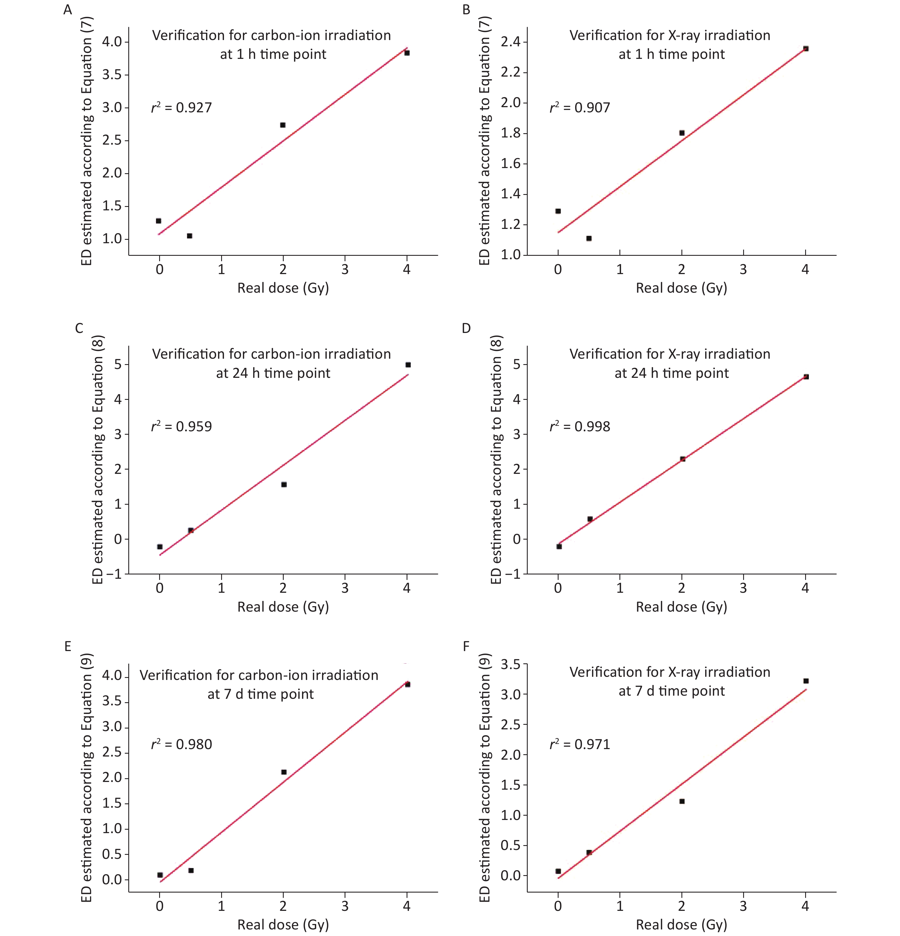
Further, the relative values of cytokine levels measured from the separate experiment were substituted into the universal Equations (7)–(9), and then a set of ED values were obtained according to each equation. Similarly, the regression analysis showed that practical radiation doses also had a positive linear correlation to the ED, in the case of either carbon-ion or X-ray radiation. The value of r2 in each correlation analysis was greater than 0.90 (Figure 7A–F), suggesting that every equation can appropriately predict the ED after an unknown quality of incident radiation. To some extent, the universal models are better than the exclusive models according to the value of r2 in each equation.
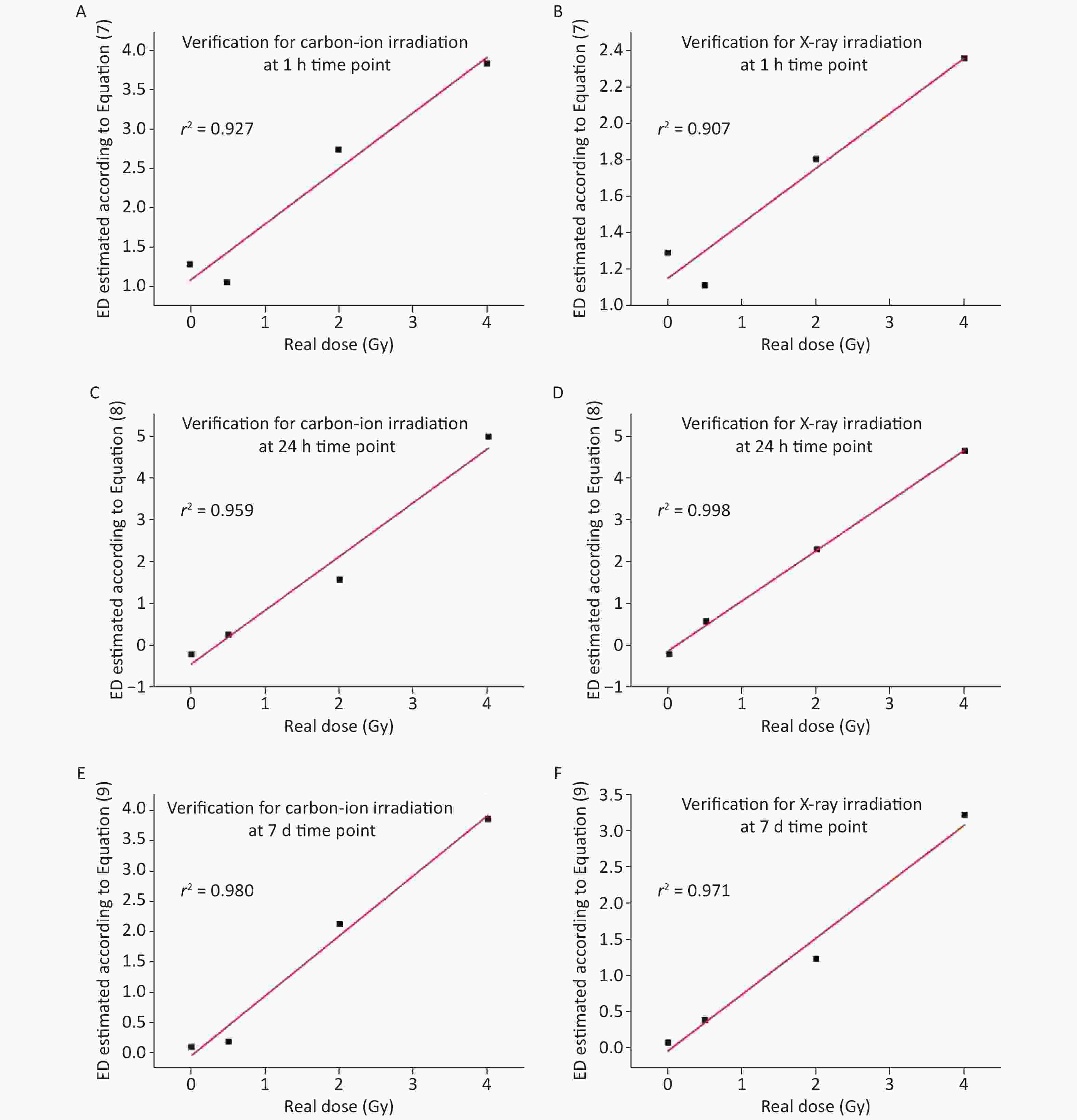
Figure 7. Verification of universal predict Equations (7)–(9) using correlation analysis. (A–B) The relationship between estimated ED and the actual carbon-ion or X-ray irradiation dose was verified by a separate experiment at 1 h post-irradiation. (C–D) The relationship between estimated ED and the actual carbon-ion or X-ray irradiation dose was verified by a separate experiment at 24 h post-irradiation. (E–F) The relationship between estimated ED and the actual carbon-ion or X-ray irradiation dose was verified by a separate experiment at 7 d post-irradiation
These results suggest that the relative levels of IL-5, IL-6 and IL-15 measured by MSD can be used to predict the ED for the incident radiation. The r2 values in each model and the cross validations suggest that these models can provide highly reliable predictions for ED after high or low LET radiation.
-
The exposure to radioactive contamination usually leads to several undesirable outcomes in humans known as acute radiation syndrome (ARS) or chronic persistent radiation sickness. This radioactive contamination includes high and low LET radiation[17-21]. Therefore, there is an urgent need to identify reliable biomarkers related to ionizing radiation and establish sensitive, rapid, minimally invasive and widely applicable detection methods to estimate the radiation dose, which is a critical factor in improving treatment efficiency.
The traditional biological methods used to estimate radiation dose include chromosome aberration analysis[22], precocious condensed chromosome fragment analysis[23], micronucleus analysis[24] and somatic mutation analysis[25]. A high-throughput method, rapid automated bisymmetry tool (RABiT), has been established by Columbia University to detect the DNA damage marker γ-H2AX[26]. However, these methods have some shortcomings. Usually, chromosome aberration analysis or micronucleus analysis require more than two days of cell culture and these experimental procedures are complex[27,28]. The analyses of some aberrations are largely affected by artificial factors or have a narrow measurement range. PCR analyses to quantify the miRNA or mRNA levels, require a more complex experimental technique[29,30]. Even the high-throughput method, RABiT, relies on blood samples from the irradiated patient and the cell culture procedure takes more than one day. Additionally, the response range between the fluorescence intensity of γ-H2AX and the irradiation dose is also narrow[31–33].
Low-dose or nonlethal environmental exposure to radiation do not cause distinct symptoms immediately, although symptoms may be observed after a few hours or days. However, these exposures can immediately and persistently alter inflammatory responses to disrupt the tissue repair processes[34]. Interestingly, changes in inflammatory cytokines can be reproducibly detected over time[35]. However, many cytokines have closely related or overlapping biological effects in vivo, and the detection of single cytokines has some limitations. Thus, it is necessary to find or develop a rapid and accurate strategy that can simultaneously measure many cytokines and quantify the levels of cytokine expression in patients after radiation exposure. Based on our experimental results and analysis, we showed that the MSD assay meets all these conditions to simultaneously detect a large number of cytokines, and we obtained more sensitive data regarding the changes in some cytokines in the plasma of irradiated mice. Among the biomarkers detected by MSD, IL-6 was detected at 0.29–1538.05 pg/mL. Some studies in which IL-6 was detected using ELISA at different irradiation conditions or time points, the minimum detectable concentration was only ≥ 50 pg/mL or ≥ 50 ng/mL[11,12]. Therefore, the MSD technique has significant advantages in measuring multiple biomarkers related to ionizing radiation and their detection precision.
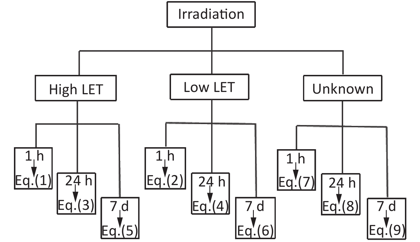
Although 21 cytokines were detected in this study after carbon-ion and X-ray irradiation, the changes in some cytokines were intricate or irregular. We found that the relative levels of IL-5, IL-6 and IL-15 expression at different time points after irradiation increased regularly with the increase in dose, regardless of the type of radiation. IL-5 is associated with the induction of terminal differentiation of late-developing B-cells into immunoglobulin-secreting cells[36]. IL-6 is involved in inflammation processes inducing an acute phase response, and plays an essential role in differentiating B-cells into immunoglobulin-secreting cells[37]. IL-15 stimulates the proliferation of T-lymphocytes and natural killer (NK) cells and plays important roles in both innate and adaptive immunity[38]. They are all involved in irradiation-induced inflammation. Therefore, according to our MSD assay, we established a set of equations for estimating the radiation ED depending on the expression of IL-5, IL-6 and IL-15 after irradiation. The cross-validation using the data of separate experiments demonstrated that our prediction equations work perfectly. In addition, both the exclusive equations for carbon-ion or X-ray irradiation and universal equations for the unknown quality of irradiation had high fitting values, and the fitting values of the equations for X-rays were higher than those for carbon-ion. As shown in Figure 8, we can easily choose a certain equation to estimate the ED according to the measured value of the relative expressions of IL-5, IL-6 and IL-15 at different time points after different or unknown quality of irradiation.
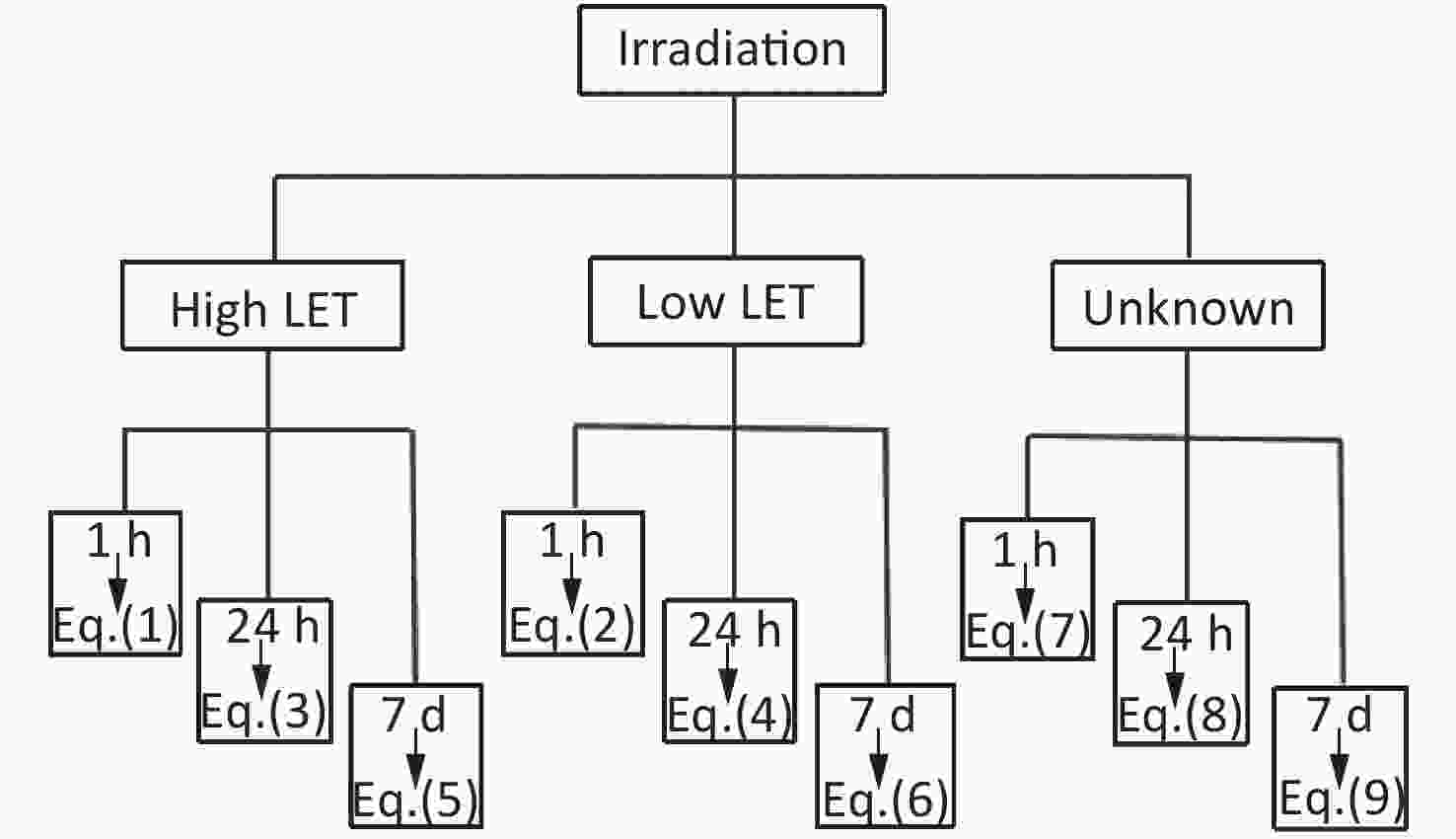
Figure 8. Schematic diagram for choosing a certain equation to estimate the ED. The blood samples of irradiated patient were collected at different time points after irradiation. The relative levels of IL-5, IL-6 and IL-15 were measured by MSD. The values of cytokine levels were substituted into a certain equation (Eq.1–9) to estimate the ED according to the irradiation condition (low, high LET or unknown quality of irradiation)
In conclusion, IL-5, IL-6 and IL-15 meet the essential characteristic criteria of radiosensitive biomarkers, and these cytokines can be used as radiation indicators depending on the MSD analysis. Our prediction models can be conveniently used to estimate the radiation ED for the incident radiation at different times, which can be used to evaluate radiation damage to improve the therapeutic approach.
-
The authors do not have any conflicts of interest.
Funds:
This study was supported by the National Natural Science Foundation of China [11635013, 11705248, U1832101]; National Key Research and Development Program of China [2017YFC0108605]; and the Science and Technology Research Project of Gansu Province [No. 145RTSA012 and 17JR5RA307]. We also appreciate the staff of the Heavy Ion Research Facility in Lanzhou for offering the 12C6+ ion [Y9HIRFL200]

 Quick Links
 DownLoad: