


-
Resveratrol, 3,5,4-trihydroxystilbene, is a natural polyphenol phytoalexin found in various plant species and red wine. Although this compound has been widely used and extensively investigated, there are still uncertainties regarding the applicable dose range and relevant toxicities in experimental animals and humans. In fact, disparate doses (5–5,000 mg/day) and variable experimental settings related to the bioactivity studies of resveratrol have been used and investigated, which might lead to variations in readouts[1].
The gut microbiota plays an important role in drug metabolism, and as a result, may alter the safety and effectiveness of a drug[2]. Specifically, the gut microbiota, in conjunction with the liver and intestinal tract contribute to resveratrol metabolism. Conversely, resveratrol also affects the gut microbiota, through which it exerts its pharmaceutical effect on certain diseases, such as cardiovascular diseases or metabolic syndrome. For example, treatment with resveratrol at 200 mg/(kg∙day) can significantly reduce body weight, visceral adipose content, and the blood levels of glucose (GLU) and lipids in high-fat diet (HFD) mice, which is believed to occur as a result of changes in the gut microbiota characterized by increased relative abundance of Bacteroidetes-to-Firmicutes, inhibited growth of Enterococcus faecalis, and boosted growth of Lactobacillus and Bifidobacterium[3]. The intake of a combination of 30 mg/(kg∙day) quercetin and 15 mg/(kg∙day) resveratrol for 10 weeks has been shown to be beneficial in HFD-induced obese rats, as a result of which HFD-related gut microbiota dysbiosis could be corrected[4]. A recent pilot, randomized, placebo-controlled clinical trial showed that 1 g/day resveratrol plus a western-style diet for 35 days is beneficial in improving insulin sensitivity and GLU homeostasis in obese men with metabolic syndrome[5].
Considering the vital roles of the gut microbiome in interfering with both health and drug metabolism, it is worth studying the impact of high-dose resveratrol on the gut microbiota profile and its relevance to host health. The aim of this study was to perform a subacute toxicity study of high-dose resveratrol (750 mg/kg) after a course of 28 consecutive daily administrations. In addition to evaluating common toxicity indicators, including mortality, body and organ weight, food intake, and blood biochemical and hematological parameters, emphasis was placed on evaluating the compositional changes in the gut microbiota.
In the current study, 10 female and 10 male Sprague Dawley (SD) rats (weighing 65–89 g, 3 weeks old) were divided into four groups: female control group (F–C), females treated with resveratrol group (F–R), male control group (M–C), and males treated with resveratrol group (M–R). Resveratrol, extracted from grapes with a purity of 98% (supplied by Hsiehs Biotech, Singapore), was dissolved in deionized water to produce the drug suspension. SD rats were purchased from Charles River Experimental Animal Technology, Beijing, China, and the experimental animal quality certificate number was 11400700260101. The rats were maintained under the following conditions: 20–23 °C, 50%–70% humidity, 12-h light/dark cycle with food and water provided ad libitum. All animal experiments were authorized by Experimental Animal Ethics Committee of Zhejiang Center for Disease Control and Prevention. According to the 450 mg/day acceptable daily intake for humans, the F–R and M–R groups were intragastrically administered 750 mg/kg of resveratrol; the F–C and M–C groups were given the same volume of deionized water for 28 consecutive days. Body weight and food intake were recorded weekly, and the food utilization rate was calculated. The rats were fasted for 12 h and then anesthetized with pentobarbital sodium (35 mg/kg) by intraperitoneal injection. The abdominal aorta blood was collected, and the liver, kidney, spleen, and testis (ovary from females) were excised. The organs were weighed, and the relative weight of the viscera body was calculated. An automatic biochemical analyzer (Hitachi 7180, Hitachi High-Tech, Tokyo, Japan) was used to measure blood biochemical parameters, including alanine aminotransferase, aspartate aminotransferase, GLU, blood urea nitrogen, urine creatinine, triglyceride, total cholesterol, total protein, and albumin/globulin. An automatic blood cell analyzer (Sysmex 1800i, Sysmex Corporation, Kobe, Japan) was used to measure hematological parameters, including the counts of white blood cells, red blood cells, lymphocytes, monocytes, granulocytes, and hemoglobin. Parts of the liver, kidney, spleen, bladder, and small intestine were fixed with 10% neutral formalin solution, embedded in paraffin, and sliced into 2–3-micron-thick sections for hematoxylin-eosin staining. Tissue histopathology was studied using a microscope. Data are expressed as the mean ± standard deviation (mean ± SD). Significant differences among groups were detected via t test using SPSS 11.5 software and P < 0.05 indicated a statistically significant difference.
During the experiment, the behavior and mortality of the rats remained unchanged. No significant changes (P > 0.05) were observed in body weight gain, feed intake, or food utilization (Supplementary Table S1, available in www.besjournal.com); absolute organ weight, relative organ weight of the liver, kidneys, spleen, or testes/ovaries over the body weight (Supplementary Table S2, available in www.besjournal.com); or abnormalities in color, shape, and size of the heart, liver, spleen, lung, kidney, stomach, intestine, and testis/ovaries (Supplementary Table S3, available in www.besjournal.com). In addition, there were no significant differences in the investigated biochemical biomarkers (Supplementary Table S4, available in www.besjournal.com) or the hematological parameters (Supplementary Table S5, available in www.besjournal.com) between groups. Taken together, we found that, in terms of mortality, behavioral changes, body and organ weights, biochemical parameters associated with liver and kidney function, lipid and glucose metabolism, and hematological parameters, the oral gavage of 750 mg/kg of resveratrol for 28 days showed no detectable subacute toxicity in either male or female rats.
Features Male Female Control Resveratrol P value Control Resveratrol P value Weight gain (g) 229.6 ± 23.1 234.9 ± 27.8 0.748 132.6 ± 16.6 156.2 ± 17.4 0.060 Total food intake (g) 859.2 ± 37.9 836.9 ± 83.0 0.600 663.0 ± 63.2 715.0 ± 51.7 0.192 Total food utilization rate (%) 26.7 ± 1.9 28.0 ± 1.3 0.229 20.0 ± 1.8 21.8 ± 0.9 0.093 Weekly food utilization rate (%) Week 1 41.6 ± 4.0 41.8 ± 2.7 0.928 39.7 ± 2.7 38.6 ± 5.8 0.711 Week 2 29.9 ± 2.7 28.2 ± 1.3 0.246 21.1 ± 2.5 24.5 ± 3.1 0.091 Week 3 21.6 ± 2.2 25.4 ± 1.1** 0.009 12.4 ± 2.9 15.1 ± 5.9 0.389 Week 4 20.0 ± 3.1 22.7 ± 2.7 0.173 11.2 ± 3.4 14.1 ± 1.8 0.131 Note. Values expressed as mean ± SD, n = 5, T test, **P < 0.01 vs. Control group. Table S1. Effects of daily oral administration of resveratrol on body weight gain, food intake and food utilization of rats
Features Male Female Control Resveratrol P value Control Resveratrol P value Liver weight (g) 8.86 ± 0.40 9.32 ± 1.22 0.446 6.43 ± 0.81 7.12 ± 0.64 0.175 Liver/body weight (%) 3.18 ± 0.18 3.29 ± 0.29 0.491 3.41 ± 0.14 3.44 ± 0.28 0.858 Kidney weight (g) 2.73 ± 0.38 2.88 ± 0.37 0.540 1.83 ± 0.16 2.01 ± 0.09 0.061 Kidney/body weight (%) 0.97 ± 0.12 1.01 ± 0.04 0.499 0.97 ± 0.02 0.97 ± 0.03 0.905 Spleen weight (g) 0.65 ± 0.15 0.75 ± 0.21 0.386 0.50 ± 0.10 0.52 ± 0.10 0.727 Spleen/body weight (%) 0.23 ± 0.04 0.27 ± 0.07 0.369 0.26 ± 0.03 0.25 ± 0.04 0.629 Testis or ovary weight (g) 3.03 ± 0.34 3.23 ± 0.40 0.423 0.13 ± 0.03 0.14 ± 0.01 0.362 Testis or ovary/body weight (%) 1.08 ± 0.03 1.14 ± 0.10 0.225 0.07 ± 0.02 0.07 ± 0.01 0.641 Note. Values expressed as mean ± SD, n = 5, T test. Table S2. Effects of daily oral administration of resveratrol on organ weights and viscera-body ratio of rats
Histopathological changes Male Female Control Resveratrol Control Resveratrol Liver Local central venous stasis 1 1 0 0 Scattered round vacuoles in the cytoplasm 2 0 0 0 Inflammatory cell infiltration in portal area 1 2 1 0 Focal inflammatory cell infiltration in lobules 2 2 2 1 Focal necrosis with inflammatory cell infiltration 1 1 0 0 Kidney Infiltration of focal inflammatory cells in renal interstitium 0 1 0 1 Infiltration of submucosal Inflammatory cells in the renal pelvis 0 0 1 1 Exudates in renal tubules 2 2 1 2 Renal cyst 0 1 0 1 Mild swelling of renal tubular epithelial cells 0 1 0 1 Spleen Mild dilatation and congestion of sinus 0 1 1 0 Bladder Focal inflammatory cell infiltration in lamina propria 0 0 1 0 Small intestine Round vacuoles occasionally appear in the lamina propria of mucosa 0 0 0 0 Note. Values represent incidences, i.e. number of rats having the lesion per group. Table S3. Effects of daily oral administration of resveratrol on the incidence of lesions observed histologically in Wistar rats
Features Male Female Control Resveratrol P value Control Resveratrol P value ALT (U/L) 58.20 ± 14.82 57.40 ± 8.90 0.920 58.60 ± 8.41 49.80 ± 5.45 0.085 AST (U/L) 315.20 ± 91.80 276.00 ± 31.74 0.409 286.40 ± 38.67 268.60 ± 17.74 0.377 GLU (mmol/L) 5.01 ± 0.49 4.60 ± 0.37 0.175 5.92 ± 1.04 5.02 ± 0.45 0.116 BUN (mmol/L) 5.00 ± 0.37 4.84 ± 1.40 0.811 6.17 ± 0.76 6.48 ± 1.03 0.603 UCR (μmol/L) 47.40 ± 3.21 43.60 ± 4.16 0.144 48.80 ± 3.03 45.80 ± 4.32 0.240 TG (mmol/L) 0.31 ± 0.21 0.34 ± 0.22 0.851 0.43 ± 0.25 0.32 ± 0.07 0.381 TC (mmol/L) 1.87 ± 0.59 1.46 ± 0.18 0.180 2.54 ± 0.58 2.25 ± 0.41 0.382 Total protein (g/L) 58.76 ± 2.99 57.86 ± 2.18 0.601 63.10 ± 2.16 63.16 ± 2.42 0.968 ALB/GLO 1.54 ± 0.09 1.64 ± 0.20 0.309 1.45 ± 0.09 1.52 ± 0.14 0.348 Note. Values expressed as mean ± SD, n = 5, T test. Table S4. Effects of daily oral administration of resveratrol on the biochemical parameters of rats.
Features Male Female Control Resveratrol P value Control Resveratrol P value WBC (×109/L) 8.22 ± 3.45 9.03 ± 4.30 0.750 8.17 ± 2.25 9.48 ± 2.90 0.448 RBC (×1012/L) 7.78 ± 1.11 7.79 ± 0.44 0.986 7.56 ± 0.98 7.14 ± 0.19 0.374 HGB (g/L) 158.80 ± 26.41 157.60 ± 5.55 0.923 155.20 ± 13.22 141.60 ± 6.43 0.072 LYM (%) 68.68 ± 3.69 71.44 ± 2.76 0.217 69.44 ± 6.48 71.82 ± 2.15 0.458 MONO (%) 1.66 ± 0.28 1.20 ± 0.46 0.094 1.70 ± 0.76 1.80 ± 0.75 0.840 GRA (%) 29.66 ± 3.51 27.36 ± 2.56 0.270 28.86 ± 5.87 26.38 ± 2.12 0.400 Note. Values expressed as mean ± SD, n = 5, T test. Table S5. Effects of daily oral administration of resveratrol on the hematological parameters of rats
We evaluated the impact of resveratrol on gut microbiota structure by employing 16S rRNA (V3-V4 region) gene sequencing technology. Fecal samples collected at the end of the experiment were sent to Suzhou Precision Gene Co., Ltd. for 16S rRNA gene sequencing. Briefly, DNA was extracted using a fecal DNA isolation kit (Tiangen Biotech Co., Ltd., Beijing, China). For each fecal sample, amplicon PCR was performed on the V3-V4 region of the 16S rRNA gene using the primer pair. The sequences for the primer pairs with barcodes are 341F:5’-CCTAYGGGRBGCASCAG-3’ (forward) and 806R:5’-GGACTACNNGGGTATCTAAT-3’ (reverse). The PCR products were mixed in equidense ratios. The amplicons were pooled in equal concentrations for sequencing on an Illumina MiSeq PE 250 sequencing platform. Raw sequencing data are available in the Sequence Read Archive database of NCBI and were connected to bioproject PRJNA541101. The raw fastq files obtained by the Illumina sequencing machine were quality-filtered using Trimmomatic, vsearch, and other tools. A high-quality sequence was used for community structure analysis using the QIIME pipeline. The operational taxonomic unit (OTU) picking method was performed using the UCLUST closed reference method, and the representative OTUs were assigned taxonomy using the UCLUST classifier, with Greenegenes used as the reference dataset. The Chao1 and Shannon indices were measured and principal component analysis (PCA) was performed; further statistical analyses were carried out using R. Linear discriminant analysis effect size (LEfSe) (
http://huttenhower.sph.harvard.edu/galaxy/root?tool_id=lefse_upload ) combined the standard tests (Kruskal-Wallis sum-rank test and Wilcoxon rank-sum test) with linear discriminant analysis (LDA) for statistical significance comparison. The version of R software used in the current study was R 3.6.1. After removing the chimera, a total of 879,409 effective reads were produced from all samples, with an average of 43,970 ± 12,237 reads in each sample (Supplementary Table S6, available in www.besjournal.com). Rarefaction curve analysis suggested that the sequencing depth included rare new phylotypes and most of the diversity (Supplementary Figure S1, available in www.besjournal.com).ERN Q30 (%) GC (%) F-C-1 20349 71.06 52.11 F-C-2 57407 85.26 52.7 F-C-3 51545 85.29 52.2 F-C-4 59032 83.17 52.4 F-C-5 20986 71.0 52.49 M-C-1 35064 83.76 52.39 M-C-2 58849 85.76 52.24 M-C-3 44484 85.03 52.72 M-C-4 38489 83.03 52.57 M-C-5 60991 83.41 52.49 F-R-1 38403 83.55 52.3 F-R-2 42060 83.59 52.89 F-R-3 58651 85.21 52.38 F-R-4 30362 81.9 53.14 F-R-5 37649 83.22 52.58 M-R-1 40105 82.85 52.89 M-R-2 55479 85.57 52.62 M-R-3 48339 84.7 52.69 M-R-4 42649 84.05 51.76 M-R-5 38516 83.35 52.18 Means ± SD 43,970 ± 12,237 82.74 ± 4.14 52.49 ± 0.32 Note: ERN: effective reads number; Q30: base sequencing error rate was 1/1000; GC%: the percentage of guanine and cytosine. Table S6. 16S rRNA gene sequencing data summary
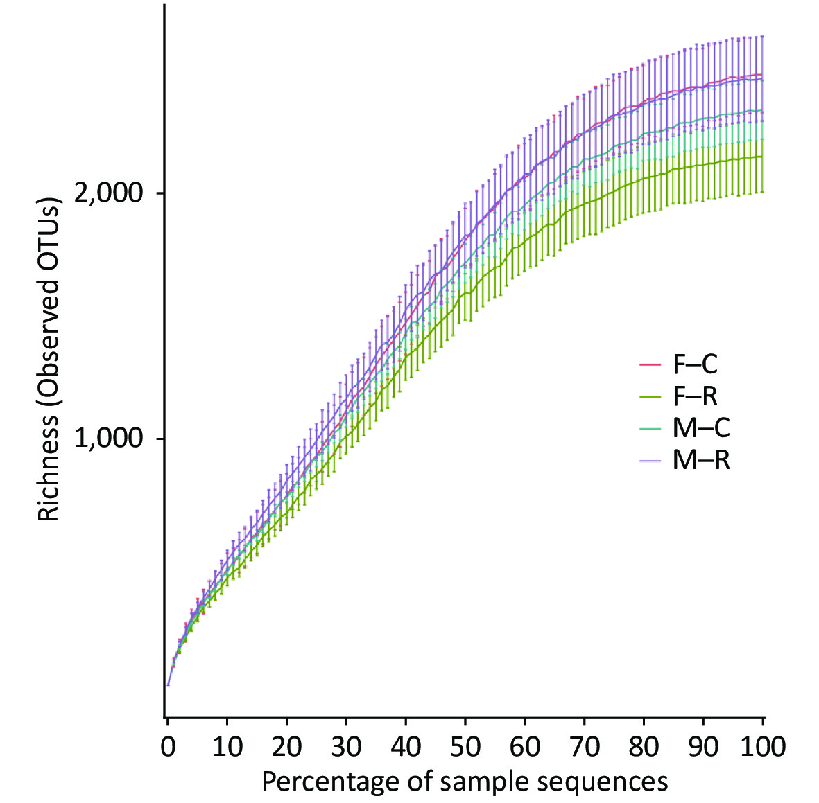
Figure S1. Percentage of sample sequences
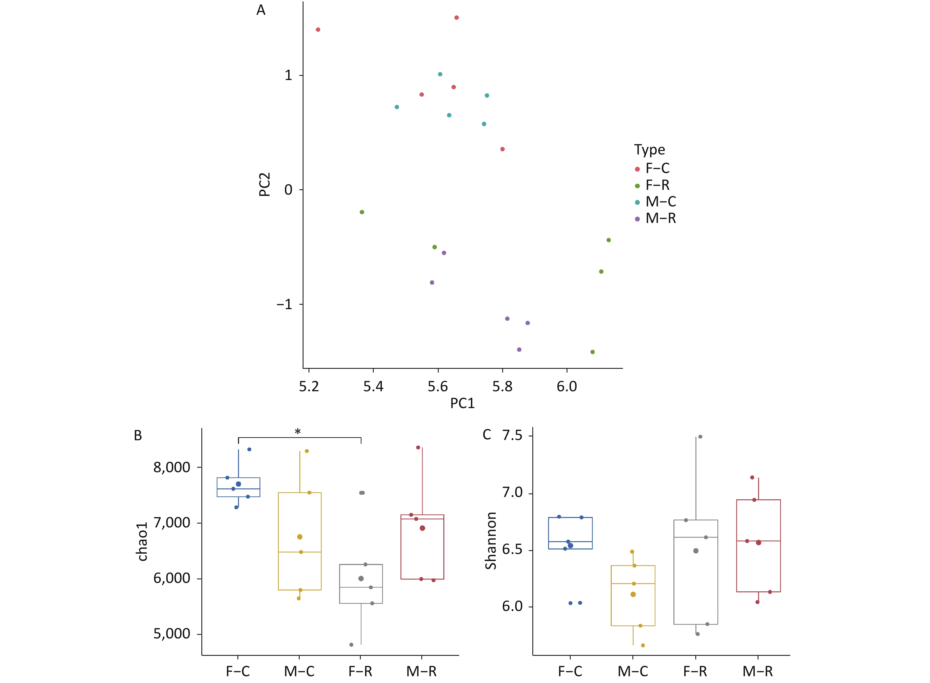
PCA showed a distinct clustering in the microbiota composition of the control groups (F–C group and M–C group) and resveratrol treatment groups (F–R group and M–R group) (Figure 1A); however, the gut microbiota of the F–C group did not significantly differ from that of the M–C group, which indicated that resveratrol reconstructed the overall structure of gut microbiota to a new state in both male and female rats.
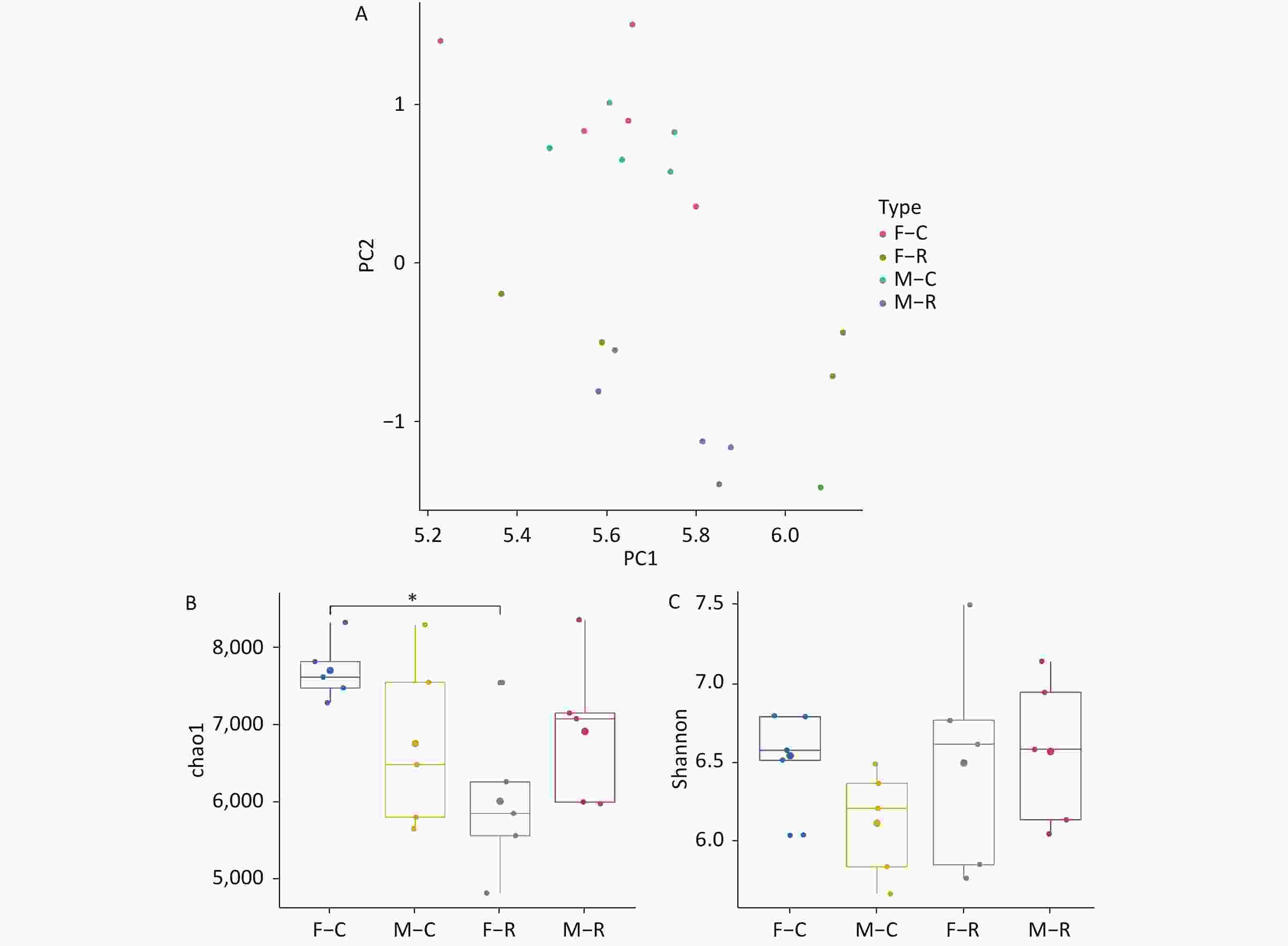
Figure 1. Resveratrol treatment reconstructed the bacterial community and changed the diversity of rat gut microbiota (n = 5). (A) Plots were generated using the principal components analysis (PCA) data. Orange: F–C; green: F–R; blue: M–C; purple: M–R. (B) Chao1 index; each dot represents a rat, data are expressed as the medians (n = 5). (C) Shannon index; each dot represents a rat, data are expressed as the medians (n = 5). *Compared with F–C group, P < 0.05.
The Chao1 and Shannon indices are indicators of gut microbiota richness and diversity, respectively, which together can reflect the healthy state of the gut microecology. The median values of each group are presented in Figure 1B and 1C. Compared with the F–C group rats, the M–C group rats showed relatively low richness and diversity of the gut microbiota. After resveratrol treatment, compared to the F–C group, while the diversity did not change, the richness of the F–R group rats decreased significantly (P < 0.05). In contrast, compared to the M–C groups, the richness and diversity of the M–R group rats were both slightly increased in response to resveratrol treatment. In addition, resveratrol significantly decreased the richness of gut microbiota in female rats (P < 0.05), but significantly increased the richness and diversity of the gut microbiota in male rats (P < 0.05). Previous studies have demonstrated that greater diversity in the gut microbiota is related to a relatively healthy gut environment[6]. With regard to richness and diversity, we speculate that 750 mg/kg of resveratrol is associated with a healthier gut microecology in male rats, but a weaker gut microecology in female rats.
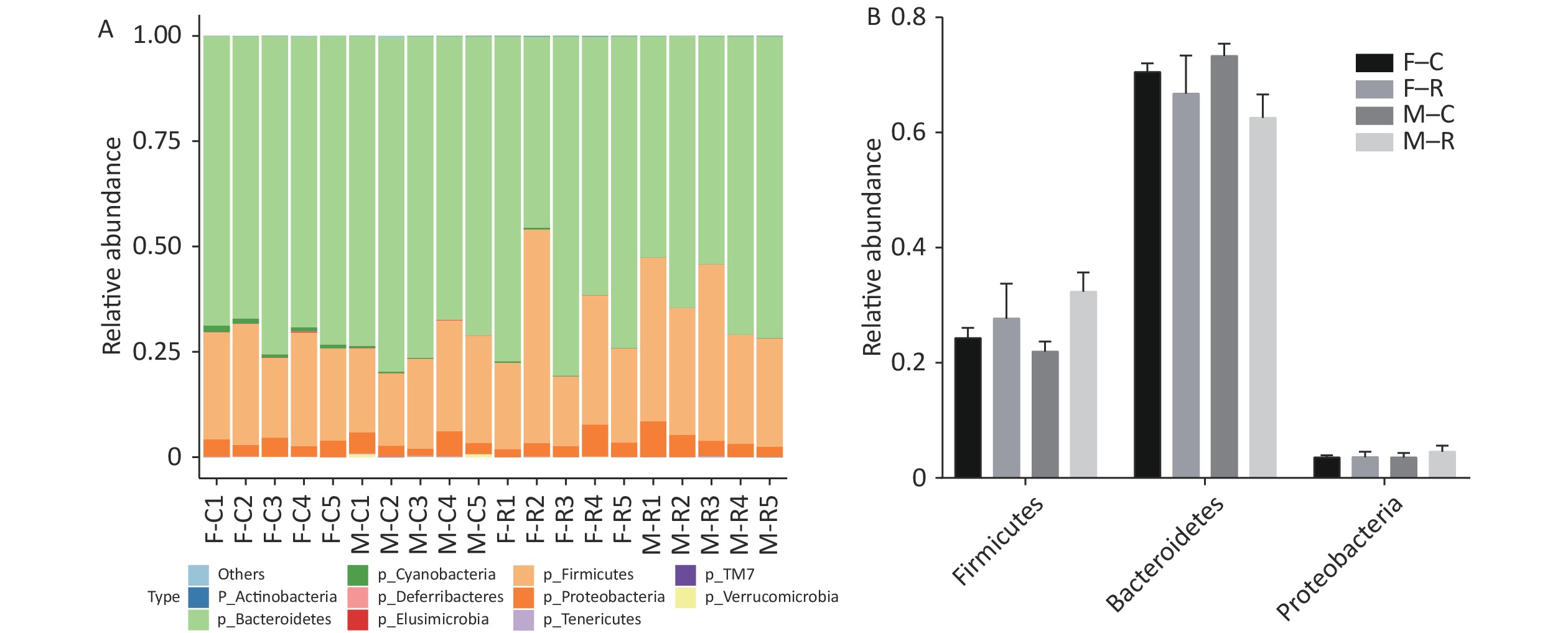
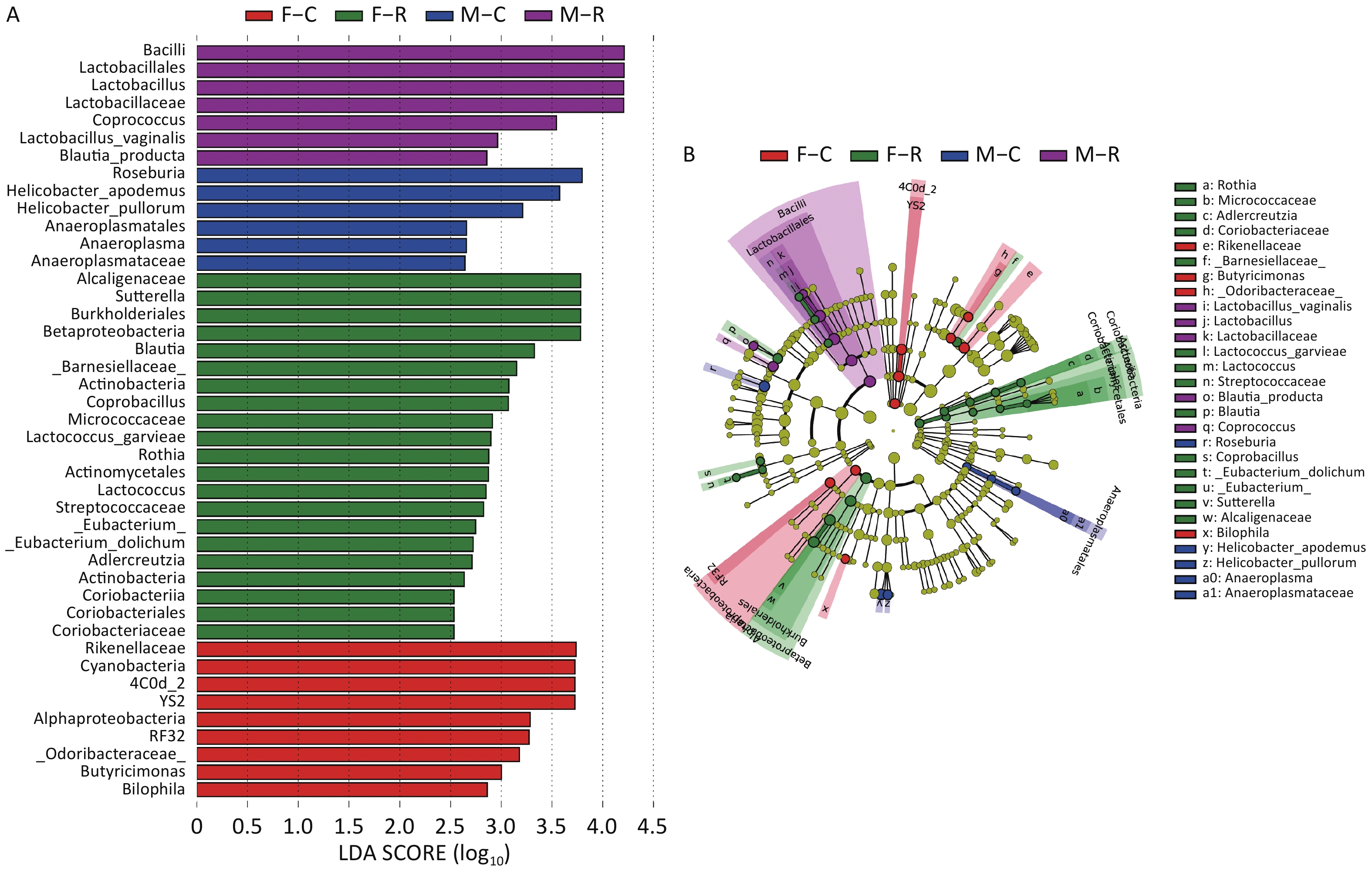
The relative abundance for each group at the phylum level is presented in Figure 2A and 2B. Bacteroidetes, Firmicutes, and Proteobacteria were the dominant bacterial phyla in the gut microbiota of rats. There was no significant difference in Bacteroidetes, Firmicutes, and Proteobacteria between all groups, suggesting that neither resveratrol nor sex had an obvious effect on gut microbiota at the phylum level. Thus, it is more likely that the differences in richness and diversity were due to the existence of specific bacteria differentially responding to resveratrol treatment in female and male rats. To address this assumption, LEfSe analysis was performed with an LDA score threshold of > 2.5 for discriminative features. Our results showed that that resveratrol was associated with enriched Alcaligenaceae, Sutterella, Burkholderiales, and Betaproteobacteria in the gut of female rats, but enriched Bacilli, Lactobacillales, Lactobacillus, Lactobacillaceae, and Coprococcus in the gut of male rats (Figure 3A and 3B). Among them, Lactobacillus is the main microorganism employed as a probiotic, which can confer a health benefit to the host, such as preventing the occurrence of gastrointestinal diseases, modulating the immune system, and maintaining the homeostasis of the gut microbiota[7]. Previous studies have indicated that probiotic-mediated prevention of food allergy was conferred by an enrichment of Coprococcus[8], which is also considered to be a short-chain fatty acid-producing bacteria[9]. In summary, we provided evidence showing that while resveratrol constructed a relatively beneficial characteristic bacterial profile for male rats, it was relatively harmful for female rats.
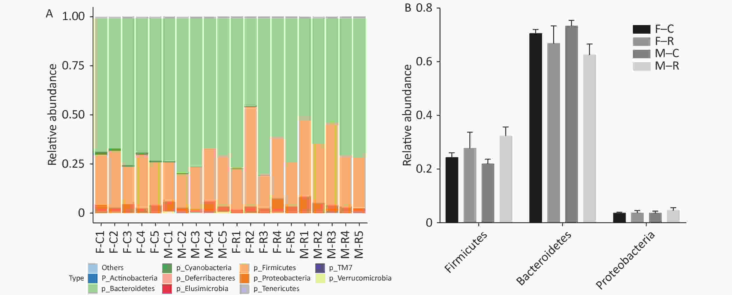
Figure 2. The relative abundance for each group at the phylum level. (A) The effect of resveratrol treatment on the relative abundance of members of the microbiota at the phylum level. The relative abundance of the 10 top-ranked phylum are presented. (B) The relative abundance of Firmicutes, Bacteroidetes, and Proteobacteria in four groups.
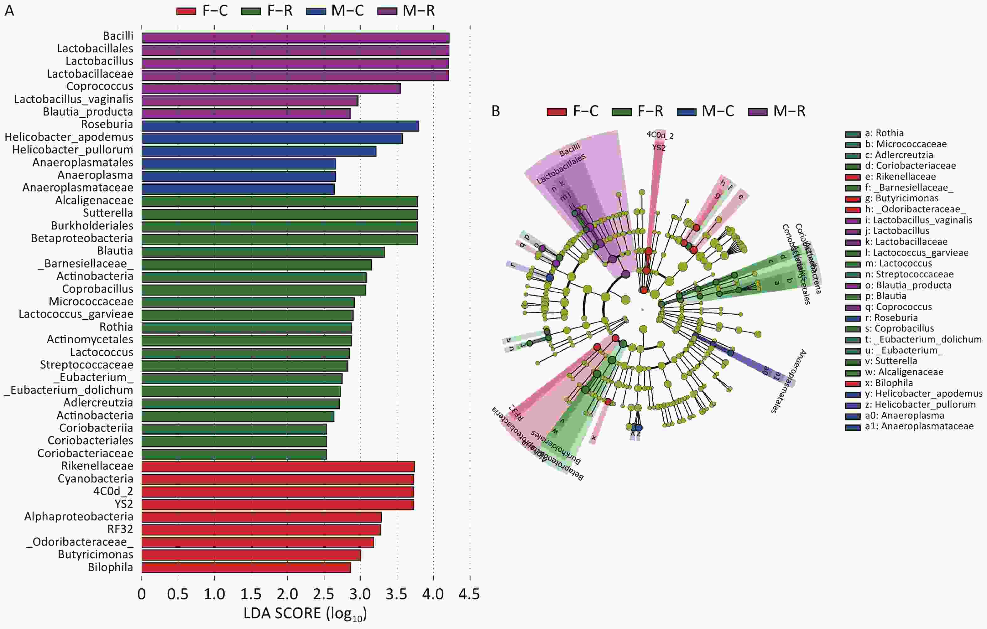
Figure 3. The characteristic bacterial taxa responding to the resveratrol treatment. (A) Linear discriminant analysis (LDA) scores were computed for differentially abundant taxa in the feces microbiomes among four groups. The LDA score indicated the effect size and ranking of each differentially abundant taxon (LDA > 2.5). (B) Taxonomic cladogram from LEfSe showed differences in feces taxa. The circles are marked in red (elevated in F–C group), green (elevated in F–R group), blue (elevated in M–C group), purple (elevated in M–R group), and yellow (non-significant changes among four groups). Letters correspond to the right taxa.
In the current study, the sample size for each group (n = 5) was relatively small, and there was an unstable appearance of gut microbiota composition in one group. Together with the fact that only one dose of 750 mg/kg resveratrol was applied, our understanding of the differences between male and female rats is limited. Further assessments with larger experimental scales to investigate dose-dependent effects are required to gain sufficient evidence on the safety of resveratrol. Because of the strong metabolic capacity of the gut microbiota, drug metabolism as well as its toxicity or efficacy could be greatly affected[10]. Accordingly, we will also conduct future studies to verify the complicated interactions between resveratrol, the gut microbiota, and the metabolism of resveratrol in the gut.
In conclusion, we revealed that while using regular evaluation methods, no toxicity to both male and female rats was detected, and 750 mg/kg of resveratrol affected the gut microecology in a sex-dependent manner. Our study indicated that further assessments are required to gain sufficient safety evidence for resveratrol, as well as other pharmaceutical drugs, regarding gut health.
Conflicts of Interest There is no conflict of interest in this study.
Acknowledgements The authors gratefully acknowledge the Suzhou precision gene Co., Ltd. for providing assistance with the data analysis of 16S rRNA gene sequencing.
HTML
Reference

 Quick Links
 DownLoad: