


-
The Chinese liver fluke, Clonorchis sinensis (C. sinensis), generally parasitizes the liver and hepatic duct causes a serious food-borne parasitic disease[1,2]. C. sinensis causes mechanical stimulation and obstruction, and its metabolites and secretions[3] lead to local expansion, hyperplasia, inflammation, and even nodules in the hepatic ducts. This results in fatty degeneration, atrophy, and necrosis of adjacent liver cells and even liver cirrhosis and cancer[4-6].
C. sinensis is mainly prevalent in China, South Korea, northern Vietnam, and parts of Russia[7,8]. Around 15 million humans have been estimated to be infected worldwide, with 85% in China[9]. The prevalence of C. sinensis is subject to the combined effects of social, environmental, and control factors, and the main reason is human ingestion of C. sinensis metacercaria[10].
Except for Xinjiang, Inner Mongolia, Gansu, Qinghai, Tibet, Ningxia, and other provinces and autonomous regions in China, C. sinensis has been spread or reported in 25 provinces and cities, with the number of patients as high as 13 million. In some areas of Hunan Province, the infection rate of C. sinensis has always been high because of the consumption of sashimi and salted fish by the local residents and cross-pollution of raw and cooked food and tool containers[11]. In 1990, C. sinensis was reported in 20 counties (cities and districts), including Qiyang County and Lengshuitan District, in Hunan Province. Approximately 2.1%–85.2% of the population in these areas were infected, and the age of the infected people ranged from 2 to 75 years[12]. The infection rate in children (less than 15 years of age) was 19.8%, and that in adults was 2.5%. In Tongdao Coungty, Huaihua, the infection rate of C. sinensis has always been high. In 2011, a survey of C. sinensis in humans was conducted in Tongdao County, and the infection rate in the population was more than 70%. However, no studies have investigated the prevalence of C. sinensis or the types of epidemic strains in this county, in fact, no C. sinensis cases have been reported in the counties and cities around Tongdao County.
Between 2016 and 2020, many surveys and studies have been conducted on human parasitic diseases in 122 counties (districts) and 14 cities (prefectures) across Hunan Province. In addition, a detailed investigation was performed on the infection status of the human population and fish metacercariae in Qiyang County, Lengshuitan District, and Tongdao County, where the infection rate was high.
The mitochondrial genome has a simple structure, maternal inheritance, lack of recombination, and rapid evolution rate; therefore, it is particularly suitable as a marker for genetic research, and it has been used by many scholars to study genetic variations between and within a variety of parasites[13,14]. Two gene fragments, partial sequences of the mitochondrial cytochrome c oxidase subunit I (Cox1) gene of C. sinensis and nicotinamide adenine dinucleotide dehydrogenase (NADH) subunit I gene, have been used to study the genetic diversity of C. sinensis in Qiyang County, Lengshuitan County[15,16], and Tongdao District. In this study, the genetic relationship after the sequencing was evaluated[17-20].
-
Between 2016 and 2020, C. sinensis was monitored in 14 cities (prefectures) and 122 counties (districts) in Hunan Province. Fecal samples (> 30 g) were collected from the subjects, and a modified Kato-Katz method was used to evaluate the infection rate and infectiosity of C. sinensis[21]. C. sinensis eggs identified in the smears were counted using microscopy[22].
Take a part out from the fresh fecal sample to cover a 8 cm × 8 cm, 80-mesh nylon silk; scrape the fine fecal sample and place it in the round table hole of the plastic quantitation plate until it is filled up, about 38.75 mm3 (4 cm × 3 cm × 1 cm); after mixing thoroughly, cover it with hydrophilic cellophane containing malachite green (mixed evenly and soaked according to the ratio of 100 mL distilled water, 100 mL pure glycerin, 1 mL malachite green (or methylene blue, 3%) and flatten it to spread the feces under the hydrophilic cellophane without overflowing the glass slide and form a uniformly thick round fecal film with a diameter of about 2 cm[22]; place the prepared modified Kato-Katz thick smear at room temperature to make it transparent before microscopic examination. If C. sinensis eggs are discovered in the smear, it is determined to be positive, and the C. sinensis eggs are counted by microscopy.
-
A preliminary investigation of the infection rate of C. sinensis was conducted among the human population and metacercariae of commercial freshwater fish in three counties (districts) south of Hunan, Tongdao County, Qiyang County, and Lengshuitan District, where the infection rates were relatively high. Small wild fish, especially crucian carp, parabramis pekinensis, cyprinuscarpio, and grass carp (often made into sashimi), were collected from fish ponds around the epidemic area. The direct compression method was used to check for fish metacercariae: A piece of muscle (about 0.5 g) was cut from the dorsal, pectoral, and caudal fins of each fish, placed between two glass slides, and squeezed until a translucent mist was obtained, and active metacercariae were detected under a microscope and counted.
-
Cats and dogs (more than 5 years of age) livers were purchased from counties and towns located to the east, west, south, north, and middle of Qiyang County, Lengshuitan District, and Tongdao County, one final host was collected from each (Figure 1) [23,24]. The animal liver and bile ducts were dissected, the C. sinensis adults were washed out with distilled water, counted, and stored in 90% ethanol.
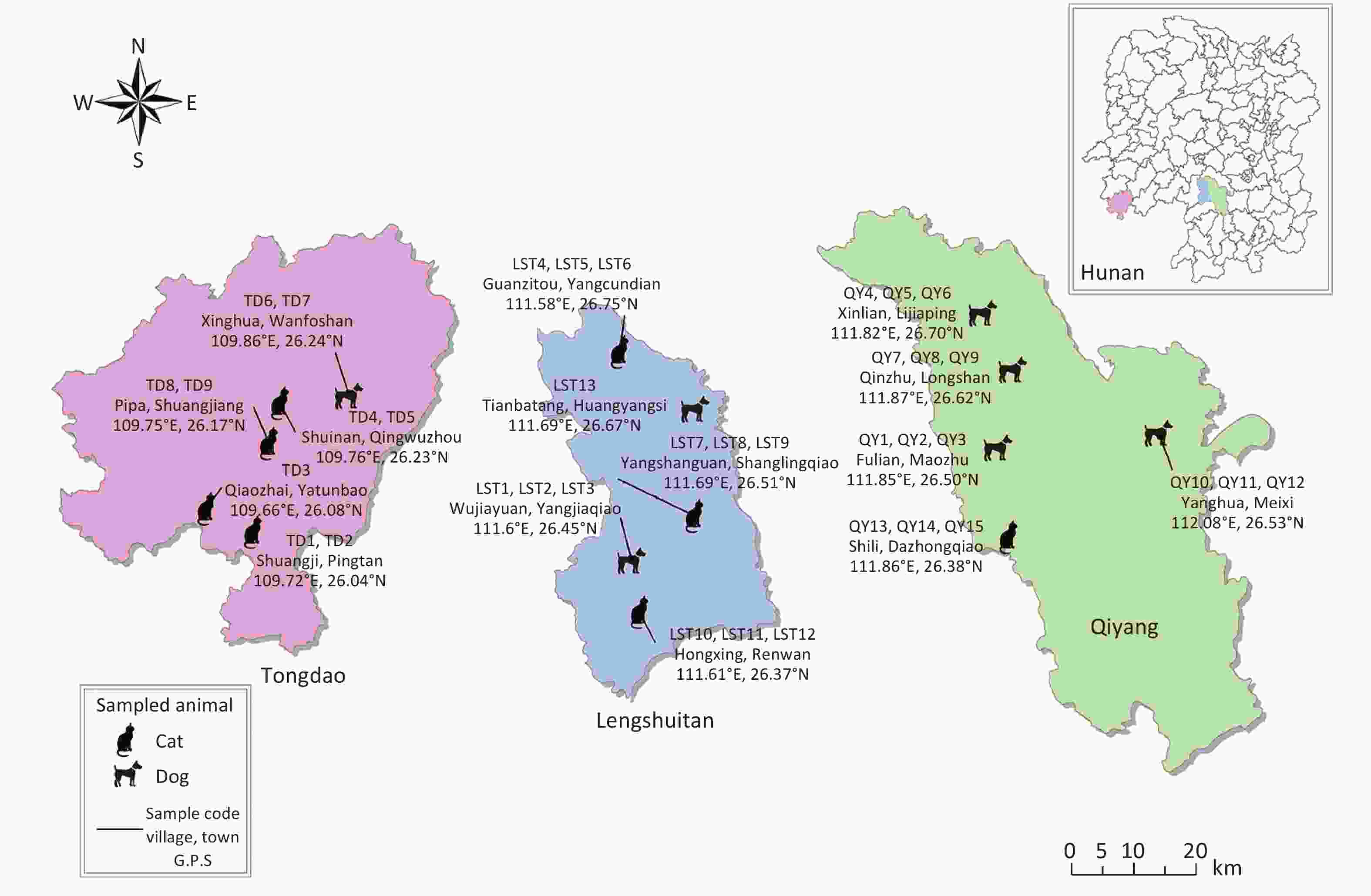
Figure 1. Sampling map for Tongdao, Qiyang, and Lengshuitan. Samples were obtained from the following towns in Tongdao: Shuangji Village of Pingtan Township (cats, sample numbers TD1 and TD2), Qiaozhai Village of Yatunpu Town (cat, sample number TD3), Shuinan Village of Jingwuzhou Town (cats, sample numbers TD4 and TD5), Xinghua Village of Wanfoshan Town (dogs, sample numbers TD6 and TD7), and Pipa Village of Shuangjiang Town (cats, sample numbers TD8 and TD9). Samples were collected from the following towns in Lengshuitan: Wujiayuan Neighborhood Committee of Yangjiaqiao Sub-district (dogs, sample numbers lST1, lST2, and lST3), Guanzitou of Yangcundian Township (cats, sample numbers lST4, lST5, and lST6), Yangshanguan Village of Shanglingqiao Town (cats, sample numbers lST7, lST8, and lST9), Hongxing Village of Renwan Street (cats, sample numbers lST10, lST11, and lST12), and Tianbatang Village of Huangyangsi Town (dog, sample number lST13). Samples were obtained from the following towns in Qiyang: Fulian Village of Maozhu Town (dogs, sample numbers QY1, QY2, and QY3), Xinlian Village of Lijiaping Town (dogs, sample numbers QY4, QY5, and QY6), Qingzhu Village of Longshan Street (dogs, sample numbers QY7, QY8, and QY9), Yanghua Village of Meixi Town (dogs, sample numbers QY10, QY11, and QY12), and Shili Village of Dazhongqiao Town (cats, sample numbers QY13, QY14, and QY15).
The C. sinensis adults were puted into the ultra-low temperature refrigerator for freeze-thaw, the adults was then cut into pieces with scissors, and the broken adults C. Sinensis were puted into ice for tissue homogenization. Nucleic acid was extracted from the homogenized tissue using the DNA Mini Kit QIAGEN Nucleic Acid Extraction Kit[25].
The partial sequences of C. sinensis Cox1 and Nad1 were used as the amplification sites for PCR[26, 27], and the amplification conditions were as follows: 5 min pre-denaturation at 94 °C, followed by 30 s denaturation at 94 °C, 30 s annealing at 55 °C, and 30 s extension at 72 °C and finally 5 min extension at 72 °C. The PCR reaction system were Forward Primer (10 μm) 1 μL, Reverse Primer (10 μm) 1 μL, 2xTaq Master Mix 10 μL, DNA 1 μL, and finally ddH2O was added to 20 μL. The amplified products were electrophoresed with the QIAxcel Advanced automatic electrophoresis apparatus (Qiagen Germany). The selected PCR products underwent recovery and purification, and they were sent to Invertrogen Biotechnology Co., Ltd (USA). For bidirectional sequencing.
The complete mitochondrial genome sequence of C. sinensis from Tongdao (
http://www.primer3plus.com/cgi-bin/dev/primer3plus.cgi ) was used to design primers COX-F (-TTGGTTATGGGGGCTTGGTG-), COX-R(-CGCTCAGATCTCAGCAGGTT-), NAD-F(-GAGCGGCTAGATCGGGTTAG-), and NAD-R (-GTGTTGCACGCGACCAAATA-). These primers were synthesized by Invertrogen Biotechnology Co., Ltd (USA). -
Excel 2019, SPSS 18.0, Instata 10.0 and other software were used for statistical analysis. In the analysis, homogeneity test of variance was conducted first. If homogeneity of variance was observed, one-way ANOVA was used for overall comparison, and then pair one-factor t-test between the two groups was conducted by Dunnett method after differences were found. If the variance is not uniform, the original data should be converted into appropriate variables to meet the homogeneity test of variance, and then the converted data will be used for statistics. If the purpose of homogeneity of variance is still not achieved after the conversion of variables, the rank sum test is used for statistics, and the difference in population comparison is found, then the pair comparison is performed by Tamhane's ST2 test, which does not require homogeneity of variance.
Pair chi-square χ2 test was used to compare the difference in the detection rate of C. sinensis infection which detected by the three methods, and Kappa test was used for consistency analysis. P < 0.05 was considered statistically significant.
-
All the obtained sequences were checked using chromatograms and Contig Express and manually corrected for scoring errors. Chromas Lite version 2.1 was used to detect the sequences. DNAMAN was used to splice the sequences in both directions.
The unique haplotype was found using DNASP 5.10, and the following organisms were searched in GenBank: C. sinensis from Russia (FJ381664), C. sinensis from Guangdong (JF729303), C. sinensis from Gansu (JF729304), C. sinensis from USA (NC_012147), Opisthorchis felineus (EU921260), Opisthorchis viverrini (JF739555), Fasciola hepatica (NC_002546), Paragonimus westermani (NC_027673), Schistosoma haematobium (DQ157222), Schistosoma japonicum (NC_002544), Trichobilharzia regenti(NC_009680), Taenia solium (NC_004022) Schistosoma mekongi (NC_002529), and the outgroup Gyrodactylus (NC_004022). Then, the Clustal W algorithm was used for alignment in MEGA 6.
Two independent genetic marker Cox1 and Nad1 were analyzed with the Bayesian method and BEAST 2 software to construct a phylogenetic tree, The maximum likelihood tree inferred with MEGA 6 with the best model (GTR+G+I) was used as in-put. Additionally, the maximum likelihood analysis (by J Modeltest v. 2.1.7) was performed with MEGA 6 and bootstrap resampled 1,000 times, for phylogroups. With every 1,000 generations recorded among 10,000,000, a burn-in of 25%, and final 10,000 trees summarized using the tree annotator. GTR+G+I was estimated to be the best fit substitution model. The maximum likelihood was estimated with MAGE 6 and 1,000 bootstrap values. The clades were collapsed using FigTree (
http://tree.bio.ed.ac.uk/software/figtree/ ) with the collapse module[28]. -
The infection rate of C. sinensis in Hunan Province between 2016 and 2020 is shown in Table 1. The infection rate in the province, except in Yongzhou and Huaihua, was less than 1.5%. The infection rate in Yongzhou and Huaihua year-by-year showed a downward trend because of prevention, intervention, health education, and other reasons. Tongdao county, Lengshuitan District, and Qiyang County were monitored ashigh infection areas in Yongzhou and Huaihua; the systematic investigation of population infections in these three regions was of great significance in controlling the spread of C. sinensis (Table 2).
Item 2016 2017 2018 2019 2020 5 years in total Sample size Infection rate (%) Sample size Infection rate (%) Sample size Infection rate (%) Sample size Infection rate (%) Sample size Infection rate (%) Sample size Infection rate (%) Changsha 2,017 0 0 0 2,156 0 3,227 0 2,017 0.05 9,417 0.01 Zhuzhou 3,205 0 1,000 0 1,000 0 3,033 0 2,413 0 10,651 0 Xiangtan 3,014 0 0 0 1,041 0 987 0 0 0 5,042 0 Hengyang 2,554 0.04 0 0 1,000 0 5,000 0 0 0 8,554 0.01 Shaoyang 4,103 0 3,014 0 2,000 0 4,002 0 0 0 13,119 0 Yueyang 9,768 0 2,013 0.15 2,000 0 4,000 0 1,000 0 18,781 0.02 Changde 4,099 0 0 0 1,000 0 3,004 0 3,000 0 11,103 0 Zhangjiajie 0 0 1,000 0 1,109 0 1,006 0 1,000 0 3,115 0 Yiyang 3,731 0 0 0 2,619 0.04 3,500 0 1,000 0 10,850 0.01 Chenzhou 7,031 0 1,029 0.29 2,052 0.05 5,023 0 4,000 0 19,135 0.02 Yongzhou 5,979 7.26 1,006 5.47 4,166 5.59 5,035 5.54 10,102 2.15 26,288 4.63 Huaihua 7,233 7.47 1,000 23.80 5,072 3.21 5,229 4.25 8,294 2.50 26,828 5.11 Loudi 4,635 0.02 0 0 3,580 0 4,500 0 1,000 0 13,715 0.01 Xiangxi 2,276 0 2,006 0.15 2,135 0 3,006 0 2,018 0 11,441 0.03 Hunan 59,645 1.64 12,068 2.50 30,930 1.29 50,552 0.99 35,844 1.19 188,039 1.38 Table 1. Infection status of C. sinensis in Hunan Province from 2016 to 2020
Region Number of examinees Number of infected people Infection rate
(%)Tongdao County 5,385 1,449 26.90 Qiyang County 3,761 713 18.96 Lengshuitan District 3,200 699 21.80 Total 12,346 2861 23.17 Table 2. Infection status of C. sinensis among populations in Tongdao, Qiyang, and Lengshuitan
The results showed that 1,449 people were infected in Tongdao County, with an infection rate of 26.90%; 713 people in Qiyang County, with an infection rate of 18.96%; and 699 people in Lengshuitan District, with an infection rate of 21.80%. Significant differences (χ2 = 82.91, P < 0.01) were observed in the infection rates in these three regions.
-
The infection of fish metacercariae by C. sinensis in Qiyang County, Lengshuitan District, and Tongdao County was investigated (Table 3). Common freshwater fish cultured in fish ponds were purchased from Tongdao County, Qiyang County, and Lengshuitan District. Differences in infectiosity invarious freshwater fish metacercariae were investigated; specifically, both infection rate and average infection rate were the highest in crucian carp (90.41% and 73.54 piece/g, respectively), followed by Parabramis pekinensis (78.26% and 46.16 piece/g, respectively), cyprinuscarpio (59.26% and 11.33 piece/g, respectively), and grass carp (25.00% and 1.83 piece/g, respectively). The infection rates of these four fishwere significantly different (χ2 = 103.56, P < 0.01); the infection rate and infectiosity were higherin Qiyang County than in the other regions.
Region Category Number of examinees (mantissa) Number of infected (mantissa) Infection rate (%) Average infectiosity (piece/g) Tongdao County Crucian 25 22 88.00 72.32 ± 38.05 Parabramispekinensis 12 10 83.33 46.08 ± 22.89 cyprinuscarpio 7 4 57.14 7.14 ± 5.82 Grass carp 4 1 25.00 0.75 ± 1.00 Qiyang County Crucian 18 17 94.44 77.78 ± 26.95 Parabramispekinensis 10 9 90.00 49.10 ± 18.56 cyprinuscarpio 8 5 62.50 10.25 ± 6.20 Grass carp 5 1 20.00 1.40 ± 2.09 Lengshuitan District Crucian 30 27 90.00 70.52 ± 33.82 Parabramispekinensis 24 17 70.83 43.28 ± 26.00 cyprinuscarpio 12 7 58.33 16.58 ± 10.17 Grass carp 7 2 28.57 2.00 ± 2.40 Total Crucian 73 66 90.41 73.54 ± 33.43 Parabramispekinensis 46 36 78.26 46.16 ± 23.21 cyprinuscarpio 27 16 59.26 11.33 ± 8.33 Grass carp 16 4 25.00 1.38 ± 1.95 Table 3. Infection of fish metacercaria in Tongdao, Qiyang, and Lengshuitan (
$\overline x$ ± s) -
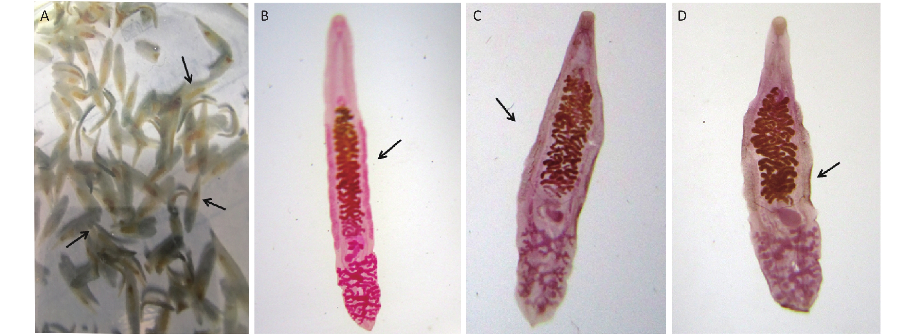
C. sinensis adults were collected from cats and dogs (age, more than 5 years) and observed under a microscope (Figure 2). The body of an adult C. sinensis long and narrow, with a flat dorsal abdomen, narrow front end, blunt and round rear end (like a sunflower seed), and no spines on the surface. The oral suckers are slightly larger than the abdominal suckers, which are located at the front 1/5 of the body. The alimentary canal is simple, with the mouth at the center of the oral sucker, the pharynx is spherical, and the esophagus is short, followed by the intestinal branches. The intestinal branches are divided into two, which reach the posterior end along both sides of the worm without confluence and with a blind end. Judging from morphology, it is the adults of C. sinensis.
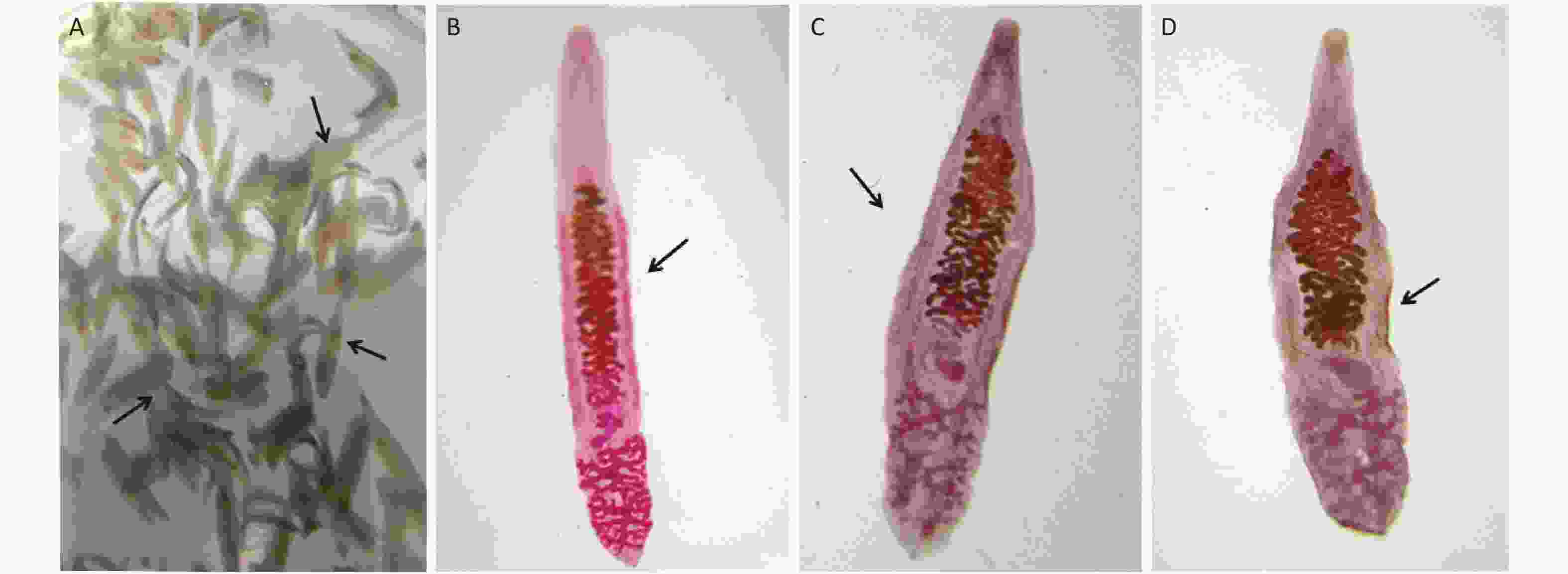
Figure 2. Adult Clonorchis sinensisobtained from hosts. (A) Naked eye of C. sinensis. (B) Clonorchis sinensisfrom Tongdaounder a microscope (magnification, 10×). (C) Clonorchis sinensisfrom Qiyang under a microscope (magnification, 10×). (D) Clonorchis sinensisfrom Lengshuitan under a microscope (magnification, 10×).
Morphological differences were detected among the C. sinensis adults from the three regions. The some adults in Qiyang County are similar to those in Lengshuitan District. The some adults of Tongdao county was more slender and brighter in color.
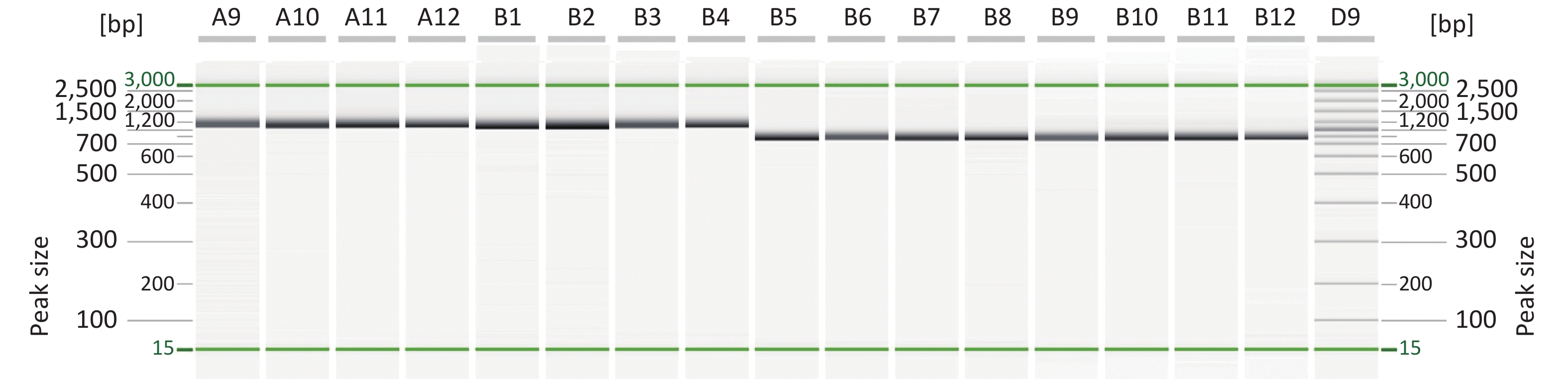
The mitochondrial genes Cox1 and Nad1 of C. sinensis were used as genetic markers for amplification. The results showed that the length of Cox1 and Nad1 are approximately 1,000 bp and 800 bp, respectively, which isconsistent with the predicted values; the electrophoresis results showed clear bands with no tailing phenomenon or unspecific bands (Figure 3).
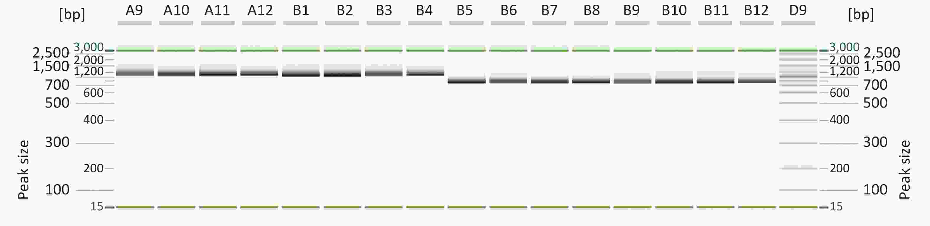
Figure 3. Electrophoresis map of Cox1 gene and Nad1 gene. Description: Lame: 3kb ladder, LameA9, A10, A11, A12, B1, B2, B3 and B4 are Cox1 gene amplification results, and Lame B5, B6, B7, B8, B9, B10, B11, B12 are Nad1 gene amplification results.
-
To verify the amplification accuracy of the two genes, the PCR products were sequenced and compared with homologous sequences from GenBank, and the amplified products were the Cox1 and Nad1 sequences of C. sinensis.
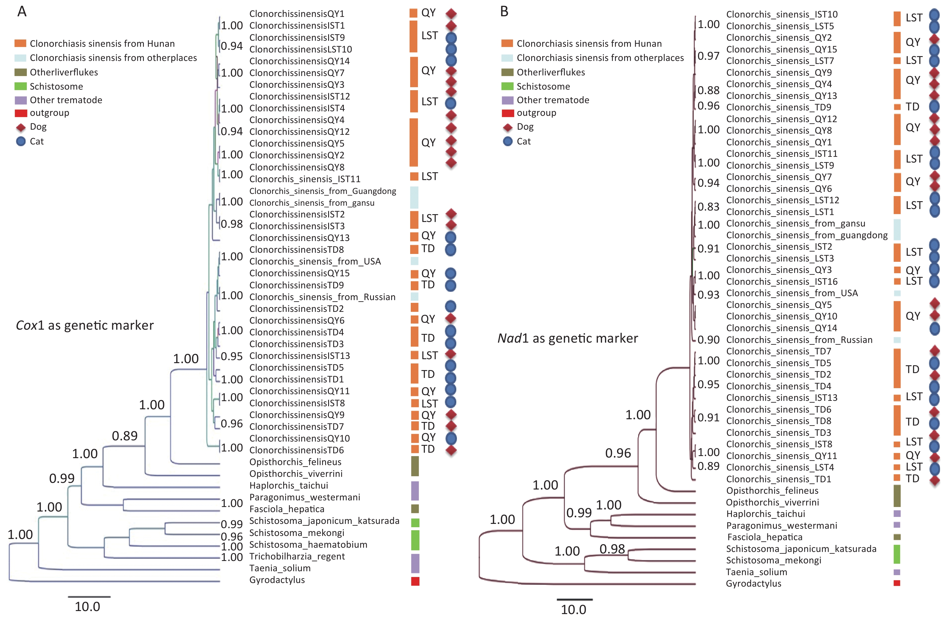
A phylogenetic tree was constructed using Cox1 as the genetic marker and Gyrodactylusas the outgroup for C. sinensis and other trematodes in Tongdao County, Qiyang County, and Lengshuitan District as well as Guangdong Province, Gansu Province, China, the United States, and Russia (Figure 4A). C. sinensisspecimens from Qiyang and Lengshuitan were clustered together without any regional differences; both exhibited high homology with those from Guangdong Province and Gansu Province, China, with a high bootstrap value. The regional clustering in Tongdao County was high and demonstrated low homology with that in Qiyang County and Lengshuitan District but high homology with that in the United States and Russia. The relationships of the other liver flukes, schistosomes, and Gyrodactylus with C. sinensis gradually decreased, which is consistent with the taxonomic records. No significant specificity was observed in the genetic relationships of the parasitic C. sinensis in different definitive hosts (dog and cat).
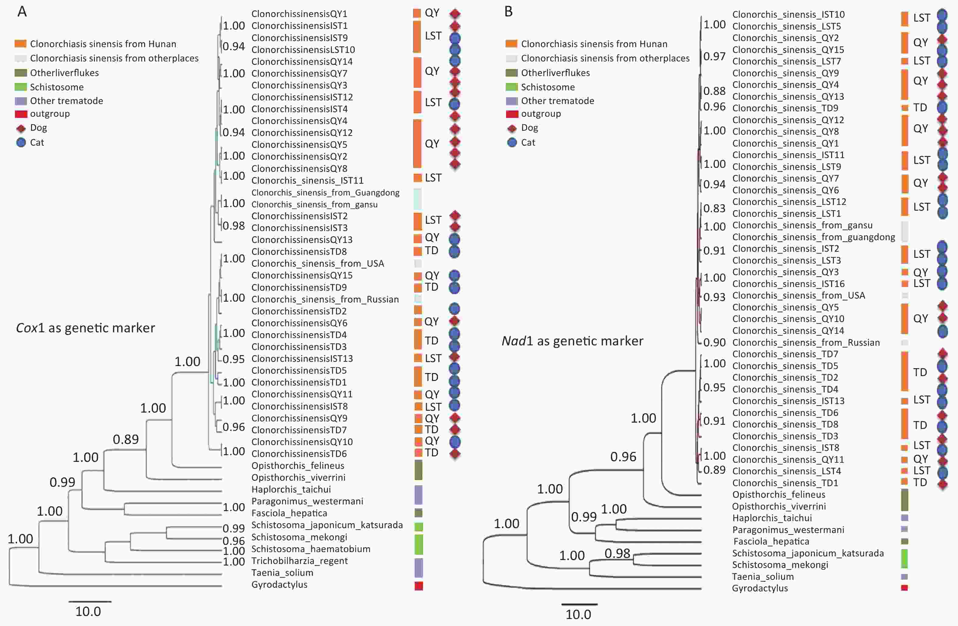
Figure 4. Phylogenetic relationships of C. sinensis with Cox1 and Nad1 as the genetic marker. (A) Phylogenetic relationships of C. sinensis with Cox1 as the genetic marker. (B) Phylogenetic relationships of C. sinensis with Nad1 as the genetic marker.
With Nad1 as the genetic marker (Figure 4B), C. sinensis from Qiyang County and Lengshuitan District showed few regional differences and high homology with those from Guangdong Province and Gansu Province, China. the regional clustering in Tongdao County was high and demonstrated high homology with that in the United States and Russia. The relationships of the other liver flukes, schistosomes, and Gyrodactylus with C. sinensisgradually decreased, which is consistent with the taxonomic records. The parasitic C. sinensis in different definitive hosts (dog and cat) showed no significant specificity in the genetic relationship. This is consistent with the phylogenetic relationship results with Cox1 as the genetic marker.
-
According to the investigation results of C. sinensis infection in the population of 122 counties (districts), 14 cities (prefectures) in the Hunan Province from 2016 to 2020, it can be seen that the infection of C. sinensis has been decreasing year by year since 2016, The high infection rates of C. sinensis among the populations in Qiyang County and Lengshuitan District were related to the high consumption of sashimi and salted fish in these areas[29]. Recently, the infection rates of C. sinensis have decreased in these areas because of improvement indietetic hygiene and scientific prevention[30], but theyare still significantly higher than the infection rates in other areas of Hunan Province. Sashimi is part of the daily diet of people in Tongdao County, Huaihua, so it is important to investigate C. sinensis infection in the population and intermediate hosts. The results of this study showed that the infection rate of C. sinensis in Tongdao County is high. Of the four fish commonly used for making sashimi, the infection rate incruciancarp was the highest, which could be attributed to its small size and phototropism. This study filled the gap of systematic report on the infection of C. sinensis between the population and the intermediate host in Tongdao County.
To study the genetic relationships of C. sinensis in Tongdao County, Qiyang County, and Lengshuitan District, the mitochondrial genes Cox1 and Nad1 were selected as markers[31] and sequenced from adult specimens collected from different areas of Tongdao County, Qiyang County, and Lengshuitan District. The sequence alignment and phylogenetic analysis showed that the parasitic species in Qiyang County and Lengshuitan Distric had a high degree of homology, which could be attributable to the similar latitudes and regional closeness. However, the parasitic species in Tongdao were different from those in Qiyang and Lengshuitan, which have no high homology with the domestic parasitic species, but similar to those in the United States and Russia. This may be related to species invasion and needs to be further studied in the future. This is the first phylogenetic analysis of C. sinensis in the high-incidence area of Hunan Province, China.
The results obtained by designing primers for Cox1 and Nad1 showed a high degree of consistency, indicating that both Cox1 and Nad1 are stable genetic markers for the phylogenetic analysis of C. sinensis. The homology analysis of C. sinensis showed that latitude and longitude and geographical clustering are important. It is suggested that C. sinensis in the Tongdao County may have the biological characteristics of high latitude species, and have similar antigenicity. It is also suggested that C. sinensis in the Tongdao County may originate from the high latitude region of Eurasia, Russia. This will lay a foundation for further exploration of its control direction, gene traceability and detection methods.
The phylogenetic analysis of C. sinensis adults detected in different definitive hosts (dogs and cats) showed that C. sinensis has no specificity in the parasitism of mammalian hosts, and it does not have different gene homology because of changes in the hosts.
A combination of molecular biology and morphology is more reliable than morphological identification. Morphological description is greatly affected by subjective factors and measurement errors. Differences in the genomes of C. sinensis explain the differences in the morphology of C. sinensis in Tongdao County, suggesting that the antigenicity of C. sinensis in Tongdao County may be closer to that of the species in high-latitude regions[32]. The results of this study have laid the foundation for further natural selection and evolutionary analyses of C. sinensis in Tongdao. C. sinensis in Tongdao may have originated from high-latitude areas, providing clues for studying the evolution and mutation adaptation of C. sinensis in high- and low-latitude areas and research on group evolution, individual differences, detection methods, control drugs, and vaccines for C. sinensis in this area.
-
The infection rate of C. sinensis was higher in Qiyang County, Lengshuitan District and Tongdao County of Hunan province, China. Mitochondrial genes Cox1 and Nad1 were used as genetic markers to study the phylogeny of C. sinensis. The results showed that the C. sinensis in Tongdao county was far from the other area of Hunan Province, which might be related to the migration and invasion of the species. Based on this method, mitochondrial genes Cox1 and Nad1 were selected as good genetic markers for the phylogenetic analysis of C. sinensis. The results of molecular genetic marker analysis were basically the same, which again confirmed the reliability of the experimental study. Taking into account the complexity of the species of C. sinensis in Hunan Province, it is recommended to reduce the infection rate of parasites in terms of controlling local transmission and preventing external sources.
-
The authors declare that they have no known competing financial interests or personal relationships that could have appeared to influence the work reported in this paper. This research did not receive any specific grant from funding agencies in the public, commercial, or not-for-profit sectors.
-
We are grateful to the Hunan Provincial Center for Disease Control and Prevention for providing C. sinensis epidemiological survey data. We thank the Xiangya School of Medicine, Central South University for their technical assistance and participation. We also thank the Centers for Disease Control and Prevention of Tongdao County, Qiyang County and Lengshuitan District for providing metacercaria and adults of C. sinensis.
Interspecies Phylogenetic Analysis of Clonorchis sinensis in High-incidence Areas of Hunan Province, China
doi: 10.3967/bes2021.121
- Received Date: 2021-05-01
- Accepted Date: 2021-08-17
-
Key words:
- Clonorchis sinensis /
- Mitochondrial genes /
- Cox1 /
- Nad1 /
- Phylogeny
Abstract:
| Citation: | LONG Xiao Lei, WANG Shi Ping, ZHOU Shuai Feng, LI Zheng Xiang, ZHUANG Shi Feng, HE Zhuo. Interspecies Phylogenetic Analysis of Clonorchis sinensis in High-incidence Areas of Hunan Province, China[J]. Biomedical and Environmental Sciences, 2021, 34(11): 881-890. doi: 10.3967/bes2021.121

|


 Quick Links
 DownLoad: