

 下载:
下载:


-
Shortwave radiation is the electromagnetic wave with a frequency of 3–30 MHz and electromagnetic wavelengths in the range of 10–100 m. The transmission mode at low power mainly depends on the peaks and troughs of its waves, which allow long-distance transmission. Shortwave radiation is widely used in many fields such as radio broadcasting, communication radar, medical treatment of osteonecrosis and so on[1-3]. While this form of radiation brings great convenience to people’s lives, it also could cause large health risks for humans. Currently, increasingly more scholars are concerned about this matter[4-5]. As the circulatory organ of the body, heart-related injuries after shortwave radiation have been infrequently reported. The heart, as the power organ of the body, continuously maintains the rhythm of systolic and diastolic blood flow in the blood vessels to guarantee the normal systemic blood supply to the system. Our previous studies have shown that the heart is one of the organs that suffers serious damage after microwave radiation[6-8]. Radiation can cause damage to cardiac function and tissue structure[6]. Currently, there are many studies on the biological effects of different wave bands of microwave radiation on heart injury[9-12]. However, shortwave-induced biological effects on non-cardiac organs have not been reported.
At present, this type of radiation is mainly used in the fields of long-distance radio communication, medical treatment, military affairs and so on[1-3]. Compared with microwaves, the application range of shortwave radiation in daily life is relatively small. Studies have shown that shortwave radiation can cause cardiac dysfunction, but the damage effect has not been reported[13-15].
Currently, the main methods of evaluating cardiac functional injury after electromagnetic wave radiation are to measure sensitive indexes such as Ca2+, CK, glutamic oxaloacetic transaminase, lactate dehydrogenase levels in the peripheral blood. Additionally, a polyphysiological recording can be used to detect heart rate, rhythm, and P and T waves. The morphological and ultrastructural changes in the heart can be observed by light and electron microscopy. It has been reported that the concentration of Ca2+ increased and the AST levels were disturbed in peripheral serum of rats after shortwave radiation. Some scholars found that workers that were exposed to radiation experienced bradycardia, sinus arrhythmia and so on[6-8,16]. However, no changes in the structure of the heart after radiation have been reported. Pathological changes in the structure are the basis of body functional disorders. Therefore, it is important to study and summarize the changes in cardiac tissue structure after shortwave radiation in order to explore related functional disorders.
The aims of this study were to establish an animal model of shortwave radiation-induced cardiac injury in rats and to detect the cardiac function and observe the changes in the heart tissue structure of rats. This work is expected to reveal the biological effects of shortwave radiation on rat heart injury by studying the relationship between function and structure, and it also provides evidence for the mechanisms of injury.
-
Animal work in this study was approved by the Beijing Institute of Radiation Medicine Animal Care and Use Committee. It was carried out on the basis of the National Institute of Health Guide for the Care and Use of Laboratory Animals. The experiments were approved by an Ethical Committee of the Academy of Military Medical Science, and the authorization number was IACUC-AMMS-2010-007.
-
One hundred male Wistar rats (weight 200–220 g, age 6–8 weeks) were provided by the Laboratory Animal Center of the Academy of Military Medical Sciences (Beijing, China) and were raised in a standard animal laboratory with free access to food and water. All experiments were conducted in accordance with the National Institutes of Health Guide for the Care and Use of Laboratory Animals. All protocols were approved by the Institutional Animal Care and Use Committee. The animals were randomly divided into the following four groups: 0 mW/cm2 high frequency shortwave exposure group, 5 mW/cm2 high frequency shortwave exposure group, 10 mW/cm2 high frequency shortwave exposure group and 30 mW/cm2 high frequency shortwave exposure group, with 25 rats per group.
-
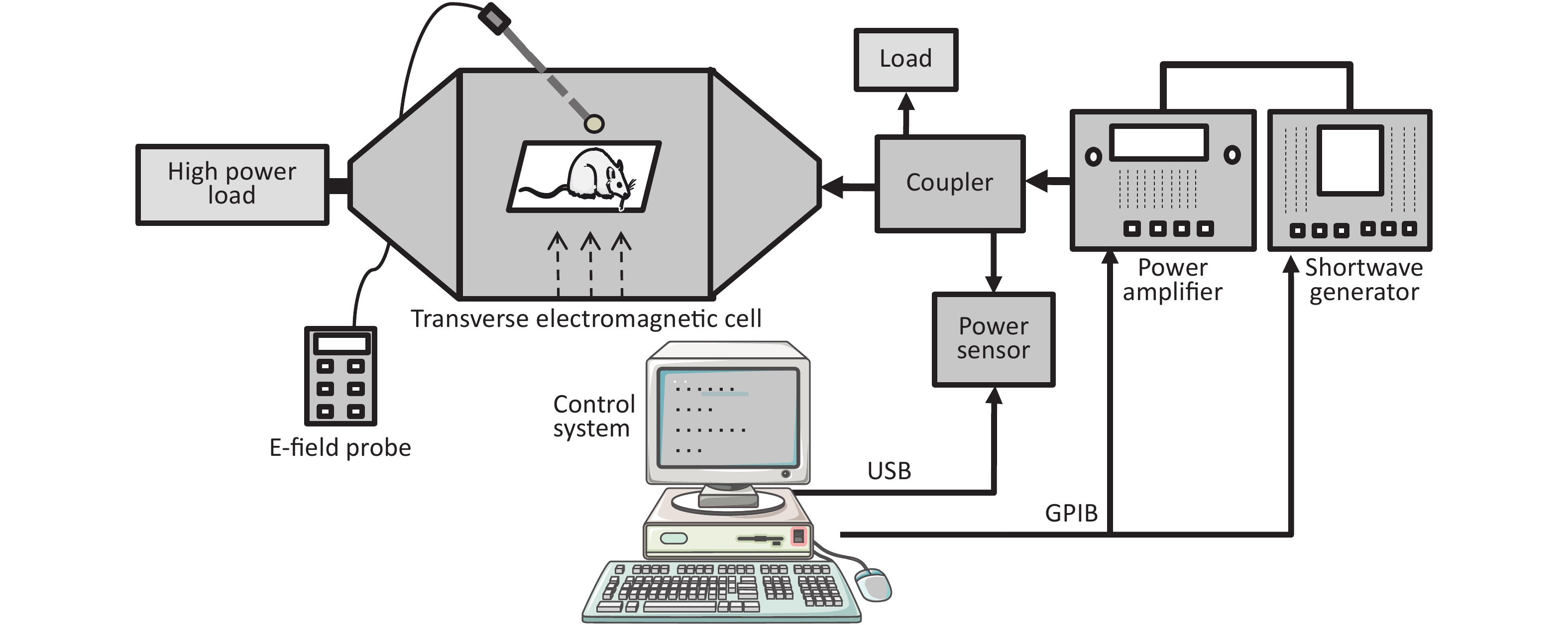
A schematic diagram of the exposure setup for high frequency shortwave exposure is shown in Figure 1. The shortwave generator (SMF100A, Rohde & Schwarz, Germany) is connected to a power amplifier (BBA100, Rohde & Schwarz, Germany), followed by a dual-directional coupler with a power sensor (NRP-Z91, Rohde & Schwarz, Germany). The forward coupling port transmits a continuous wave signal with a standard power density (e.g., 10 mW/cm2) at the frequency point of 27 MHz into a transverse electromagnetic cell (117486, ETS Lindgren, USA), which is terminated with a high power load to make the reflection coefficient valid. The electric field strength in the electromagnetic cell is measured with an E-field probe (HI-6053, ETS Lindgren, USA) and transferred into the power density unit. The control system was designed by China Academy of Information and Communications Technology, Beijing, China.

Figure 1. The shortwave radiation device and a schematic diagram of the radiation method[19].
During the exposure, the rats were restrained in a closed Plexiglas cage with holes of 1.5 cm in diameter to facilitate breathing and were placed in the middle of the electromagnetic cell. The whole bodies of the rats were exposed to shortwave with an average power density of 5, 10, or 30 mW/cm2, respectively, for 6 min only 1 time. Correspondingly, the average specific absorption rates (SARs) of the rat hearts were 1.16 × 10⁻⁴, 2.33 × 10⁻⁴, and 6.99 × 10⁻⁴ W/kg, respectively. The SAR distribution was calculated by the finite domain time difference (FDTD) method, which has been reported in a previous study[17-19]. The rats in the Con underwent a sham exposure (the rats were placed in the shortwave radiation device without turning on the machine, and the radiation dose was 0 mW/cm2 for 6 min). Our experiment run under a ‘Not blindnes’ condition.
-
The supernatant temperatures of experimental animals were monitored using an infrared temperature sensor (FLIR A40, USA) before and after shortwave exposure.
-
An automated blood biochemical analyser (Coulter JTIR, USA) was used to detect the concentration of Ca2+, AST, CK, and LDH with 5 rats in each group at each time point. The rats in each group were anaesthetized by intraperitoneal injection of 1% sodium pentobarbital (30 mg/kg) on the 1st, 7th, 14th, and 28th day after radiation. Three millilitres of blood was taken from the main abdominal vein, and the sera were prepared and tested using a computer.
-
The electrocardiograms (ECG) of the rats were recorded by a polyphysiological recording and analysis system (BIOPAC company, USA). After intraperitoneal injection of 1% sodium phenobarbital (0.5 mL for 5 min), the rats were under mild anaesthesia (pain reflex). On 1, 7, 14, and 28 d after shortwave exposure, the electrocardiograms (ECG) was recorded. The operation process was as follows: hair was cut from the limbs, which were disinfected and degreased. The electrodes were divided into red (R) right upper limbs, black (RF) right lower extremities, and green (LF) left lower extremities. The needle electrodes were pierced into the subcutaneous layer of the corresponding limbs and connected to the ECG biologic amplifier with a sensitivity of 2,000 Hz. The electrocardiograms (ECGs) of the standard II lead were recorded in a resting state 3 minutes. After storage, changes in the heart rate, P-H, R-H, and T-H were analysed and calculated.
-
Morphological changes in the rat heart were observed via HE staining. The rats in each group were anaesthetized by intraperitoneal injection of 1% sodium pentobarbital (30 mg/kg) 1, 7, 14, and 28 d after radiation (5 rats in each group at each time point). The hearts were placed in 10% neutral buffered formalin for 1 week. After dehydration, clearing, wax impregnation and embedding, the paraffin-embedded tissues were sectioned at a thickness of 5 µm. Following HE staining procedures, histological sections of the hearts were observed, and hippocampal images were captured using a light microscope (Leica, Germany).
-
The ultrastructural changes in the rat heart were observed by electron microscopy. The rats in each group were sacrificed after intraperitoneal injection of 1% sodium pentobarbital (30 mg/kg) on the 7th and 14th day after radiation (5 rats in each group at each time point). The fresh heart tissues were fixed for 2 h in 2.5% glutaraldehyde, 2 h in 1% osmium acid, dehydrated with an ethanol gradient, transitioned in acetone, embedded in Epon 812 resin, and localized in semi-thin sections. The ultrathin 70 nm sections were stained with lead-uranium. A Philip-CM120 transmission electron microscope was used to observe and photograph the samples.
-
The data were presented as the mean ± standard deviation (SD), and statistical analyses were performed using SPSS 19.0 software (IBM, USA). The data were analysed by one-way ANOVA variance analysis, LSD and Dunnett’s t-test.
-
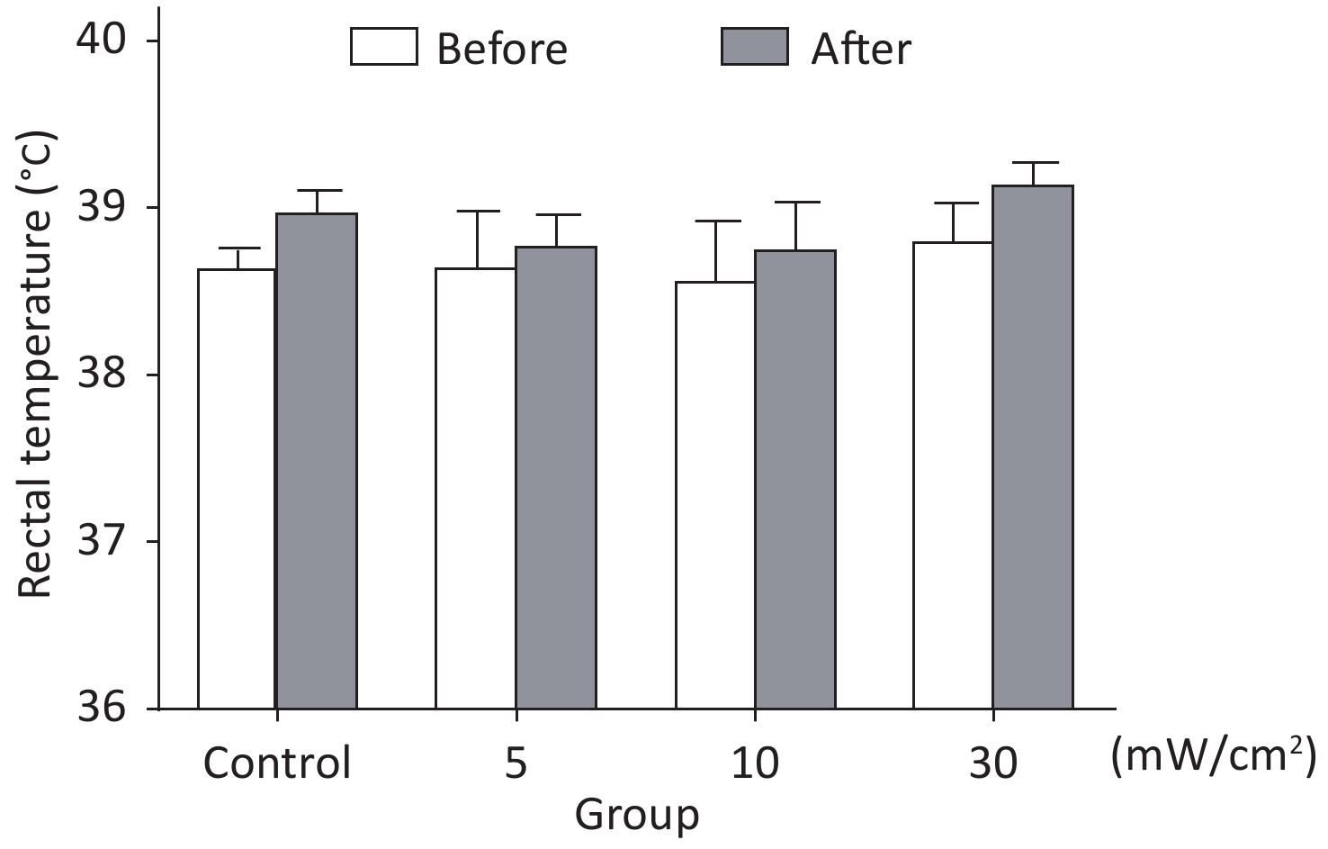
We measured the rats’ shell temperatures (n = 4) before and immediately after shortwave exposure by infrared temperature sensor. According to the results, temperatures were increased less than 1 ℃ after exposure (Figure 2). That means the thermal effects could be compensated by the physiological temperature regulation of organism and the effects discussed in this paper was basically non-thermal effects.
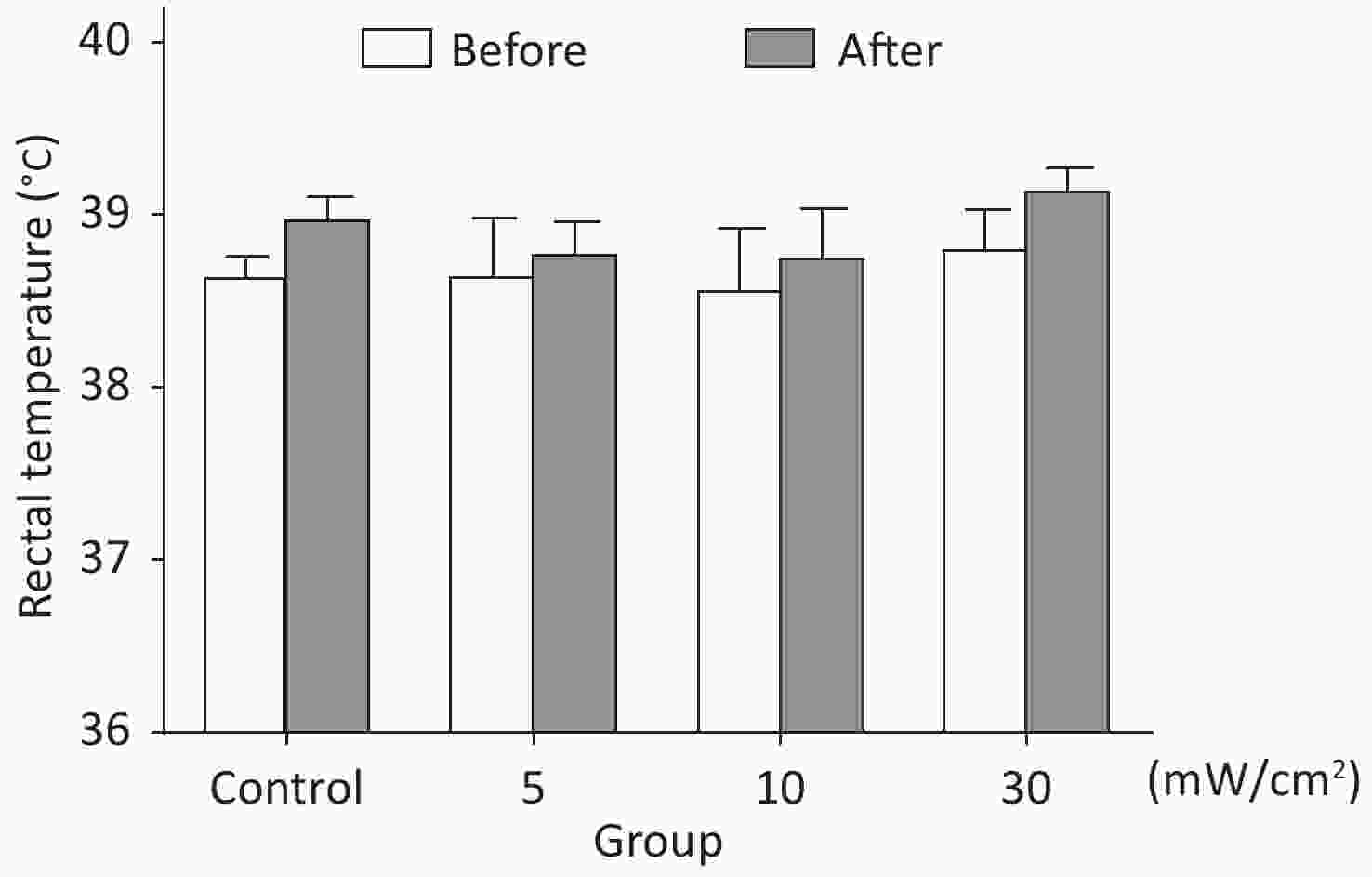
Figure 2. Rats’ shell temperatures before and immediately after shortwave radiation.
-
Ca2+ is an important cation in maintaining and regulating myocardial contraction and contraction. Myocardial enzymes are widely used in the clinical diagnosis and evaluation of cardiac function. Therefore, we detected the concentration of Ca2+, AST, CK, and LDH in the peripheral blood of rats using a blood biochemical analyser to assess cardiac function after shortwave radiation. Compared with the 0 mW/cm2 group, the concentration of Ca2+ in the peripheral blood of the 5, 10, and 30 mW/cm2 groups was significantly higher than control level 1 d after shortwave exposure. The concentration of Ca2+ in the peripheral blood of the 30 mW/cm2 group was obviously higher than control level 7 d after shortwave exposure (Figure 3A). Compared with the 0 mW/cm2 group, the AST content in the peripheral blood of the 10 and 30 mW/cm2 groups was significantly higher than control level 7 d after radiation (Figure 3B). Compared with the 0 mW/cm2 group, the CK content in the peripheral blood of the 30 mW/cm2 group was significantly higher than control level 1, 7, and 28 d after radiation (Figure 3C). Compared with the 0 mW/cm2 group, the LDH content in the peripheral blood of the 30 mW/cm2 group was significantly increased compared to levels 1, 7, and 28 d after exposure (Figure 3D).
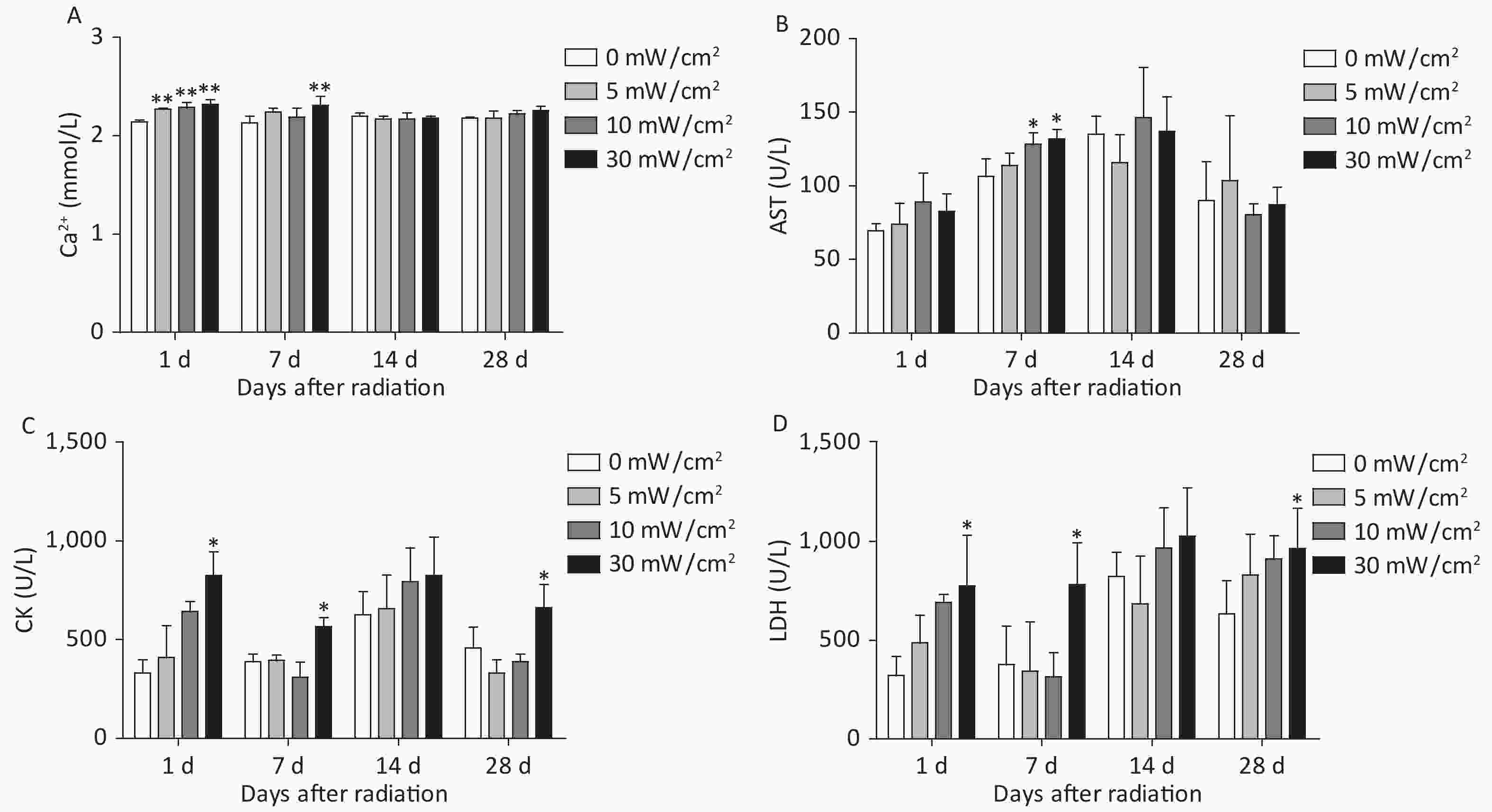
Figure 3. Changes in the Ca2+ concentration and myocardial enzymes in the peripheral serum of rats after shortwave radiation. (A) Five rats were taken from each group at each time point. Blood was taken from the abdominal main vein after anaesthesia. Changes in the Ca2+ concentration in the peripheral blood were detected by an automatic biochemical analyser on 1 d, 7 d, 14 d, and 28 d after radiation. (B–D) Five rats were taken from each group at each time point. The contents of AST, CK, and LDH in the peripheral blood were detected by an automatic biochemical analyser on 1 d, 7 d, 14 d, and 28 d after radiation. Compared with the 0 mW/cm2 group, *P < 0.05, **P < 0.01.
These results demonstrated that shortwave exposure could increase the concentration of Ca2+ and the AST, CK, and LDH content in the peripheral serum of rats. Abnormal cardiac function was positively correlated with radiation dose. Because structural damage is the material basis of functional abnormalities, structural changes were assessed next.
-
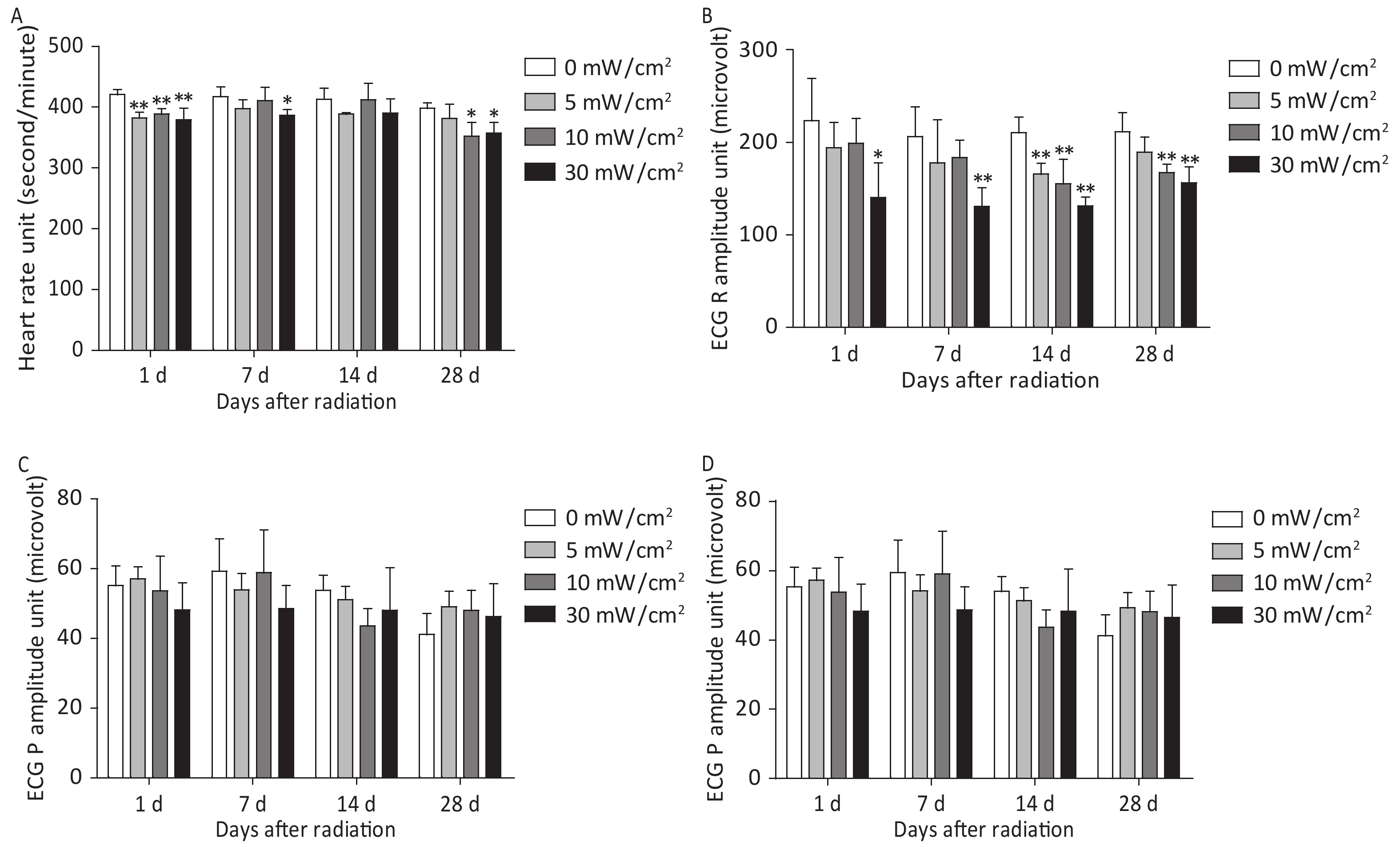
Electrocardiogram (ECG) is a vital method to record the electrical activity of the heart and is an important means to evaluate heart function. Therefore, we used an animal polygraph to analyse changes in the heart rate and the P-H, R-H, and T-H. Compared with the 0 mW/cm2 group, the heart rate significantly decreased in the 5, 10, and 30 mW/cm2 groups 1 d after exposure. The heart rate in the 30 mW/cm2 group obviously decreased 7 d after exposure. On 28 d after exposure, the heart rate of the rats in the 10 and 30 mW/cm2 groups decreased significantly (Figure 4A). Compared with the 0 mW/cm2 group, the amplitude of the ECG R wave in the 30 mW/cm2 group decreased significantly on 1 d and 7 d after exposure. Moreover, the amplitude of the ECG R wave in the 5, 10, and 30 mW/cm2 groups decreased 14 d after exposure, while the 10 and 30 mW/cm2 group decreased 28 d after exposure (Figure 4B). Compared with the 0 mW/cm2 group, the amplitude of the ECG P wave in the 5, 10, and 30 mW/cm2 groups did not change significantly (Figure 4C). Compared with the 0 mW/cm2 group, the amplitude of the ECG T wave in the 5, 10, and 30 mW/cm2 groups did not change significantly (Figure 4D).

Figure 4. Changes in the electrocardiogram in rats after shortwave radiation. (A–D) At each time point, 5 rats were taken from each group. After mild anaesthesia, the hair of the left forelimb and the right hind limb was shaved. Needle fishhook electrodes (pierced subcutaneously) were placed on the inner skin of the left forelimb and the right hind limb. Changes in the heart rate, R wave, P wave and T wave amplitude were recorded by a multi-channel physiological recorder on 1 d, 7 d, 14 d, and 28 d after radiation. Compared with the 0 mW/cm2 group, *P < 0.05, **P < 0.01.
The above results demonstrated that shortwave radiation can cause an abnormal increase in rat ECGs. Abnormal cardiac function was positively correlated with radiation dose. The results of serum biochemistry and electrocardiogram analysis showed that shortwave exposure could cause abnormal cardiac function in rats. These abnormalities may be due to disordered energy metabolism in the myocardial cells, cell membrane damage, Ca2+ overload, and decreased myocardial fibre contractility. Therefore, observing heart tissue structure and ultrastructure can not only clarify the biological effect of shortwave radiation but can also provide the basis for the injury mechanism.
-
Normal morphological structure is the premise and the basis to maintaining the physiological function of the heart; thus to clarify the effect of radiation on heart damage, we used HE staining. After shortwave exposure, the basic pathological changes, dynamic changes and dose-response relationship of heart injury were compared among the different radiation treatment groups.
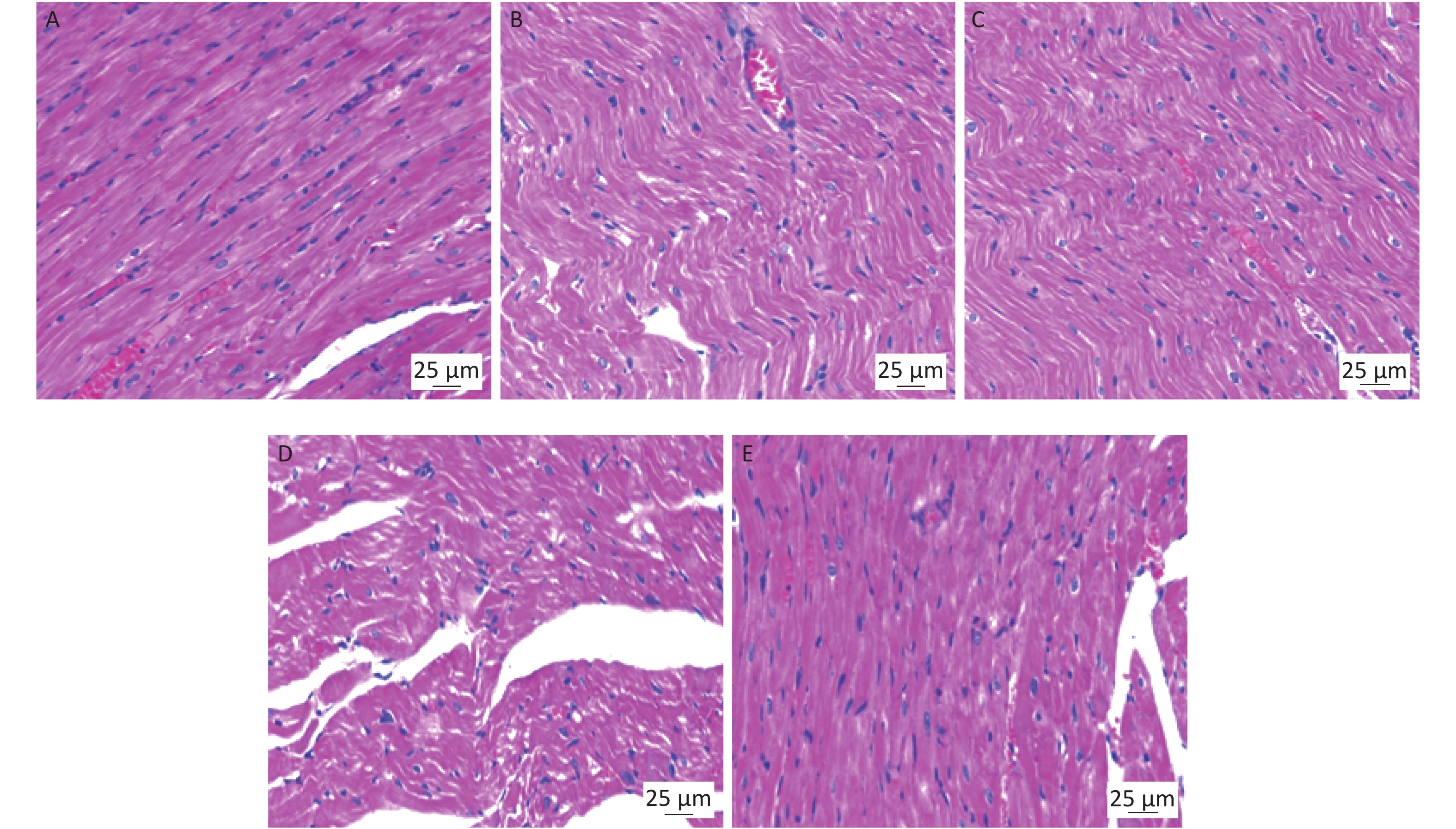
The basic pathological changes in cardiac tissue in rats were characterized by a disordered arrangement of myocardial fibres, cytoplasmic coagulation and interstitial oedema after different doses of shortwave exposure. The damage described above was most significant 7 d after radiation. Taking the results from 7 d after radiation as an example, we observed changes in the cardiac tissue structure in each group. The cardiac tissue of the rats in the 0 mW/cm2 group showed normal cardiac tissue structure, which revealed that the myocardial fibres were well arranged, and the cardiac myocytes were well karyotyped (Figure 5A). Cardiac tissue injury was not significant in the 5 mW/cm2 group, where the myocardial fibres were arranged regularly and the perinuclear space of the individual myocardial cells were widened occasionally (Figure 5B). Group 10 mW/cm2 showed a disordered arrangement of myocardial muscle fibres and increased numbers of eosinophils (Figure 5C). The 30 mW/cm2 group showed myocardial muscle fibre waviness, cytoplasm condensation, and interstitial oedema (Figure 5D). Heart injury was most significant in the 30 mW/cm2 group. Therefore, the 30 mW/cm2 group was used to observe the cardiac pathological changes after shortwave exposure. In the 0 mW/cm2 group, the heart tissue of rats showed normal cardiac structure (Figure 6A). In the 30 mW/cm2 group, the myocardial fibres showed disordered arrangement 1 d after radiation (Figure 6B) and showed eosinophilic enhancement, wavy arrangement of the myocardial fibres and widening of the perinuclear space of the myocardial cells 7 d after radiation (Figure 6C). After 14 d, the myocardial fibres were loose and disordered (Figure 6D). The arrangement of the myocardial fibres was regular. Occasionally, the perinuclear space of individual myocardial cells widened 28 d after radiation (Figure 6E).
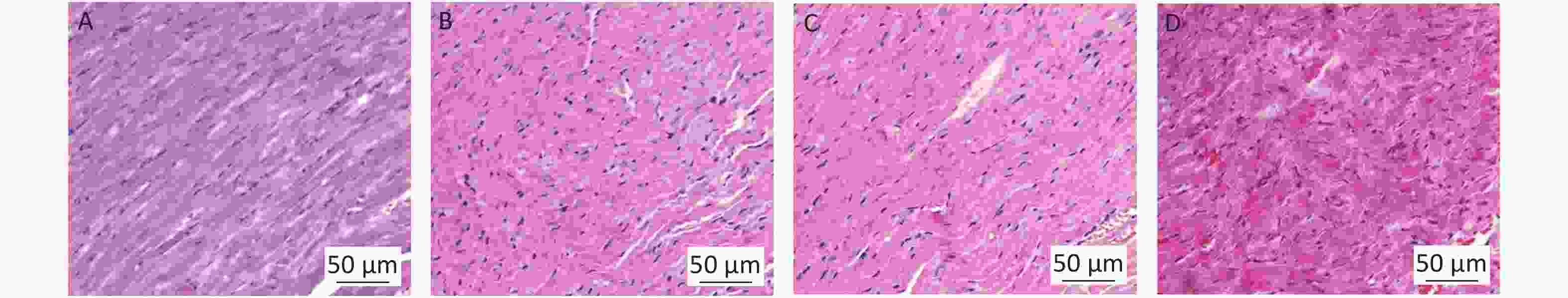
Figure 5. The basic pathological changes associated with cardiac injury in rats after shortwave radiation (scale bar = 50 µm). On the 7 d after radiation, five rats in each group were euthanized after anaesthesia. (A) The cardiac structure in the 0 mW/cm2group; (B) The cardiac structure in the 5 mW/cm2 group; (C) The cardiac structure in the 10 mW/cm2 group. (D) The cardiac structure in the 30 mW/cm2 group.
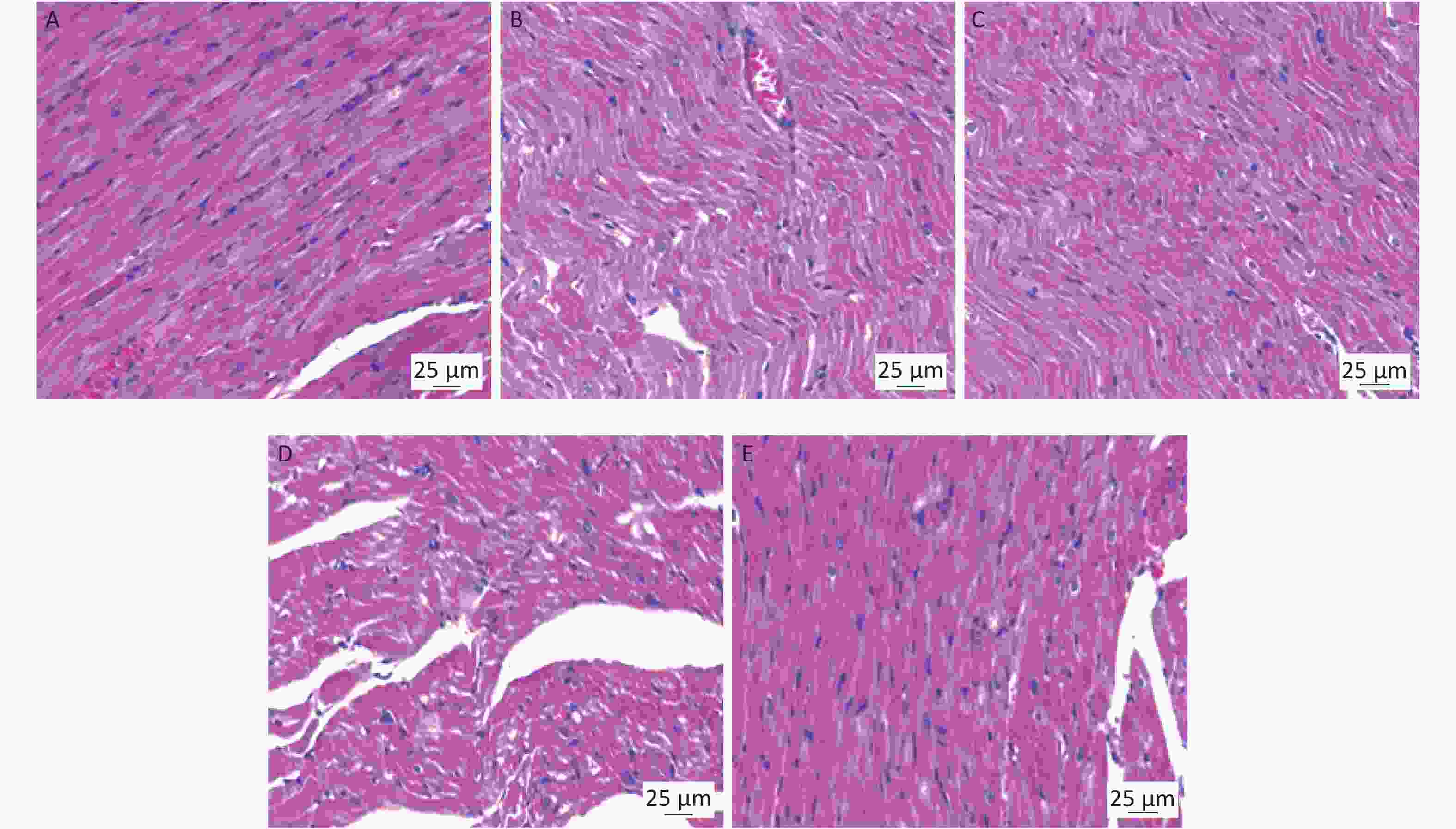
Figure 6. Changes in the cardiac tissue structure in rats exposed to 30 mW/cm2 shortwave radiation (scale bar = 25 µm). On 1 d, 7 d, 14 d, and 28 d after radiation, five rats in each group were euthanized after anaesthesia. (A) The cardiac structure in the 0 mW/cm2 group. (B) The changes in the cardiac tissue structure in rats of the 30 mW/cm2 group 1 d after radiation. (C) The changes in the cardiac tissue structure in the 30 mW/cm2 group 7 d after radiation. (D) The changes in the cardiac tissue structure in the 30 mW/cm2 group 14 d after radiation. E: The changes in the cardiac tissue structure in the 30 mW/cm2 group 28 d after radiation.
The above results demonstrate that shortwave exposure could cause cardiac structural damage in rats. The 30 mW/cm2 group showed the most obvious damage to the cardiac tissue structure. No obvious changes in the cardiac structure were observed in the 5 mW/cm2 group. A significant dose-response relationship in the above changes was observed, as the 10 mW/cm2 group displayed injuries between those of the 5 and 30 mW/cm2 groups. Myocardial fibre dysfunction was the most obvious injury feature. Combined with the results of the electrocardiogram, myocardial fibre weakness may be an important cause of heart rate and R wave amplitude decreases in the rats. The recovery 28 d after radiation and the damage degree was positively correlated with the radiation dose.
-
The observation of ultrastructure is a comprehensive exploration of the cellular sublevel. To clarify the injury effect and to explore the possible injury mechanism, we compared the basic pathological changes and dose-effect relationship of ultrastructural injury of the heart in different radiation groups via transmission electron microscopy.
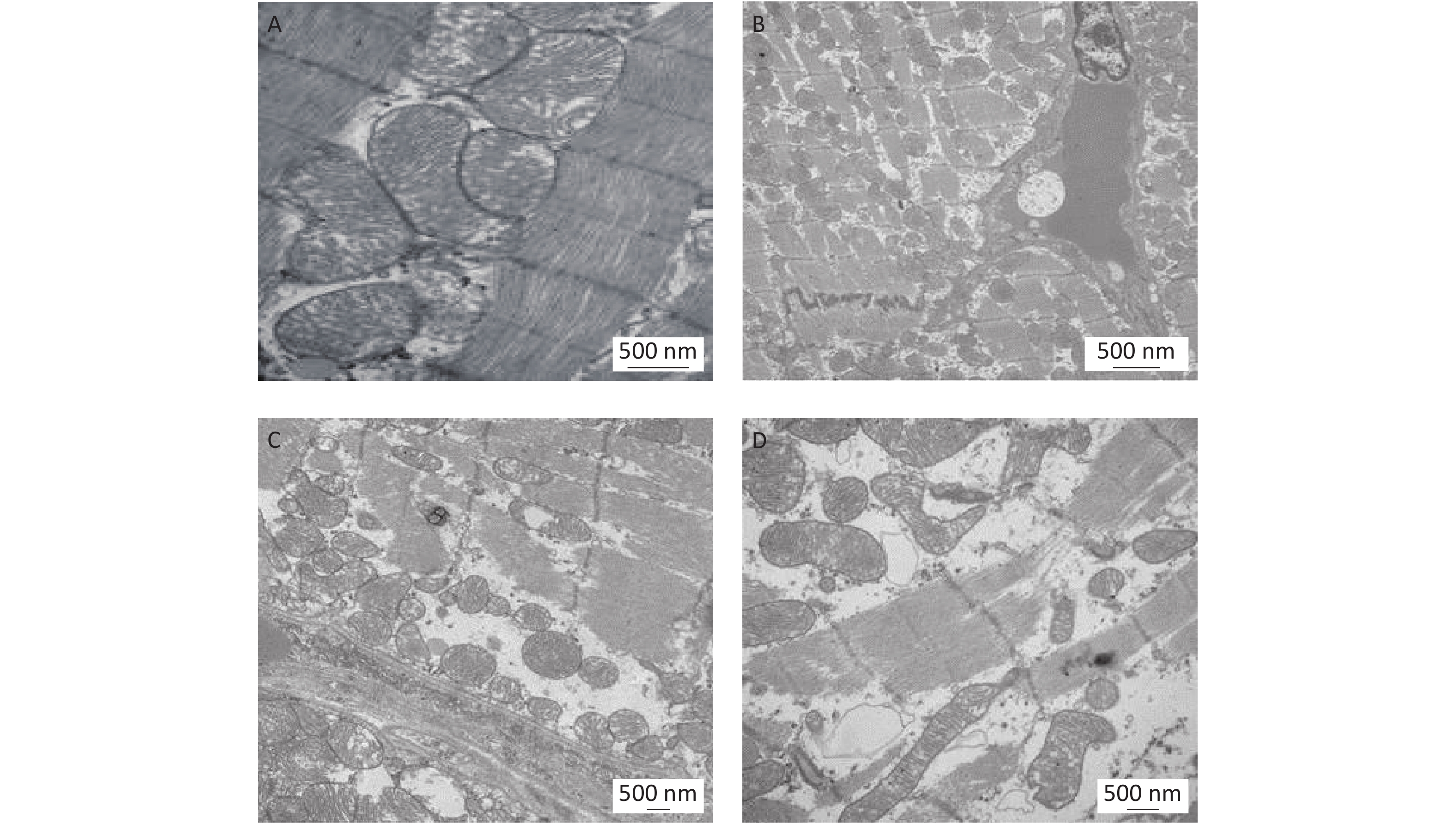
According to the HE results, at 7 d, the damage to the heart tissue structure was most significant after shortwave exposure. We therefore chose 7 days after radiation as the observation time for changes in the ultrastructure of the heart in each group over the different doses of shortwave exposure. In the 0 mW/cm2 group, normal cardiac ultrastructure was observed. The myocardial fibres were well arranged, the light and dark bands were clear, and the mitochondrial internal cristae were dense (Figure 7A). Ultrastructural damage was mild, myocardial fibres were well arranged, and some mitochondria were cavitated in the 5 mW/cm2 group (Figure 7B). In the 10 mW/cm2 group, the intercalated discs were blurred, and the mitochondria were cavitating inside (Figure 7C). In the 30 mW/cm2 group, the intercalated disc structure was blurred, the myocardial fibres were disordered, the light and dark bands were not clear, and many mitochondria were cavitated and swollen (Figure 7D). Therefore, the 30 mW/cm2 group served as an example of the basic pathological changes in the myocardial ultrastructure after radiation with the following characteristics. The myocardial fibres were in a disordered arrangement and were focally dissolved and broken, and the light and dark bands were blurred (Figure 8A). The mitochondria were cavitating and swollen, with cristae partly broken, dissolved and absent (Figure 8B). Many lysosomes and autophagic bodies were present (Figure 8C). The intercalated discs of the tight junctions between the cardiac myocytes were obscure (Figure 8D). The nuclei of the myocardium were abnormal, and the perinuclear spaces were widened significantly (Figure 8E). Interstitial oedema and widened endothelial cell nuclei were observed (Figure 8F).
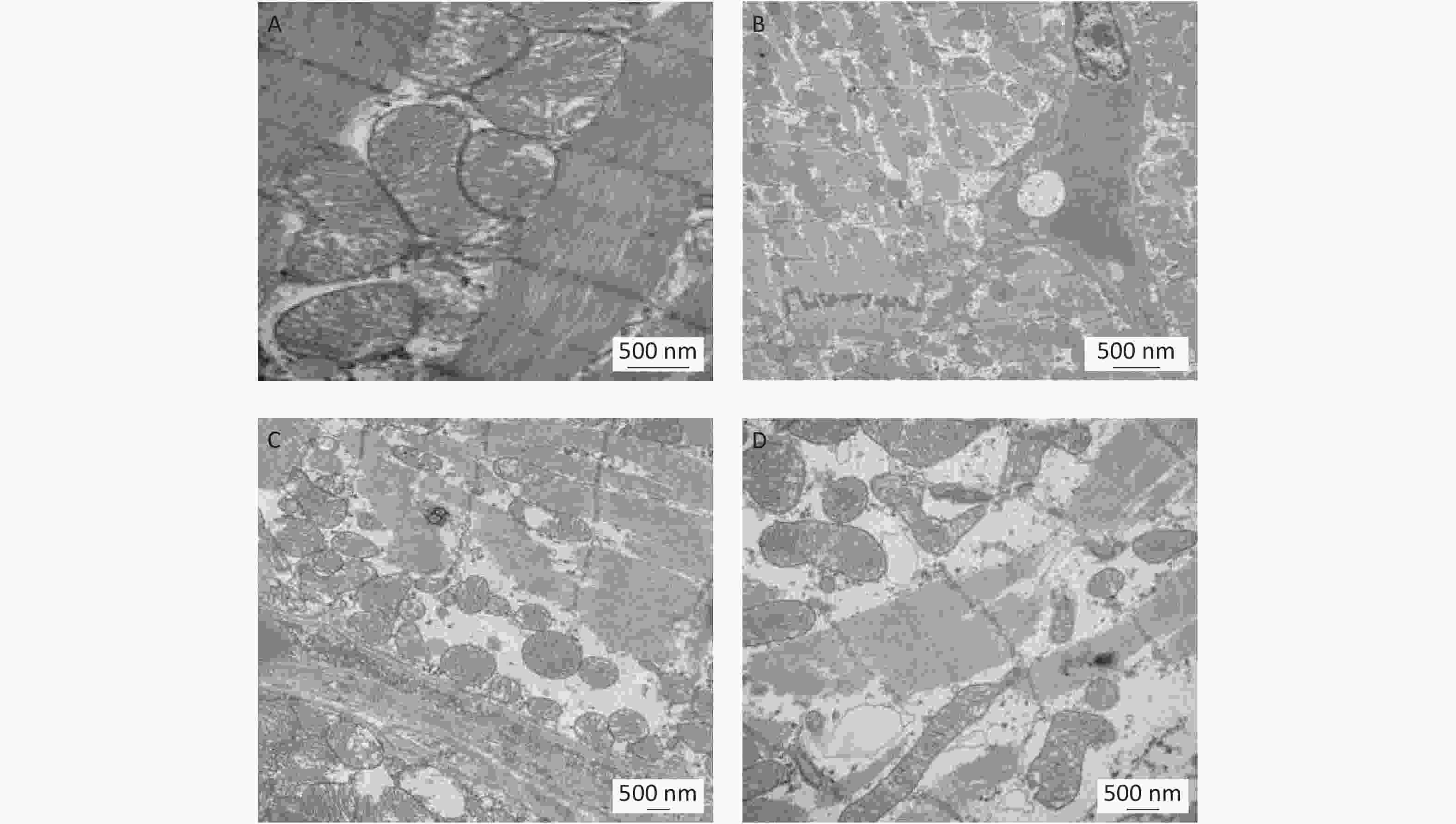
Figure 7. The basic pathological changes associated with ultrastructural damage in the rat heart after shortwave radiation (TEM scale bar = 500 nm). On 7 d after radiation, three rats in each group were euthanized after anaesthesia. (A) The ultrastructure in the 0 mW/cm2 group. (B) Ultrastructural changes in the 5 mW/cm2 group. (C) Ultrastructural changes in the 10 mW/cm2 group. (D) Ultrastructural changes in the 30 mW/cm2 group.

Figure 8. Changes in the cardiac tissue ultrastructure in rats exposed to 30 mW/cm2 shortwave radiation (TEM scale bar = 500 nm). On 1 d, 7 d, 14 d, 28 d after radiation, three rats in each group were euthanized after anaesthesia. (A) Changes in the myocardial fibres. (B) Mitochondrial changes. (C) The changes of the intercalated disc. (D) Autophagy changes. (E) Changes in the myocardial cells. (F) Interstitial changes.
These results indicate that shortwave exposure can induce ultrastructural damage in the heart of rats. After different doses of shortwave radiation, the injury of the 30 mW/ cm2 group was the most significant. Compared with the normal control group, the ultrastructure of cardiac tissue in the 5 mW/cm2 group showed no significant change. The degree of injury in the 10 mW/cm2 group was between that of the 5 and 30 mW/cm2 groups, showing a significant dose effect relationship. The main injuries were an irregular arrangement of myocardial fibres, mitochondrial cavitation and autophagic bodies. Combined with the results of the cardiac function test, mitochondria structural damage may be an important cause of energy transfer disorders in myocardial fibres. The dysfunction of energy transmission can directly lead to functional damage in cardiac muscle cells, leading to cardiac dysfunction. The above degree of injury is positively correlated with the radiation dose.
-
As the most important power organ of circulatory system, the heart is the main organ that maintains the normal blood supply of every system[16]. The rhythmic contraction and relaxation maintains the pumping of blood in the vascular system, thus guaranteeing normal blood supply to the organs of the body[7,16]. At present, people come in little contact with shortwave radiation in daily life compared with microwave radiation. Therefore, relatively few studies on the damaging effects of shortwave radiation on organisms have been reported, especially for the heart for which the biological effects are unknown. The results of prior investigations reported multiple long-term effects after short-wave contact, including HF-related symptoms, headache, insomnia, sinus arrhythmia, sexual dysfunction, endocrine disorders, immune dysfunction and other symptoms[15,17-18]. Animal experiments showed that shortwave exposure could cause functional damage to the central nervous system of mice, increase the concentration of calcium and AST in rats, decrease the secretion of adrenaline in rats, and decrease the immune function of mice. All these studies indicated that shortwave exposure can lead to damage in multiple organ systems[17-19]. The damaging effects of shortwave exposure have not been reported in terms of the heart in recent years. Therefore, our study aimed to examine and observe cardiac structure and function in rats following shortwave exposure. To this end, the changes in cardiac structure and function in rats following shortwave exposure at different power densities were compared. Finally, we aimed to identify the sensitive targets and the effects of the damage.
Ca2+ is a crucial cation required to maintain and adjust myocardial systolic and diastolic function, while Ca2+ overload or inadequate intake can lead to a variety of outcomes including electrical signal disorder, cell necrosis, and apoptosis[20-21]. Myocardial enzymes are widely used in the clinical diagnosis and evaluation of cardiac function. When myocardial cells are damaged, many myocardial enzymes are released into the blood[20]. ECG not only reflects the changes in myocardial electrical conduction but also reflects the pacing function of the sinoatrial node. Therefore, the concentration of Ca2+ and the content of myocardial enzymes in the peripheral blood could be detected using a blood biochemical analyser[20-21]. Changes in the electrical signals in the myocardium and sinoatrial node were detected by electrocardiogram. These experimental methods were used to evaluate the effects of shortwave exposure on cardiac function[22-25]. Here, we found that the cardiac function was abnormal in rats exposed to different power densities of shortwave radiation, and the damage was most significant on the 7th day after radiation followed by a trend towards recovery on the 28th day. The main manifestations included significantly increased concentrations of Ca2+, CK, AST, and LDH in the peripheral blood, and decreased the R wave amplitude and heart rate. This research began with the detection of electrophysiology and sensitive blood biochemical indicators followed by the analysis of cardiac function after radiation with different power density waves. The degree of injury was positively correlated with the radiation dose.
The observation of the structure and ultrastructure of the heart is a comprehensive exploration of the heart from the level of the tissue down to the subcellular level of the cardiomyocyte[26]. Microscopy is generally accepted as the best method to observe tissues and cells in which changes in organization of ultrastructure can be correlated with functional changes[26-28]. Then, the significance and mechanism of the disease could be discussed, which would help to clarify the occurrence and development of the disease and further improve the understanding of disease theory, diagnosis level, and guide the practice of prevention and treatment[22, 29]; however, no studies have reported the changes of cardiac structure and ultrastructure induced by shortwave radiation. In our study, we used light and electron microscopy to observe the rat heart structure. From cardiac tissue structure to ultrastructure, cardiomyocytes to intracellular organelles, the observation of myocardial contractility to the evaluation for mitochondrial energy metabolism, we observed the structural changes comprehensively and systematically. This study demonstrated that different high frequency shortwave power density exposure damaged the rat heart tissue structure and ultrastructure, with the most significant damage occurring 7 d after radiation followed by a trend towards recovery on 28 d. The main injury manifested as disordered myocardial fibres, widened myocardium perinuclear spaces, mitochondrial cavitation and swelling, significantly more autophagic bodies, and interstitial oedema. These results illustrate the characteristics and changes associated with cardiac structural damage after different power densities of high frequency shortwave exposure. Furthermore, the degree of injury was positively correlated with radiation dose.
In conclusion, changes in tissue organizational structure are the basis of organ dysfunction. Shortwave radiation can induce disordered myocardial fibre arrangement and abnormal mitochondrial cristae in rats. The damage may be caused by decreased R wave amplitude and heart rate. The concentration of Ca2+ increased significantly, and myocardial enzyme levels may be the leading cause of autophagy in myocardial cells. In summary, our study clarified the biological effect of cardiac injury in rats caused by high frequency shortwave exposure. The presence of increased numbers of autophagic bodies and increased mitochondrial dysfunction were typical pathological changes displayed after high frequency shortwave exposure. In view of these detrimental effects in rats, people who work in high frequency shortwave exposure environment may also suffer from radiation damage on cardiac function, and attentions need be pained on shortwave-related occupations. Therefore, future studies are planned to determine the possible heart injury on the workers exposed to shortwave and provide effective medical prevention. These data provide further support to study the underlying damage mechanism.
-
The authors would like to offer special thanks to the editor and referees for their comments and suggestions, which greatly improved the substance and presentation of the paper.
-
ZHANG Jing and PENG Rui Yun conceived the experiments and designed the study. YU Chao and YAO Bin Wei conducted the shortwave radiation. XU Xin Ping conducted the ECG and image analysis. WANG Hui, ZHAO Li, WANG Hao Yu, and HAO Yan Hui analysed the Blood biochemistry and ECG data. DONG Ji maked the pathological section of HE staining. ZHANG Jing wrote the manuscript. All the authors reviewed the manuscript.
-
The authors declare no competing financial and any non-financial competing interests.
doi: 10.3967/bes2020.079
Dose-dependent Cardiac Dysfunction and Structural Damage in Rats after Shortwave Radiation
-
Abstract:
Objective To detect the effects of shortwave radiation on dose-dependent cardiac structure and function in rats after radiation and to elucidate the mechanism of shortwave radiation induced cardiac injury to identify sensitive indicators and prophylactic treatment. Methods One hundred Wistar rats were either exposed to 27 MHz continuous shortwave at a power density of 5, 10, and 30 mW/cm2 for 6 min or undergone sham exposure for the control (the rats had to be placed in the exposure system with the same schedules as the exposed animals, but with an inactive antenna). The Ca2+, glutamic oxaloacetic transaminase (AST), creatine kinase (CK) and lactate dehydrogenase (LDH) content in the peripheral serum of the rats were detected by an automatic blood biochemical analyser. The electrocardiogram (ECG) of standard lead II was recorded by a multi-channel physiological recording and analysis system. The cardiac structure of rats was observed by light and electron microscopy. Results The results showed that the 5, 10, and 30 mW/cm2 shortwave radiation caused a significant increased in the levels of Ca2+, AST, CK, and LDH in the peripheral serum of rats. The cardiac structure was damaged by radiation and showed a disordered arrangement of myocardial fibres, the cavitation and swelling of myocardial mitochondria. These injuries were most significant 7 d after radiation and were not restored until 28 d after radiation. Conclusion Shortwave radiation of 5, 10, and 30 mW/cm2 can damage rat cardiac function, including damage to the tissue structure and ultrastructure, especially at the level of the myocardial fibres and mitochondria. Shortwave radiation at 5, 10, and 30 mW/cm2 induced damage to rat heart function and structure with a dose-effect relationship, i.e., the greater the radiation dose was, the more significant the damage was. -
Key words:
- Shortwave /
- Rat heart /
- Function /
- Structure /
- Damage effect /
- Dose dependence
-
Figure 1. The shortwave radiation device and a schematic diagram of the radiation method[19].
Figure 3. Changes in the Ca2+ concentration and myocardial enzymes in the peripheral serum of rats after shortwave radiation. (A) Five rats were taken from each group at each time point. Blood was taken from the abdominal main vein after anaesthesia. Changes in the Ca2+ concentration in the peripheral blood were detected by an automatic biochemical analyser on 1 d, 7 d, 14 d, and 28 d after radiation. (B–D) Five rats were taken from each group at each time point. The contents of AST, CK, and LDH in the peripheral blood were detected by an automatic biochemical analyser on 1 d, 7 d, 14 d, and 28 d after radiation. Compared with the 0 mW/cm2 group, *P < 0.05, **P < 0.01.
Figure 4. Changes in the electrocardiogram in rats after shortwave radiation. (A–D) At each time point, 5 rats were taken from each group. After mild anaesthesia, the hair of the left forelimb and the right hind limb was shaved. Needle fishhook electrodes (pierced subcutaneously) were placed on the inner skin of the left forelimb and the right hind limb. Changes in the heart rate, R wave, P wave and T wave amplitude were recorded by a multi-channel physiological recorder on 1 d, 7 d, 14 d, and 28 d after radiation. Compared with the 0 mW/cm2 group, *P < 0.05, **P < 0.01.
Figure 5. The basic pathological changes associated with cardiac injury in rats after shortwave radiation (scale bar = 50 µm). On the 7 d after radiation, five rats in each group were euthanized after anaesthesia. (A) The cardiac structure in the 0 mW/cm2group; (B) The cardiac structure in the 5 mW/cm2 group; (C) The cardiac structure in the 10 mW/cm2 group. (D) The cardiac structure in the 30 mW/cm2 group.
Figure 6. Changes in the cardiac tissue structure in rats exposed to 30 mW/cm2 shortwave radiation (scale bar = 25 µm). On 1 d, 7 d, 14 d, and 28 d after radiation, five rats in each group were euthanized after anaesthesia. (A) The cardiac structure in the 0 mW/cm2 group. (B) The changes in the cardiac tissue structure in rats of the 30 mW/cm2 group 1 d after radiation. (C) The changes in the cardiac tissue structure in the 30 mW/cm2 group 7 d after radiation. (D) The changes in the cardiac tissue structure in the 30 mW/cm2 group 14 d after radiation. E: The changes in the cardiac tissue structure in the 30 mW/cm2 group 28 d after radiation.
Figure 7. The basic pathological changes associated with ultrastructural damage in the rat heart after shortwave radiation (TEM scale bar = 500 nm). On 7 d after radiation, three rats in each group were euthanized after anaesthesia. (A) The ultrastructure in the 0 mW/cm2 group. (B) Ultrastructural changes in the 5 mW/cm2 group. (C) Ultrastructural changes in the 10 mW/cm2 group. (D) Ultrastructural changes in the 30 mW/cm2 group.
Figure 8. Changes in the cardiac tissue ultrastructure in rats exposed to 30 mW/cm2 shortwave radiation (TEM scale bar = 500 nm). On 1 d, 7 d, 14 d, 28 d after radiation, three rats in each group were euthanized after anaesthesia. (A) Changes in the myocardial fibres. (B) Mitochondrial changes. (C) The changes of the intercalated disc. (D) Autophagy changes. (E) Changes in the myocardial cells. (F) Interstitial changes.
-
[1] Zong M, Yu HQ, Wang YY, et al. Observation on the effect of ultrashort wave combined with hyperbaric oxygen in the treatment of femoral head necrosis. Chin J Phys Med Rehabil, 2019; 41, 119-1201. [2] Liu L, Chen Y, Wang BL. Effects of ultrashort wave on the adhesion, proliferation and osteogenesis of mesenchymal stem cells. Chin J Phys Med Rehabil, 2019; 41, 241-5. [3] Zhou AQ, Zhang YN, Yang GB. Research and implementation of a shortwave navigation and positioning simulation platform for passive positioning. Environ Sci Technol, 2019; 16, 1-5. [4] Altpeter ES, Roosli M, Battaglia M, et al. Effect of short-wave (6-22 MHz) magnetic fields on sleep quality and melatonin cycle in humans: the Schwarzenburg shut-down study. Bioelectromagnetics, 2006; 27, 142−50. doi: 10.1002/bem.20183 [5] Israel M, Vangelova K, Ivanova M. Cardiovascular risk under electromagnetic exposure in physiotherapy. Environment, 2007; 27, 539−43. doi: 10.1007/s10669-007-9065-0 [6] Pan MH, Peng RY, et al. Research progress on the effects of microwave radiation on the heart. Chinese Journal of Radiation Medicine and Protection, 2004; 24, 458-60. (In Chinese) [7] Zhang XY, Peng RY, Gao YB, et al. Study on the protective effect of radiation on rat heart injury induced by microwave radiation. Chinese Stereology and Image Analysis, 2012; 17, 167-72. (In Chinese) [8] Zhang J, Peng RY, Xiong L, et al. AduoLa Fuzhenglin Down regulates Microwave induced Expression of β1-adrenergic Receptor and Muscarinic Type 2 Acetylcholine Reception Myocardial Cells of Rats. Biomed and Environ Sci, 2014, 27, 204-7. [9] Kantz J, Muller J, Hadeler KP, et al. Insensitivity of cardiovascular function to low power cm-mm-microwaves. Internat J of Environ Health Res, 2005; 5, 207-15. [10] Pakhomov AG, Dubovick BV, Degtyariovig IG, et al. Microwave influence on the isolated heart function: I. Effect of modulation. Bioelectromagnetics, 1995; 16, 241−9. doi: 10.1002/bem.2250160406 [11] Zhu W, Shen N, Xu J, et al. Energy metabolism and apoptotic effect of microwave radiation on rat mycardial cells. Chin J of Pathophy, 2015; 31, 647-51. [12] Zhong HX, Xu, Liu Hm, et al. Morphological changes of heart and liver induced by high power microwave(HPM) radiation in rats. CPH, 2010; 26, 1559-60. [13] ao GD, Bai C. ECG observation of lying sitting position of ultrashort wave operators. Journal of Zhe Jiang University: Medical Edition, 1990; 19, 7-9. (In Chinese) [14] Xu PJ, Chen J, Jing S, et al. Study on the effect of short wave radiation on human body. Journal of Preventive Medicine of Chinese People's Liberation Army, 1998; 6, 11-5. (In Chinese) [15] Zhang P, Yuan ZQ, Li F, et al. The effect of ultrashort wave radiation on human serum enzymes. Journal of Preventive Medicine of Chinese People's Liberation Army, 2001; 26, 10-2. (In Chinese) [16] Zhong XH, Ye L, Shi J, et al. Research progress of microwave induced cardiac injury. Environ and Occup Med, 2014; 28, 70−72. [17] Yu C, Bai YX, Zhao L, et al. The effects of short wave radiation on neurobehavior and oxidative stress in hippocampus of rats. The Seventh National Congress of the Chinese society of toxicology and the sixth forum of young and middle-aged scholars of the Chinese Society of Toxicology, 2018; 147-8. (In Chinese) [18] Bai YX, Yu C, Yao BW, et al. Study on the effect of short wave radiation on apoptosis and oxidative stress of rat testicular tissue. Chinese Stereology and Image Analysis, 2017; 3, 98-103. (In Chinese) [19] YYu C, Bai YX, Xu XP, et al. Behavioral Abnormality along with NMDAR-related CREB Suppression in Rat Hippocampus after Shortwave Exposure. Biomed and Environ Sci, 2019; 32, 153-61. [20] Zhang J, Zhang B, Wang H, et al. Quantitative study of the long-term effects of microwave radiation on rat heart injury. Chinese Stereology and Image Analysis, 2017; 22, 1-9. (In Chinese) [21] Wang H, Zhang J, Hu SH, et al. Real-Time Microwave Exposure Induces Calcium Efflux in Primary Hippocampal Neurons and Primary Cardiomyocytes. Biomed and Environ Sci, 2018; 31, 561-71. [22] Zhang X, Zhao L, Peng RY. The compound Chinese medicine “Kang Fu Ling” protects against high power microwave-induced myocardial injury. Plos One, 2014; 9, e101532. doi: 10.1371/journal.pone.0101532 [23] Wei J, Sun J, Xu H, et al. Effects of extremely low frequency electromagnetic fields on intracellular calcium transients in cardiomyocytes. Electromagn Biol Med, 2015; 34, 77−84. doi: 10.3109/15368378.2014.881744 [24] Wang DW, Peng RY, Xie YF, et al. Study on the life tolerance and vital signs of rats with hypoxia combined with anorexia and anorexia in simulated deep burying population Study on serum biochemical indicators. Disaster Medicine and Rescue (Electronic Edition), 2017; 2. [25] Akinwusi P O , Oboro V O , Adebayo R A , et al. Cardiovascular and electrocardiographic changes in Nigerians with a normal pregnancy. Cardiovascular J Afr, 2011; 22, 71−5. doi: 10.5830/CVJA-2010-043 [26] Liu YQ, Zhao L, Gao YB, et al. Dynamic Expression of Hyperpolarization-activated Cyclic Nucleotide-gated Cation Channel 4 Involved in Microwave Induced Pacemaker Cell Injuries. Biomed and Environ Sci, 2015; 28, 823−8. doi: 10.1016/S0895-3988(15)30113-6 [27] Peng RY, Liu YQ, Gao YB, et al. Anatomical location, histology and ultrastructural observation of sinoatrial node in normal adult rats. Chinese Medical Journal of MinKang, 2015; 14, 146-7. (In Chinese) [28] Liu YQ, Zhao L, Peng RY. Pathological Changes in the Sinoatrial Node Tissues of Rats Caused by Microwave Radiation. Biomed and Environ Sci, 2015; 28, 13-24. [29] Ding CY, Lu ZZ. Effects of microwave radiation on some physiological functions of workers. Journal of Preventive Medicine of Chinese People's Liberation Army, 2013; 6, 454-6. (In Chinese) -

 点击查看大图
计量
- 文章访问数: 1109
- HTML全文浏览量: 443
- PDF下载量: 135
- 被引次数: 0

 Quick Links