

 下载:
下载:


-
Glyphosate-based herbicides (GBHs) are used for weeding purposes in orchards and rubber plantations, as well as in the cultivation of tea mulberry and genetically modified crops. In recent years, GBH has become the most widely used organic phosphorus pesticide. Glyphosate and its metabolites can easily chelate metals, resulting in the formation of persistent residues that are frequently detected in natural environments. Recently, an increasing number of past studies have indicated that glyphosate exposure may be a risk factor for mental disease, liver, and tumorigenesis[1,2]. Therefore, the widespread use of GBHs poses potential public health risks.
Within agricultural populations in Sri Lanka, glyphosate application via spraying is associated with chronic kidney disease of unknown etiology[3]. Occupational exposure to glyphosate can induce kidney toxicity. In rodents, GBHs induce histopathological injury characterized by glomerular damage, degeneration of tubular epithelial cells, and inflammation and necrosis of renal tubules[4]. Furthermore, GBH exposure is associated with increased serum levels of biochemical markers of kidney function, including bilirubin, urea, and creatinine. Oxidative stress (OS) plays a key role in the mechanism of glyphosate-induced nephrotoxicity. Specifically, Glyphosate or GBHs induce reactive oxygen species (ROS) generation and promote malondialdehyde (MDA) production, while lowering the activity of antioxidant enzymes and substances in the kidney[5]. Nevertheless, the mechanism of xenobiotic toxicity is complex and a systematic investigation of the specific metabolic regulatory mechanism of GBH-induced nephrotoxicity has not yet been reported.
Environmental metabolomics has been widely used to investigate the effects of environmental toxins on aquatic organisms and mammals. Metabolomics has two advantages in investigating the interactions between xenobiotics and health. First, it allows for the screening of biomarkers via high-throughput and sensitive data analysis. Second, it allows the use of enriched biochemical pathways related to the identified metabolites to depict a network of toxic mechanisms. In the current study, metabolomics was used to illustrate the toxic effects of GBH exposure on the kidneys and the associated biological mechanisms.
In this study, male Kunming mice were exposed to different doses of a GBH (0, 50, 250, and 500 mg/kg per day) in the normal control (NC) group, low-dose GBH (LG) group, medium-dose GBH (MG) group and high-dose GBH (HG) group for 30 days, respectively. Alterations in renal pathological and functional parameters, oxidative stress, and metabolomics were assessed as described in
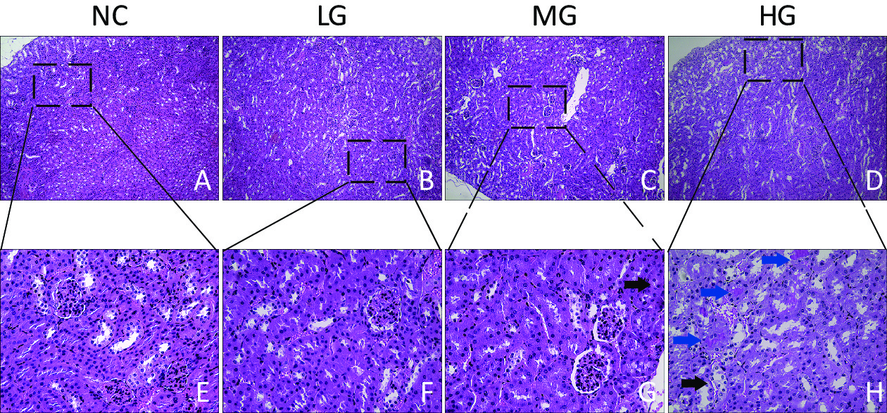
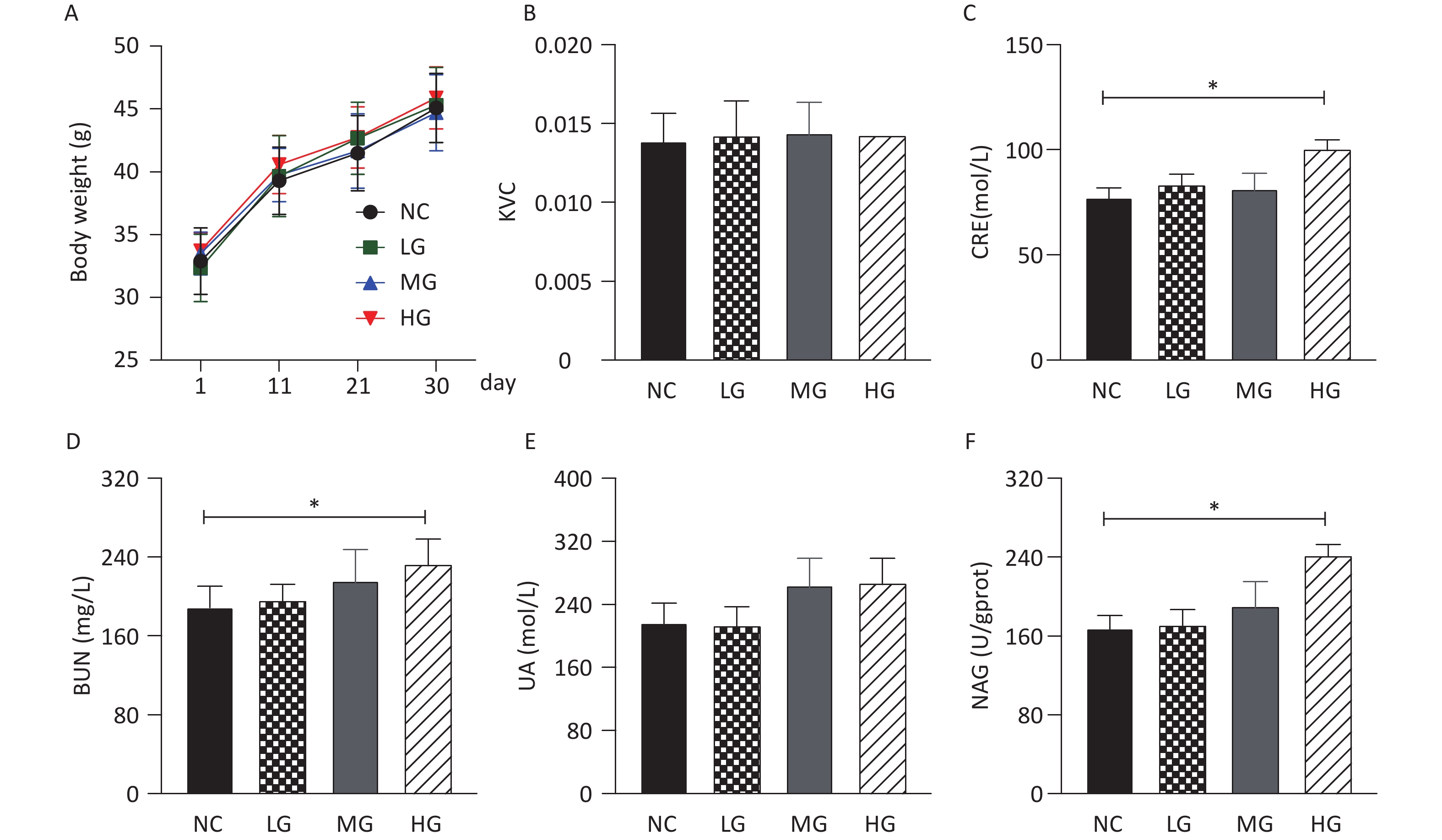
Supplementary Methods (available in www.besjournal.com). The pathological changes and kidney function parameters were evaluated to assess the nephrotoxic effects of the GBH. As shown in Supplementary Figure S1 (available in www.besjournal.com), GBH administration resulted in no significant differences in body weight, kidney viscera coefficient, and uric acid (UA) level between mice in the NC, LG, MG, and HG groups. Mice in the HG group showed significantly higher creatinine (CRE), urea nitrogen (BUN) and β-N-acetylaminoglucosidase (NAG) levels than those in the NC group (P < 0.05). Moreover, when compared with the NC group, the renal epithelial cells of mice in the MG group showed slight edema, whereas those of mice in the HG group exhibited renal tubular epithelial water degeneration, swelling, and abundant protein deposition in the renal tubules (Figure 1). These observations indicated a correlation between GBH treatment and nephrotoxicity.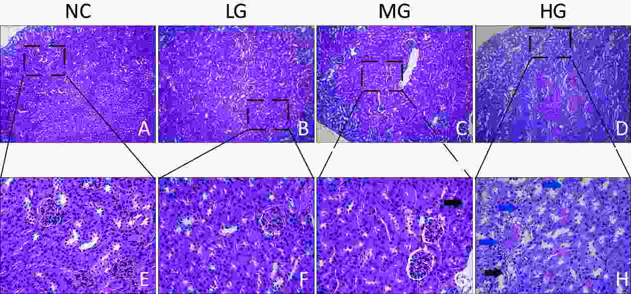
Figure 1. Effect of GBH exposure on histopathology in kidneys of male mice. A, B, C, and D, 100×; E, F, G, and H, 400×. Black arrow: water degeneration; blue arrow: protein deposition in renal tubules. NC, normal control group; LG, low dose group; MG, middle dose group; HG, high dose group.
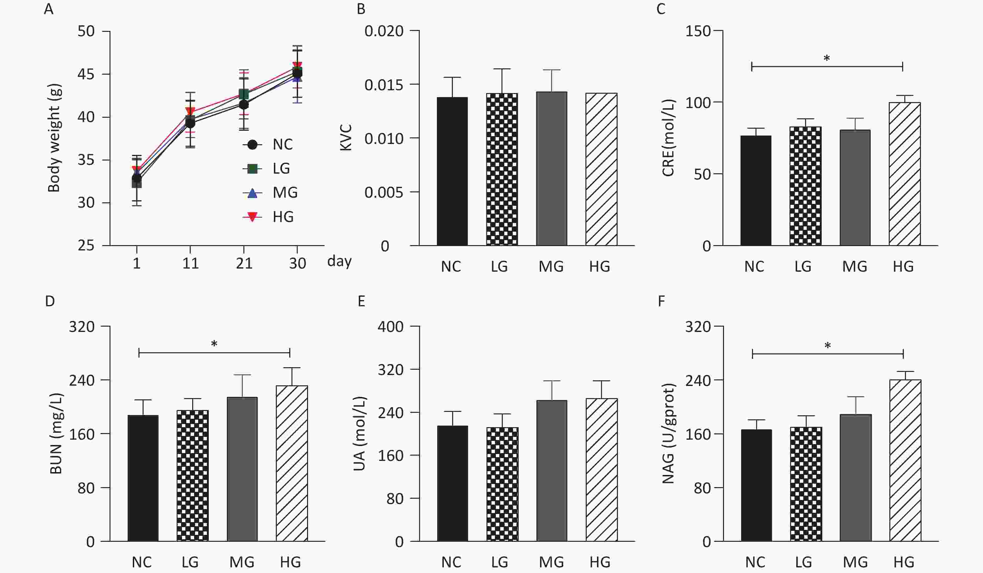
Figure S1. The effects of GBH exposure on body weight and kidney function parameters in the serum. A, body weight; B, kidney viscera coefficient; C, CRE; D, BUN; E, UA; F, NAG. Compared with the control group, *P < 0.05; n = 6. NC, normal control group, 0 mg/kg per day; LG, low dose group, 50 mg/kg per day; MG, middle dose group, 250 mg/kg per day; HG, high dose group, 500 mg/kg per day. Data are presented as means ± SD.
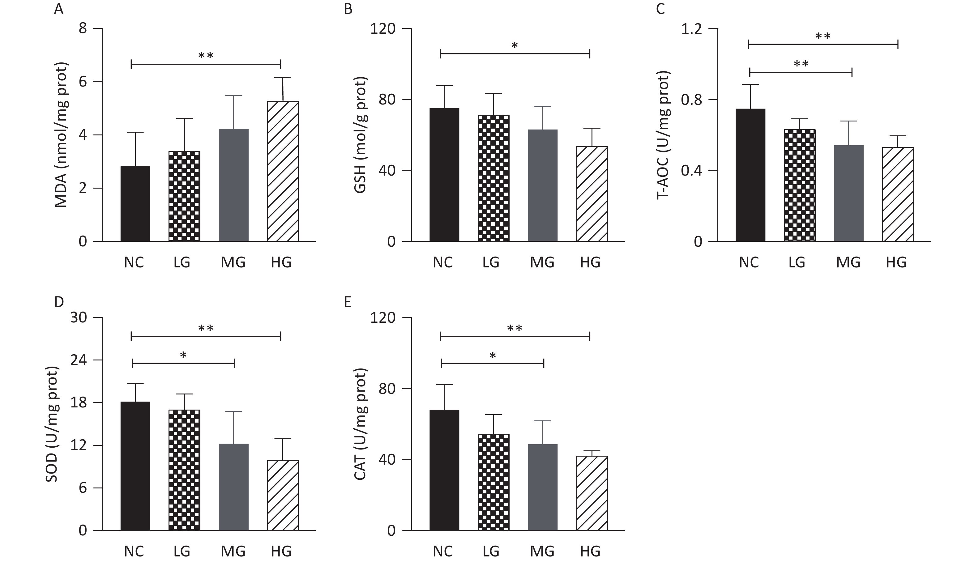
Oxidative stress, induced by ROS overproduction, is a major pathogenic factor in GBH-induced nephrotoxicity. Generally, ROS are generated via an endogenous metabolism and are beneficial for cellular signal transduction and internal balance. However, excessive ROS generation disrupts the oxidative-antioxidant system and damages DNA, proteins, and lipids. As shown in Supplementary Figure S2 (available in www.besjournal.com), the MG and HG groups showed lower glutathione (GSH), total antioxidant capacity (T-AOC), catalase (CAT), and superoxide dismutase (SOD) levels than the NC group (P < 0.05), whereas an increase in the level of MDA (P < 0.05), one of the final product of lipid peroxidation, was observed in the HG group. These observations indicate that the oxidant-antioxidant balance is disrupted in the kidneys. However, the mechanism underlying the interaction between renal pathogenesis and dysfunction owing to OS remains unclear. Therefore, in order to provide clarification on this matter, we performed renal metabolomics to compare differences in metabolite composition and metabolic pathways between the NC and HG groups.
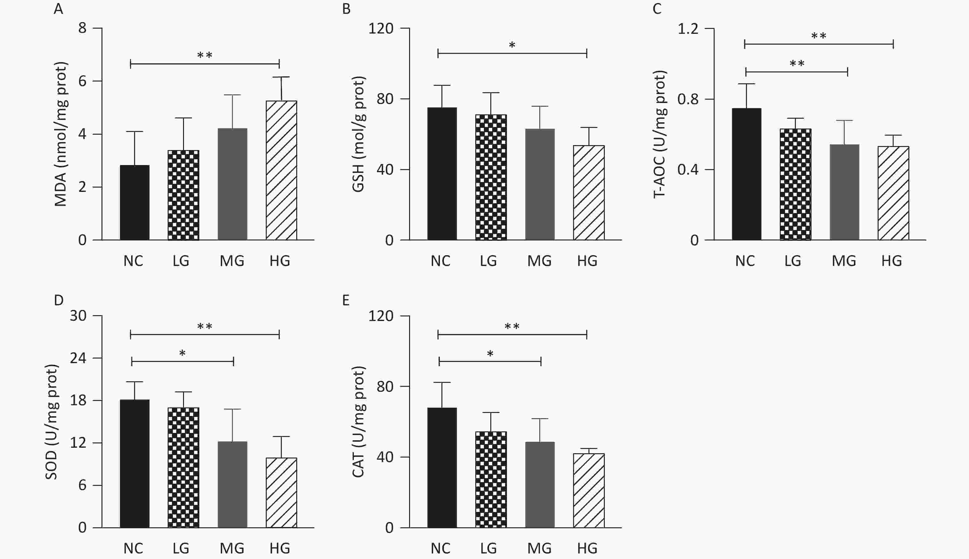
Figure S2. The effect of GBH exposure on histopathology in kidneys of male mice. A-H, HE-stain magnification; A B, C and D, 100×; E, F, G and H, 400×. Black arrow: water degeneration; blue arrow: protein deposition in renal tubules. n = 6. NC, normal control group, 0 mg/kg per day; LG, low dose group, 50 mg/kg per day; MG, middle dose group, 250 mg/kg per day; HG, and high dose group, 500 mg/kg per day.
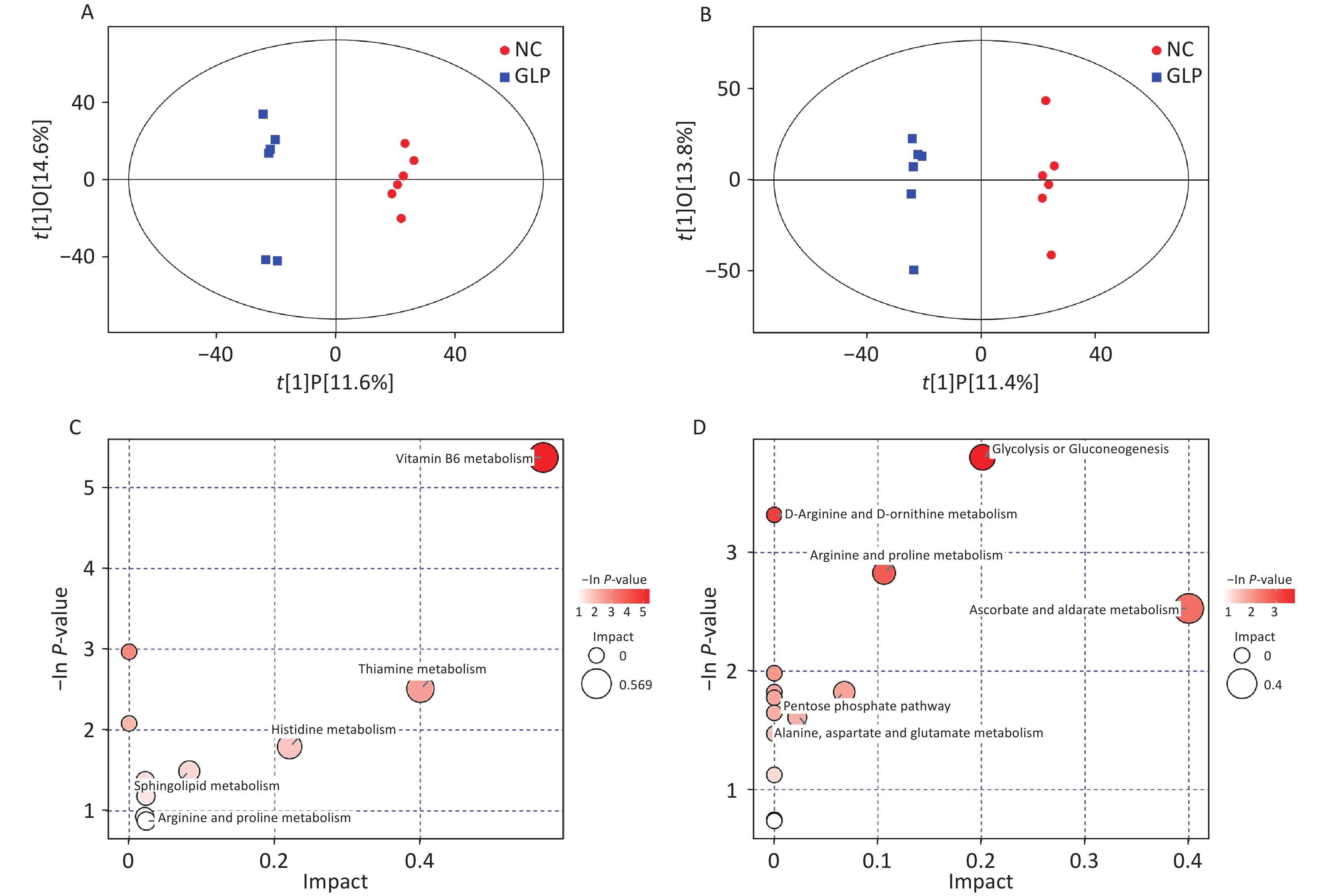
As shown in Figures 2A and B, a distinct separation was observed between the HG and NC groups in the positive and negative ion modes. The permutation test results suggested that the OPLS-DA model used in the present study was stable (Supplementary Figure S3, available in www.besjournal.com). Furthermore, 44 and 22 significantly altered metabolites (SCMs) were identified according to P < 0.05, and the value of the variable importance in the projection (VIP) was > 1 in the positive and negative ion modes (Supplementary Figure S4, Supplementary Tables S1 and S2, available in www.besjournal.com). To clarify the effects of the GBH on metabolism, all SCMs were mapped to the Kyoto Encyclopedia of Genes and Genomes databases (KEGG). Ten and fourteen KEGG pathways were enriched in the positive and negative ion modes, respectively (Figure 2C and D, Supplementary Tables S3 and S4, available in www.besjournal.com). VB6 plays a critical role in the synthesis of GSH by activating nuclear factor E2-related factor 2 (Nrf2) signaling[6]. In the present study, the GBH decreased the levels of pyridoxal and pyridoxamine, perturbed the VB6 metabolic pathway, and inhibited GSH generation, suggesting that GBH induces kidney injury by blocking the antioxidant capacity of GSH via the VB6 metabolic pathway. A previous study showed that L-arginine improves antioxidant capacity by promoting the expression of antioxidant enzymes and decreasing MDA levels[7]. Moreover, L-arginine reduces renal OS caused by excessive NO generation by activating the Nrf2 signaling pathway[8]. In this study, we observed that GBH administration reduced the levels of L-arginine and argininosuccinic acid and disrupted arginine and proline metabolic pathways. These observations suggest that arginine metabolism disorder is a critical factor in GBH-induced renal OS.
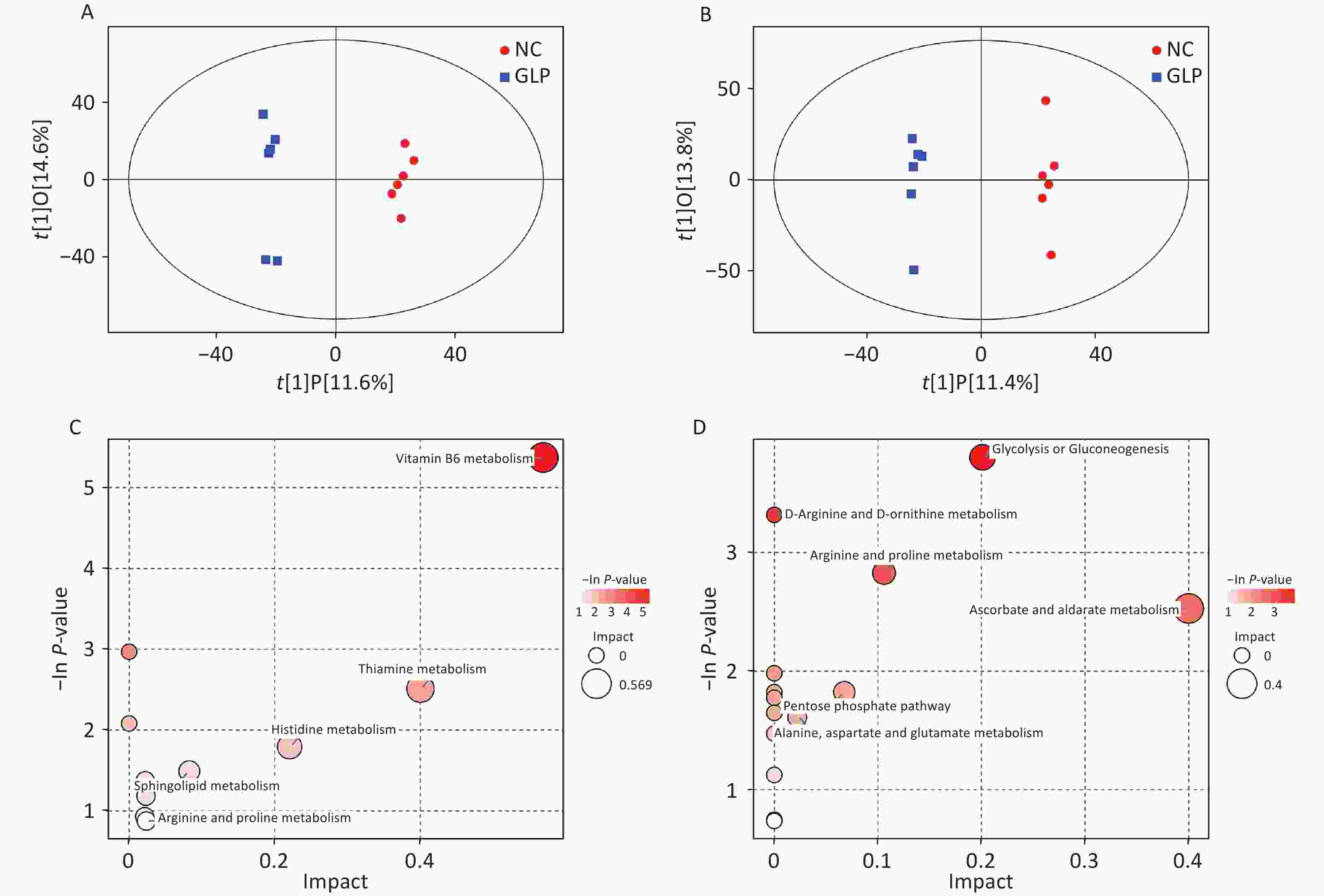
Figure 2. Effects of the GBH on global metabolomics profiles and metabolic pathways in the kidney. (A) and (B), OPLS-DA score plots from the positive and negative ion modes, respectively. (C) and (D), bubble chart of metabolic pathway analysis between HG and NC groups in the positive and negative ion modes, respectively. Each bubble in the bubble chart represents a metabolic pathway.
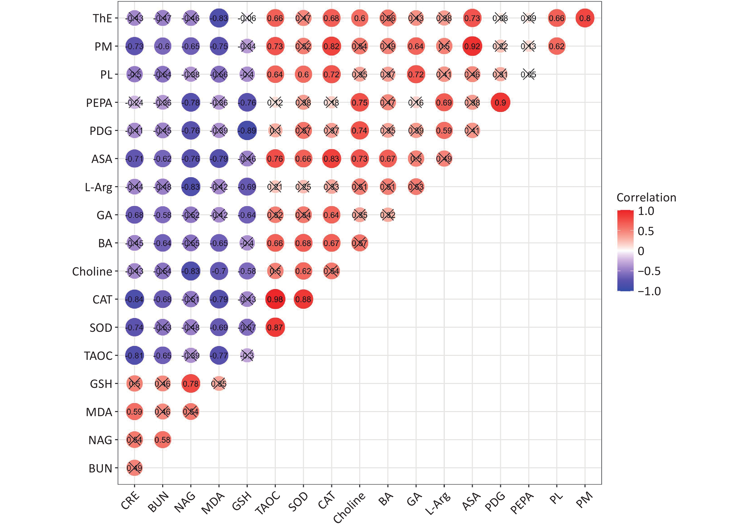
Nitric oxide, which is synthesized from L-arginine by nitric oxide synthase (NOS), plays an important role in regulating renal blood flow and osmotic pressure[9]. The decrease in L-arginine levels observed in this study suggests that GBH may disturb renal osmotic pressure. The evidence shows that in the kidneys, choline and its metabolite, betaine aldehyde, are two osmotic protective agents for renal cells because they facilitate the reabsorption of water from the renal tubules[10]. In this study, GBH exposure reduced the levels of choline and betaine aldehyde, further disturbing the glycine, serine, and threonine metabolic pathways and confirming the toxic effects of GBH on renal osmotic pressure. Moreover, significant correlations were observed between the levels of metabolites involved in osmotic pressure dysfunction and renal function indicators, the levels of metabolites involved in antioxidant capacity and OS indicators, and the levels of metabolites involved in antioxidant capacity and renal function, OS, and renal function indicators (Supplementary Figure S5, available in www.besjournal.com). These findings demonstrate that the GBH induces pathological and functional renal damage by triggering OS and osmotic pressure dysfunction.
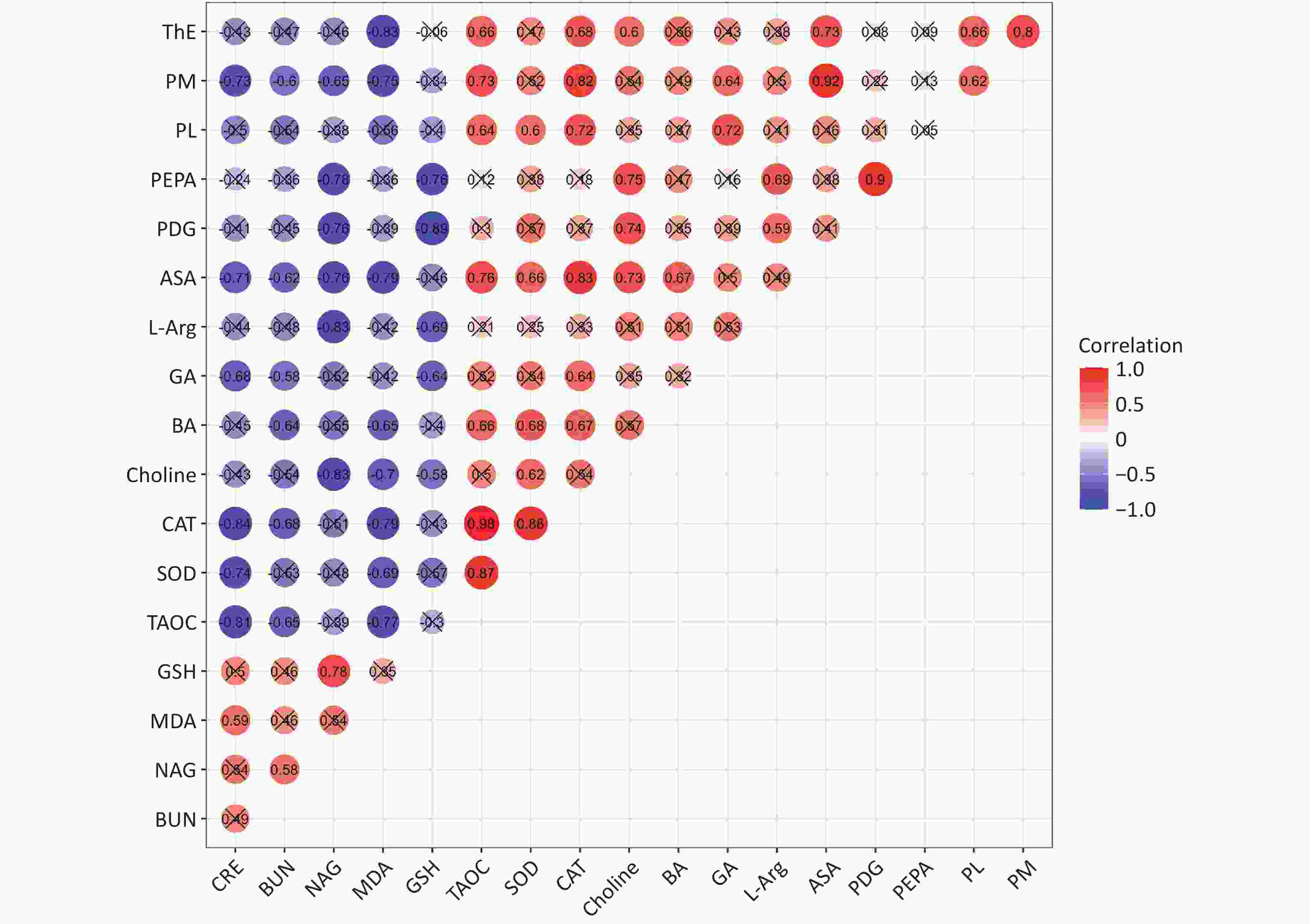
Figure S5. Correlation analysis between kidney function indexes and oxidative stress indicators and SCMs. Red indicates that the correlation coefficient greater than 0, and blue indicates that the correlation coefficient lower than 0; the number represents the value of the correlation coefficient; the dot without × indicates that there is statistical difference, the dot with × indicate that there is no statistical difference of correlation. PM, pyridoxamine; PL, pyridoxal; ASA, argininosuccinic acid; GA, D-Glucuronic acid; BA, betaine aldehyde; PDG, 2-phospho-D-glyceric acid; PEPA, phosphoenolpyruvic acid; L-Arg, L-arginine; CRE, creatinine; BUN, urea nitrogen; NAG, β-N-acetylaminoglucosidase; MDA, malondialdehyde; GSH, glutathione; CAT, catalase; T-AOC, total antioxidant capacity; SOD, superoxide dismutase. n = 6.
Glucose utilization for energy production in renal cells is necessary for maintaining normal kidney function. In general, glucose is metabolized to pyruvate by a series of enzymes. Thereafter, pyruvate is utilized in the mitochondria to generate energy via the tricarboxylic acid cycle and oxidative phosphorylation. Thiamine is a vitamin that acts as a cofactor for many enzymes involved in aerobic glucose oxidation such as pyruvate dehydrogenase and alpha-ketoglutarate dehydrogenases. In this study, the GBH perturbed glycolysis, gluconeogenesis, and thiamine metabolism in the kidneys, indicating that GBH administration induced renal glucose utilization and potential energy metabolism disorders.
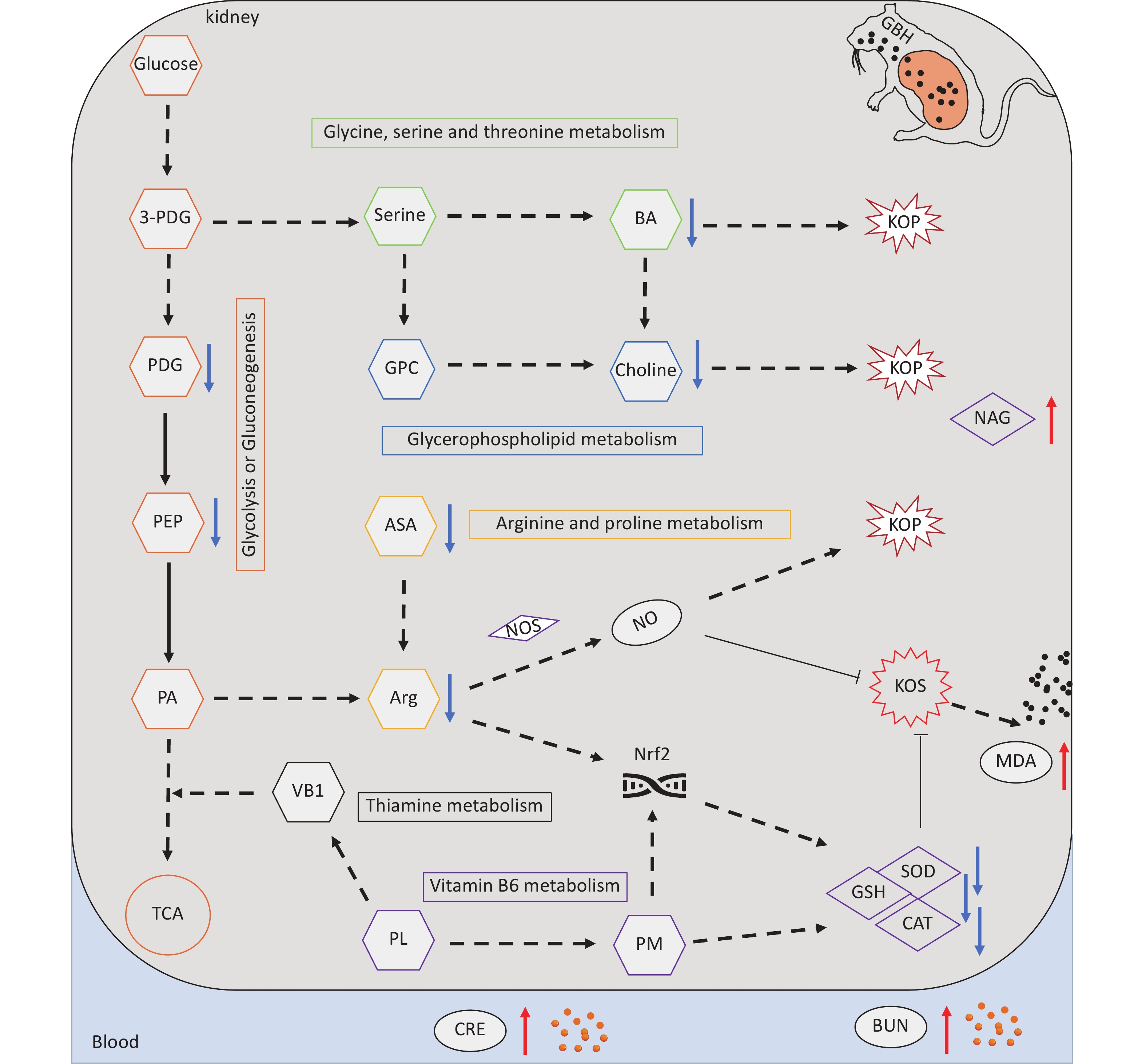
In conclusion, our results revealed that exposure to GBH induces renal tubular epithelial injury and dysfunction by inducing OS and disrupting metabolite balance in the kidneys. Notably, GBH administration disrupted VB6 and L-arginine metabolism, energy metabolism, and choline metabolism, thus promoting renal OS and osmotic pressure imbalance, resulting in pathological renal injury and dysfunction (Figure 3). However, this study has some limitations. First, the GBH dose administered to the mice in the HG group exceeded the actual exposure levels within the population. Secondly, GBH contains other auxiliary components, and we did not investigate whether the nephrotoxicity observed in this study was induced by glyphosate alone or by the combined effect of glyphosate and auxiliary components. Therefore, further studies focusing on actual exposure conditions and the effects of auxiliary components are necessary to identify new aspects of glyphosate- and GBH-induced nephrotoxicity health risks.
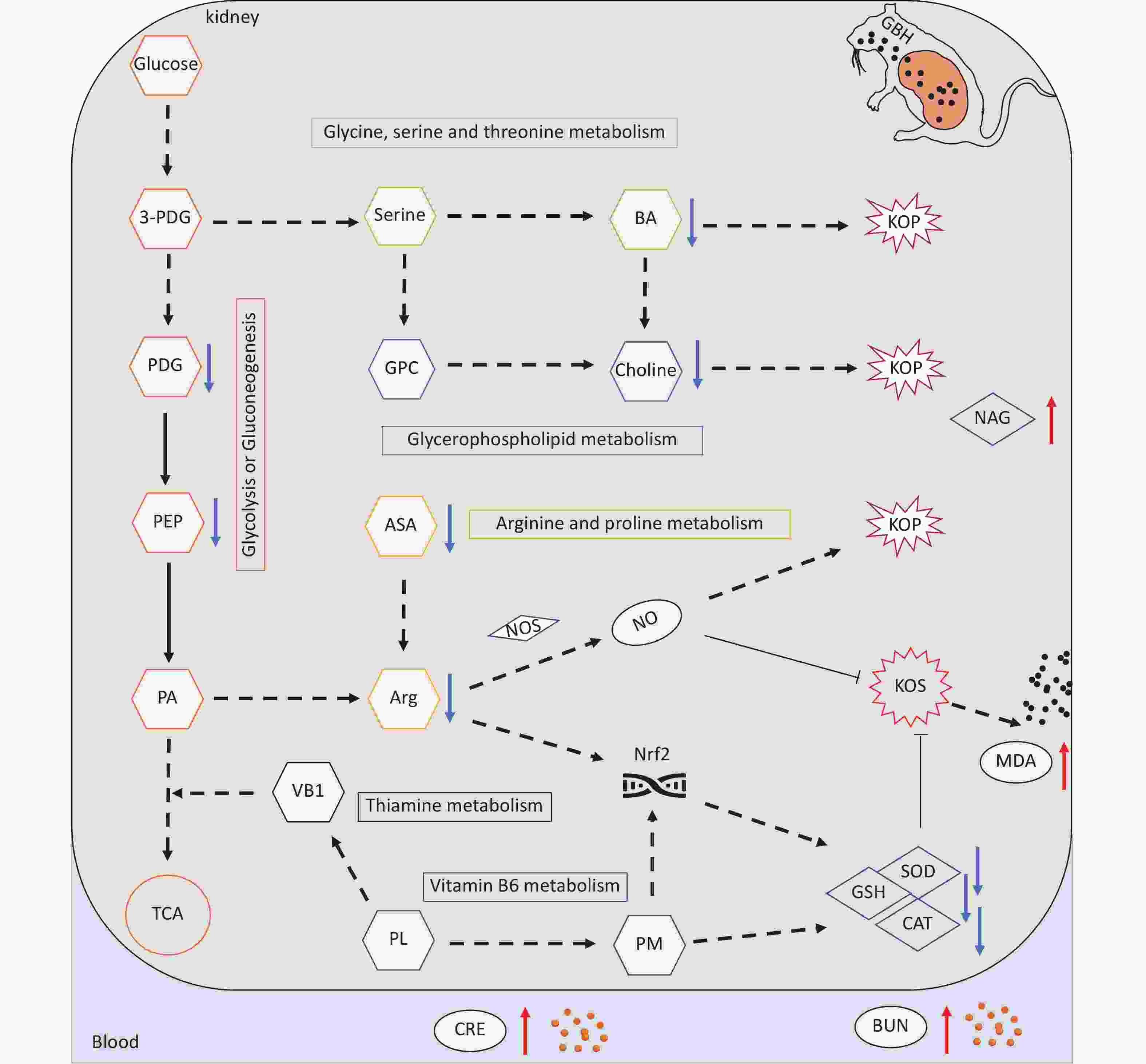
Figure 3. Mechanism of GBH-induced nephrotoxicity. Rectangle, metabolism pathways; hexagon, metabolites; diamond, enzymes. When compared with the control group, the red arrow depicts upregulation and blue arrow depicts downregulation. Black solid line arrow, direct reaction; dotted line arrow, multi-step reaction. PL, pyridoxal; PM, pyridoxamine; GA, D-Glucuronic acid; PDG, phospho-D-glyceric acid; PEPA, phosphoenolpyruvic acid; PA, Pyruvate; TCA, tricarboxylic acid cycle; Arg, L-arginine; ASA, argininosuccinic acid; BA, betaine aldehyde; NOS, nitric oxide synthase; Nrf2, nuclear factor erythroid2-related factor 2; KOS, kidney oxidative stress; KOP, kidney osmotic pressure; GSH, glutathione; CAT, catalase; SOD, superoxide dismutase; CRE, creatinine; BUN, urea nitrogen; NAG, β-N-acetylaminoglucosidase; MDA, malondialdehyde.
全文HTML
 24317+.pdf
|

|

 Quick Links