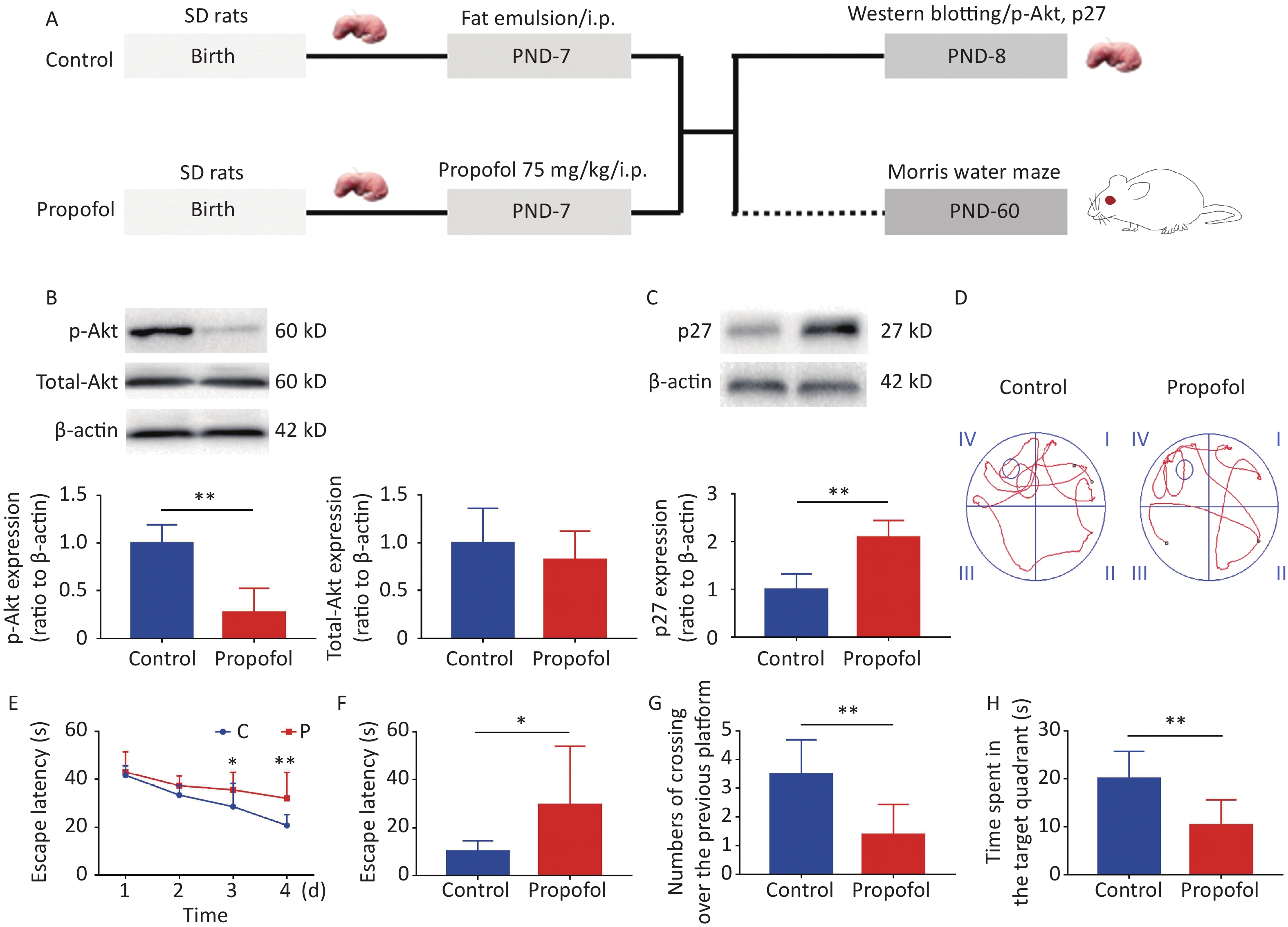
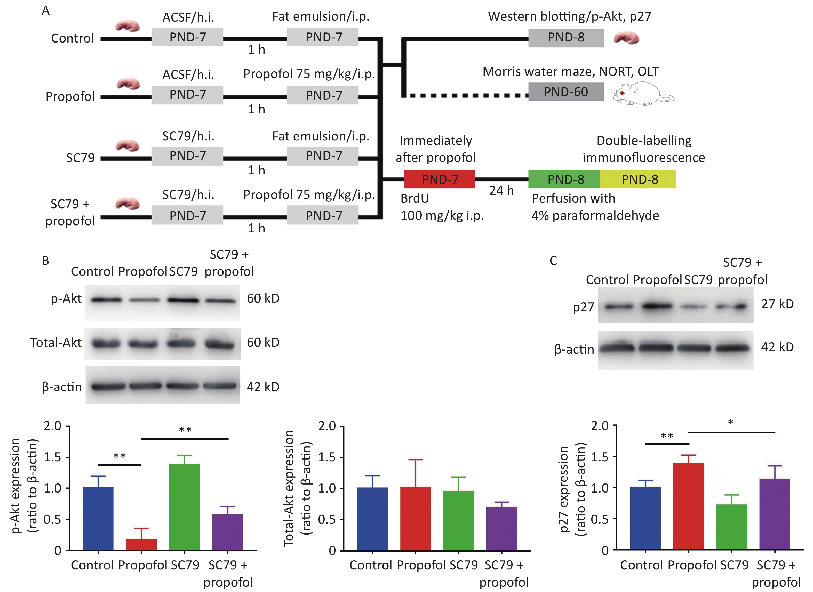
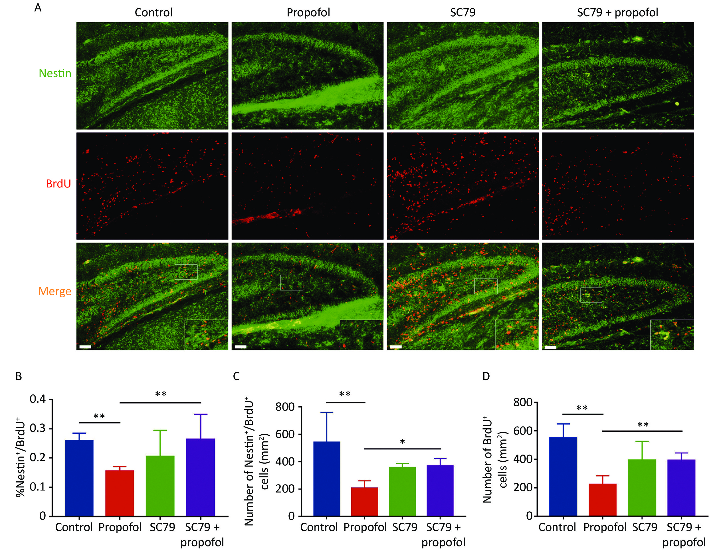
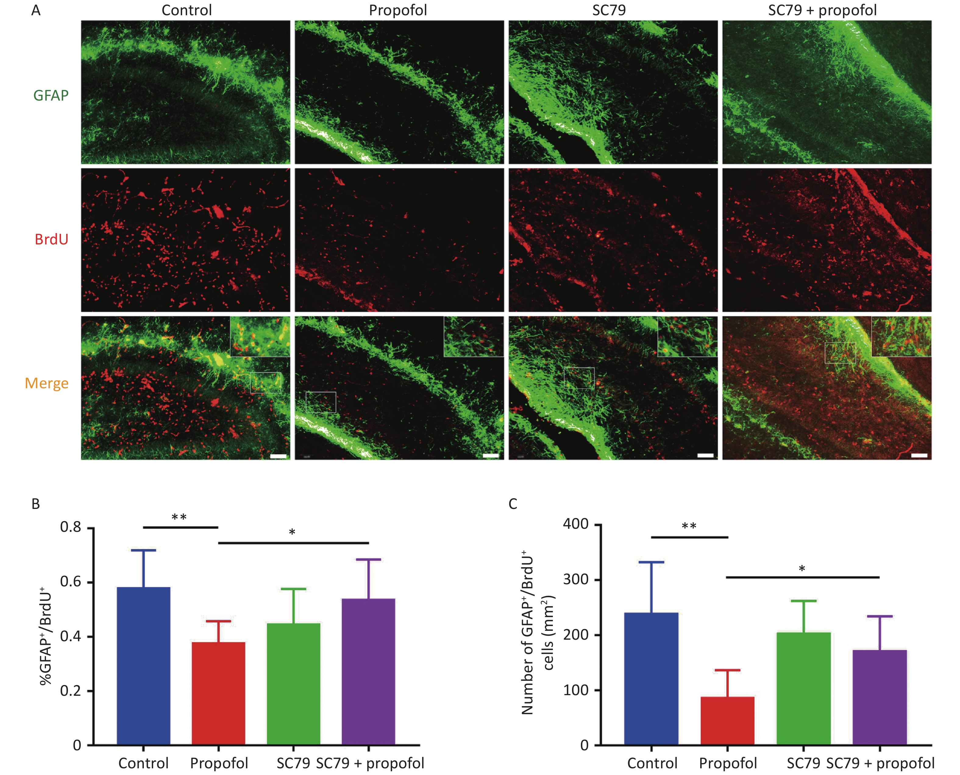
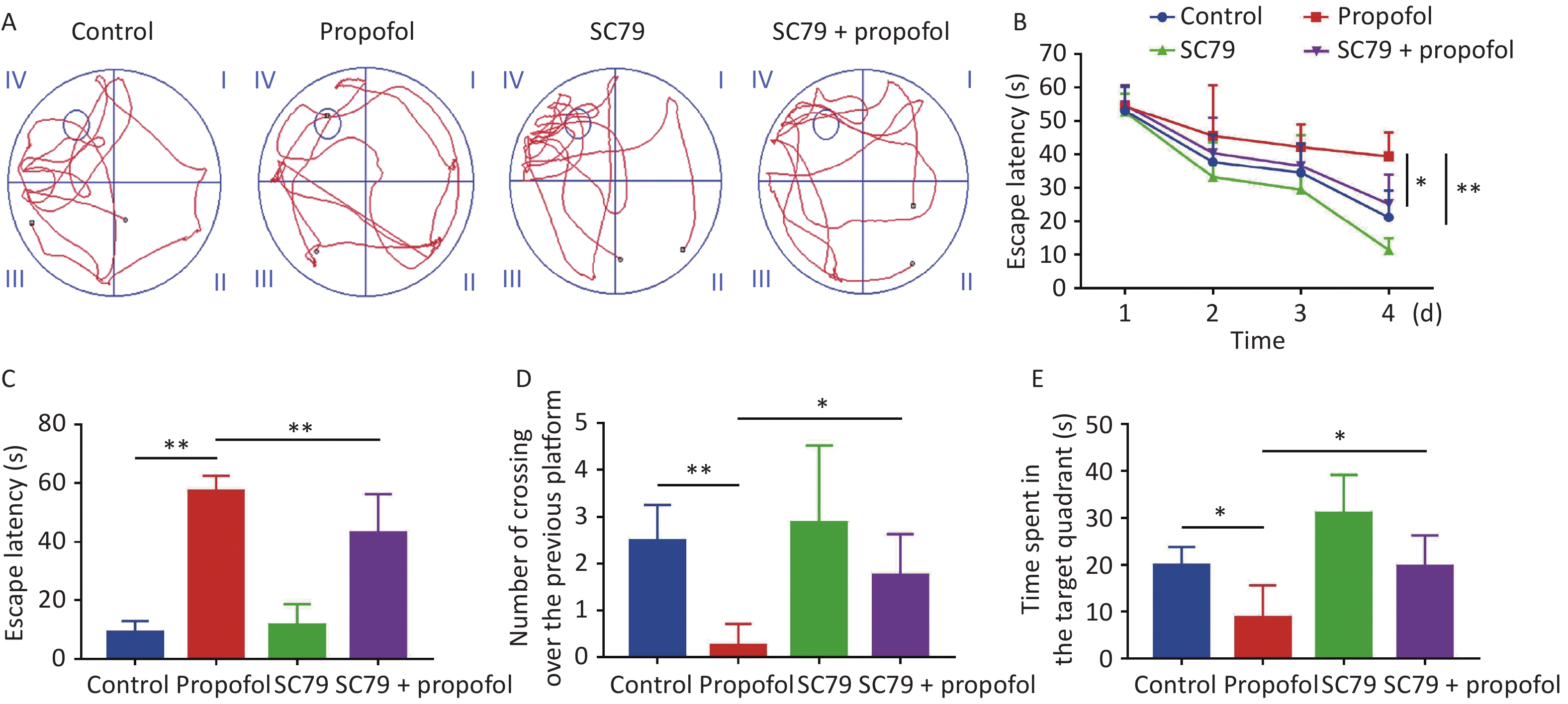
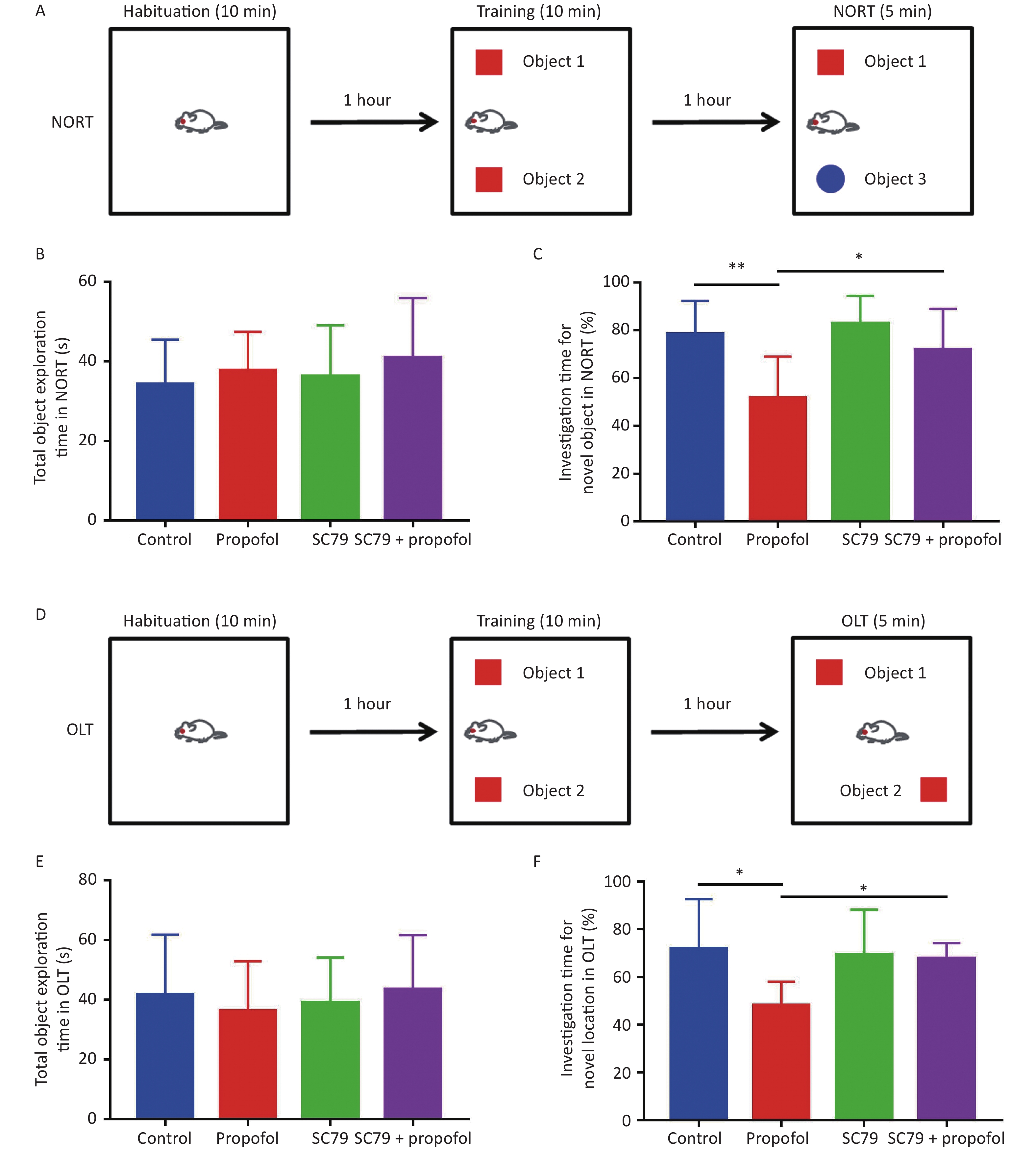
Objective Neonatal exposure to propofol has been reported to cause neurotoxicity and neurocognitive decline in adulthood; however, the underlying mechanism has not been established. Methods SD rats were exposed to propofol on postnatal day 7 (PND-7). Double-immunofluorescence staining was used to assess neurogenesis in the hippocampal dentate gyrus (DG). The expression of p-Akt and p27 were measured by western blotting. The Morris water maze, novel object recognition test, and object location test were used to evaluate neurocognitive function 2-month-old rats. Results Phosphorylation of Akt was inhibited, while p27 expression was enhanced after neonatal exposure to propofol. Propofol also inhibited proliferation of neural stem cells (NSCs) and decreased differentiation to neurons and astroglia. Moreover, the neurocognitive function in 2-month-old rats was weakened. Of significance, intra-hippocampal injection of the Akt activator, SC79, attenuated the inhibition of p-AKT and increase of p27 expression. SC79 also rescued the propofol-induced inhibition of NSC proliferation and differentiation. The propofol-induced neurocognition deficit was also partially reversed by SC79. Conclusion Taken together, these results suggest that neurogenesis is hindered by neonatal propofol exposure. Specifically, neonatal propofol exposure was shown to suppress the proliferation and differentiation of NSCs by inhibiting Akt/p27 signaling pathway.
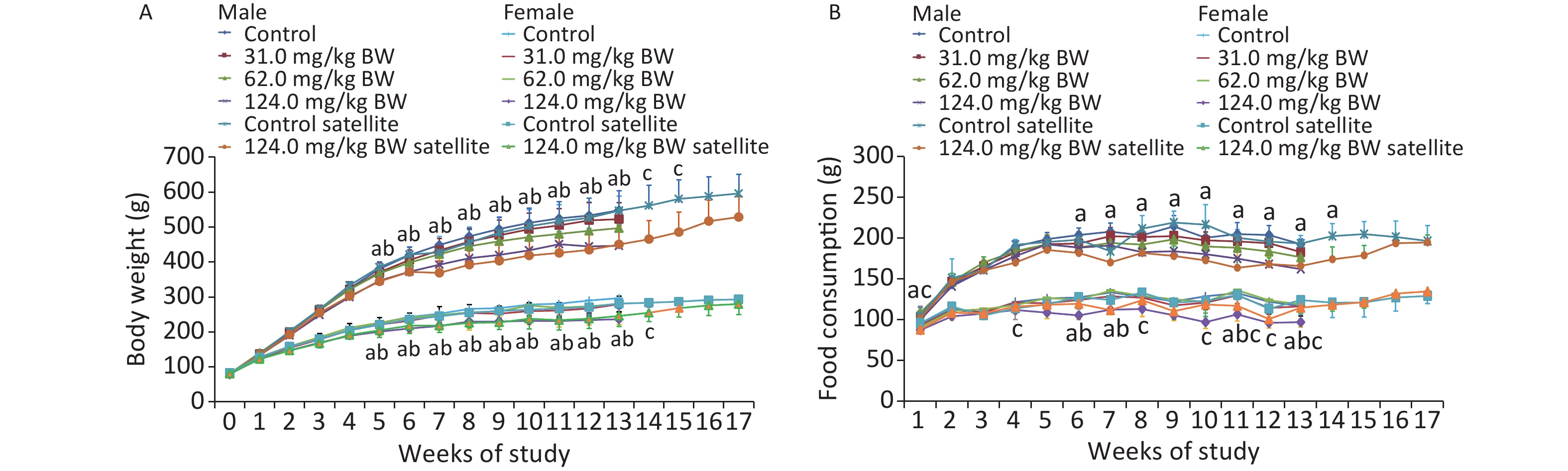
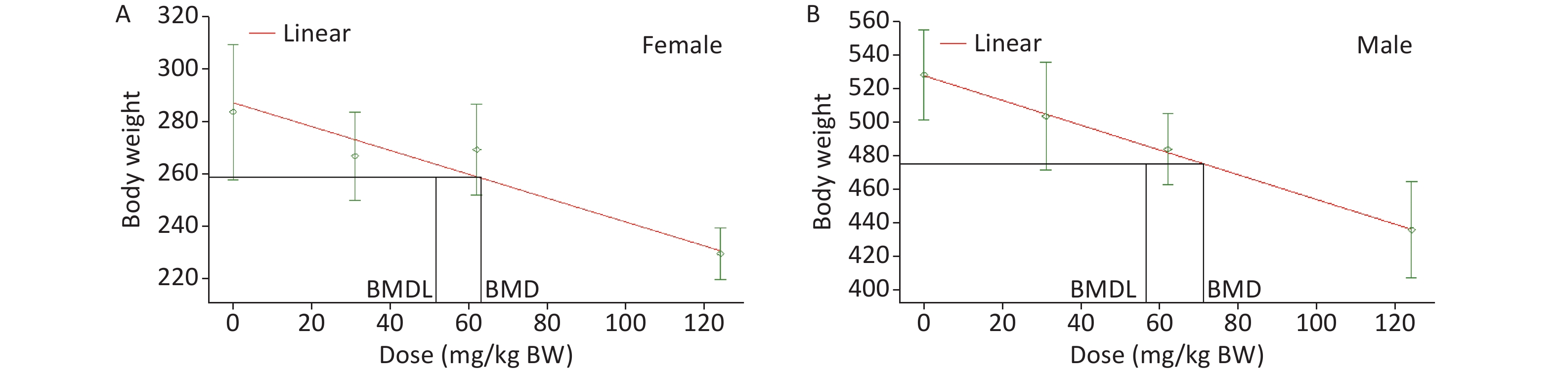
Objective The present study was undertaken to evaluate the subchronic oral toxicity of sodium dehydroacetate (DHA-Na) and to determine the point of departure (POD), which is a critical factor in the establishment of an acceptable dietary intake. Methods DHA-Na was administered once daily by gavage to Sprague–Dawley rats at dose levels of 0.0, 31.0, 62.0, and 124.0 mg/kg BW per day for 90 days, followed by a recovery period of 4 weeks in the control and 124.0 mg/kg BW per day groups. The outcome parameters were mortality, clinical observations, body weights, food consumption, hematology and clinical biochemistry, endocrine hormone levels, and ophthalmic, urinary, and histopathologic indicators. The benchmark dose (BMD) approach was applied to estimate the POD. Results Significant decreases were found in the 62.0 and 124.0 mg/kg BW groups in terms of the body weight and food utilization rate, whereas a significant increase was found in the thyroid stimulating hormone levels of the 124.0 mg/kg BW group. Importantly, the 95% lower confidence limit on the BMD of 51.7 mg/kg BW was modeled for a reduction in body weight. Conclusion The repeated-dose study indicated the slight systemic toxicity of DHA-Na at certain levels (62.0 and 124.0 mg/kg BW) after a 90-day oral exposure.
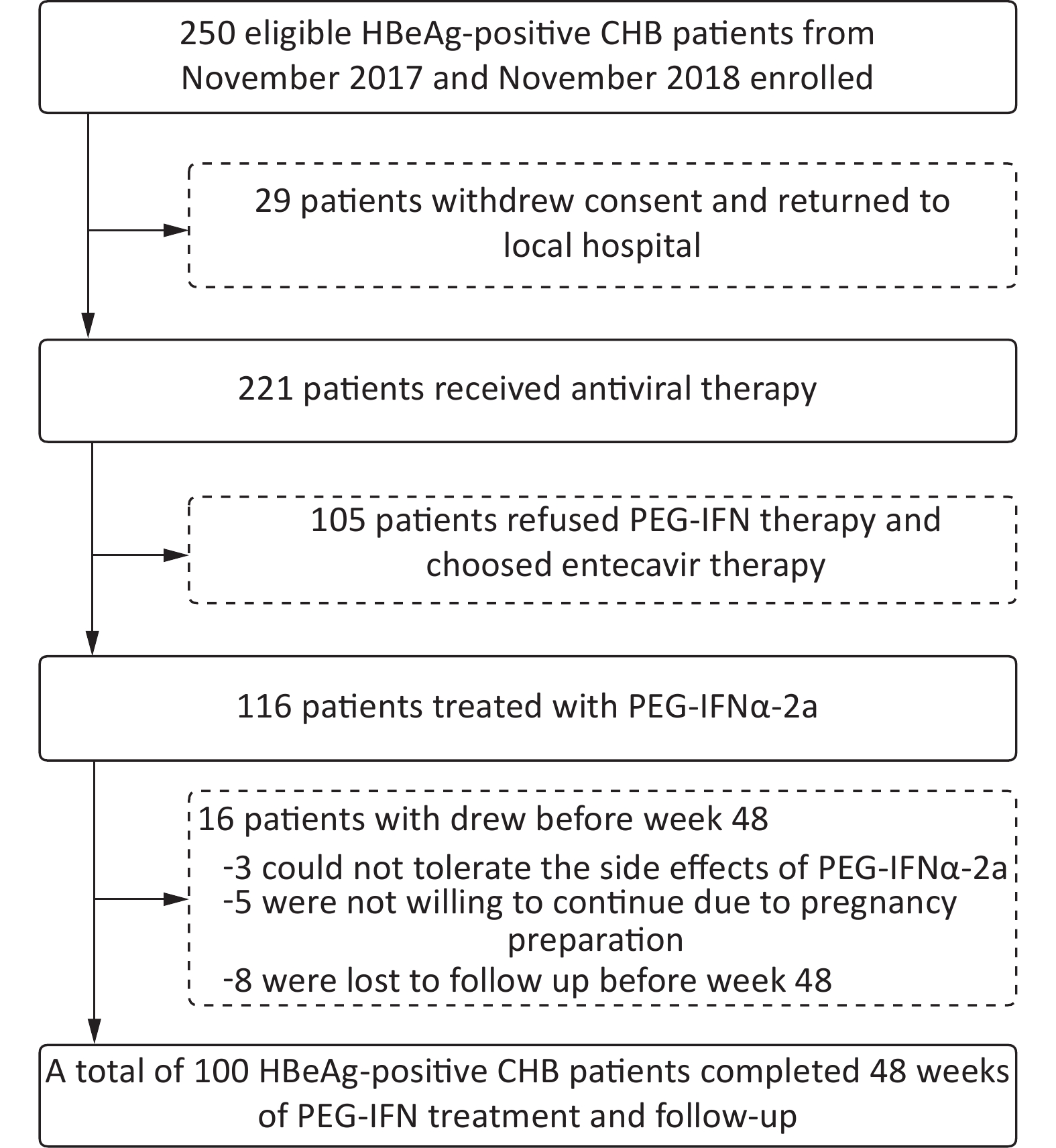
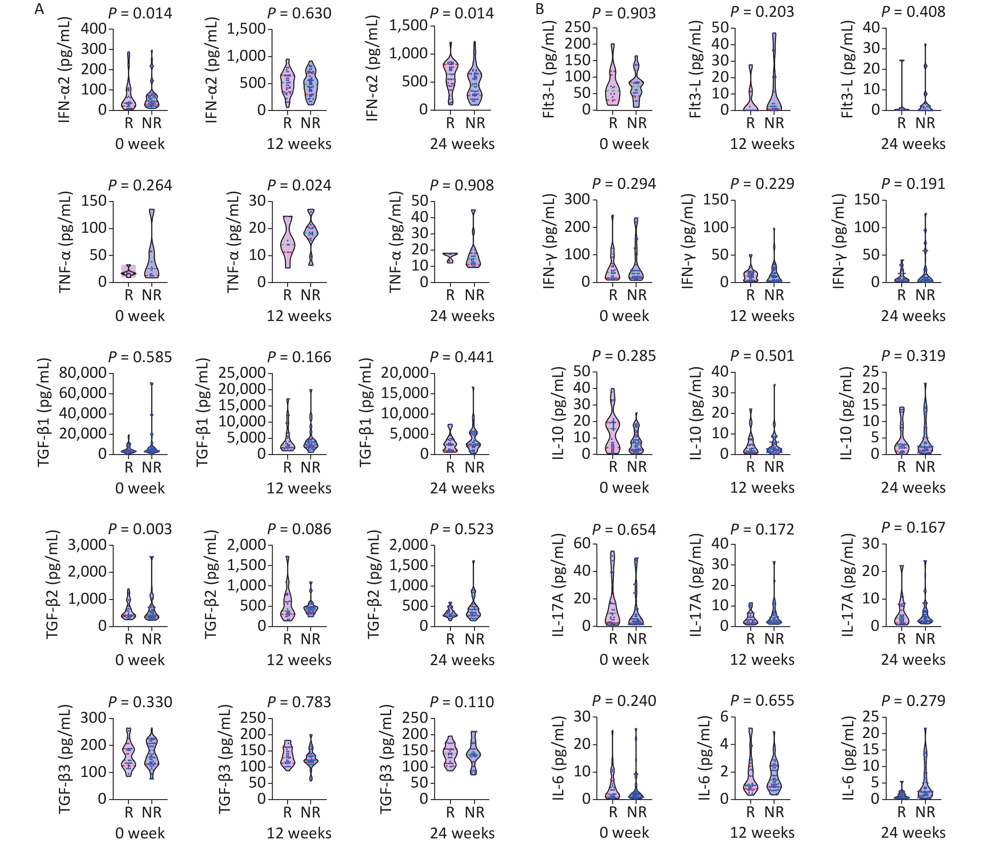
Objective This study aimed to investigate whether cytokine profiles and virological markers might add value in monitoring the effects of peginterferon (PEG-IFN) therapy for hepatitis B e-antigen (HBeAg) positive chronic hepatitis B (CHB). Methods HBeAg positive patients with CHB were treated with PEG-IFN for 48 weeks. Clinical biochemical, and HBV serological indexes, as well as cytokines, were detected at baseline and every 12 weeks. Results A total of 116 patients with CHB were enrolled in this study; 100 patients completed the 48-week treatment and follow-up, of whom 38 achieved serum HBeAg disappearance, 25 achieved HBeAg seroconversion, 37 showed HBsAg decreases ≥ 1 log10 IU/mL, 9 showed HBsAg disappearance, and 8 became HBsAb positive. The cytokine levels at baseline and during treatment were similar between the HBeAg disappearance group and non-disappearance group. The disappearance of HBeAg was independently associated with HBeAg levels at weeks 12 and 24, and with the HBeAg decline at week 24 (P < 0.05). The HBsAg response was independently associated with HBsAg, the HBsAg decline, HBeAg, the HBeAg decline at week 12, and HBsAg at week 24 (P < 0.05). Conclusion There was no significant correlation between the response to interferon (IFN) and cytokines during PEG-IFN treatment. The changes in virological markers predicted the response to IFN after 48 weeks.
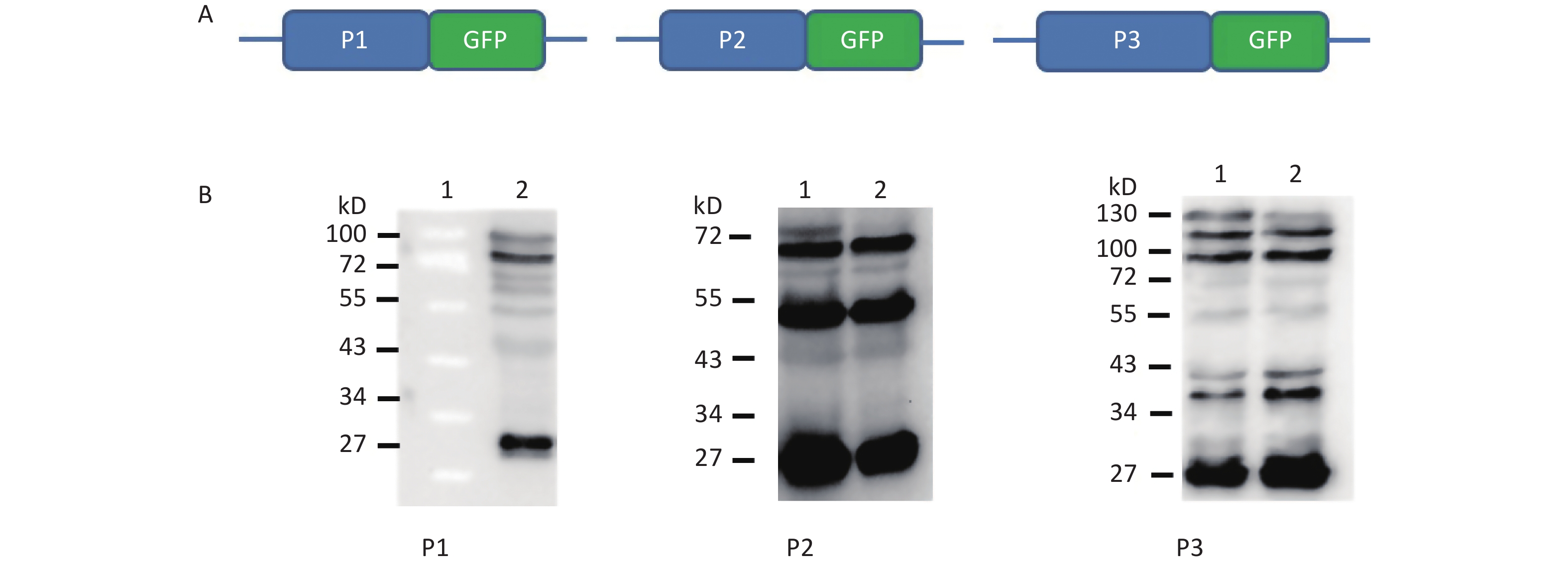
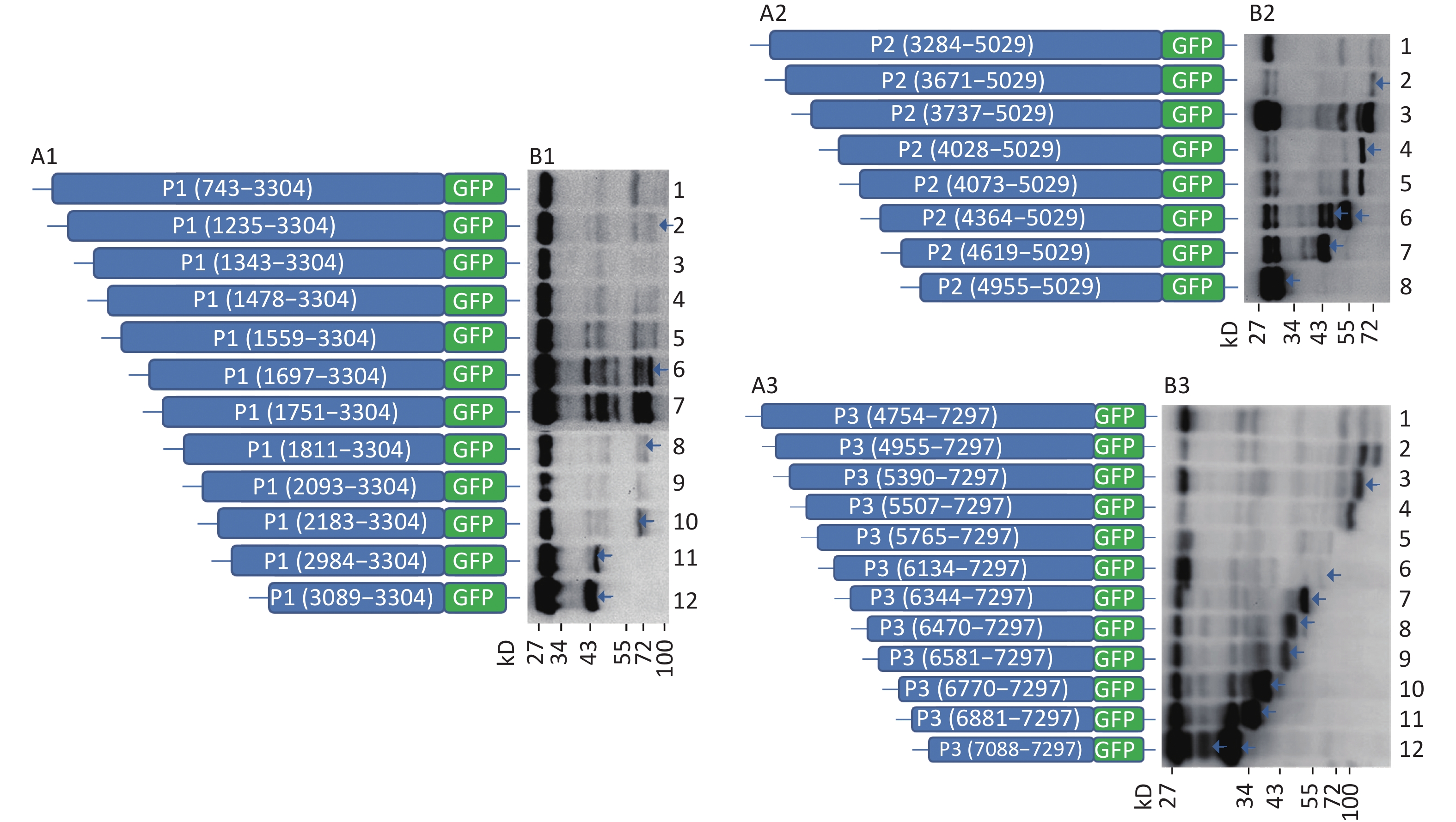
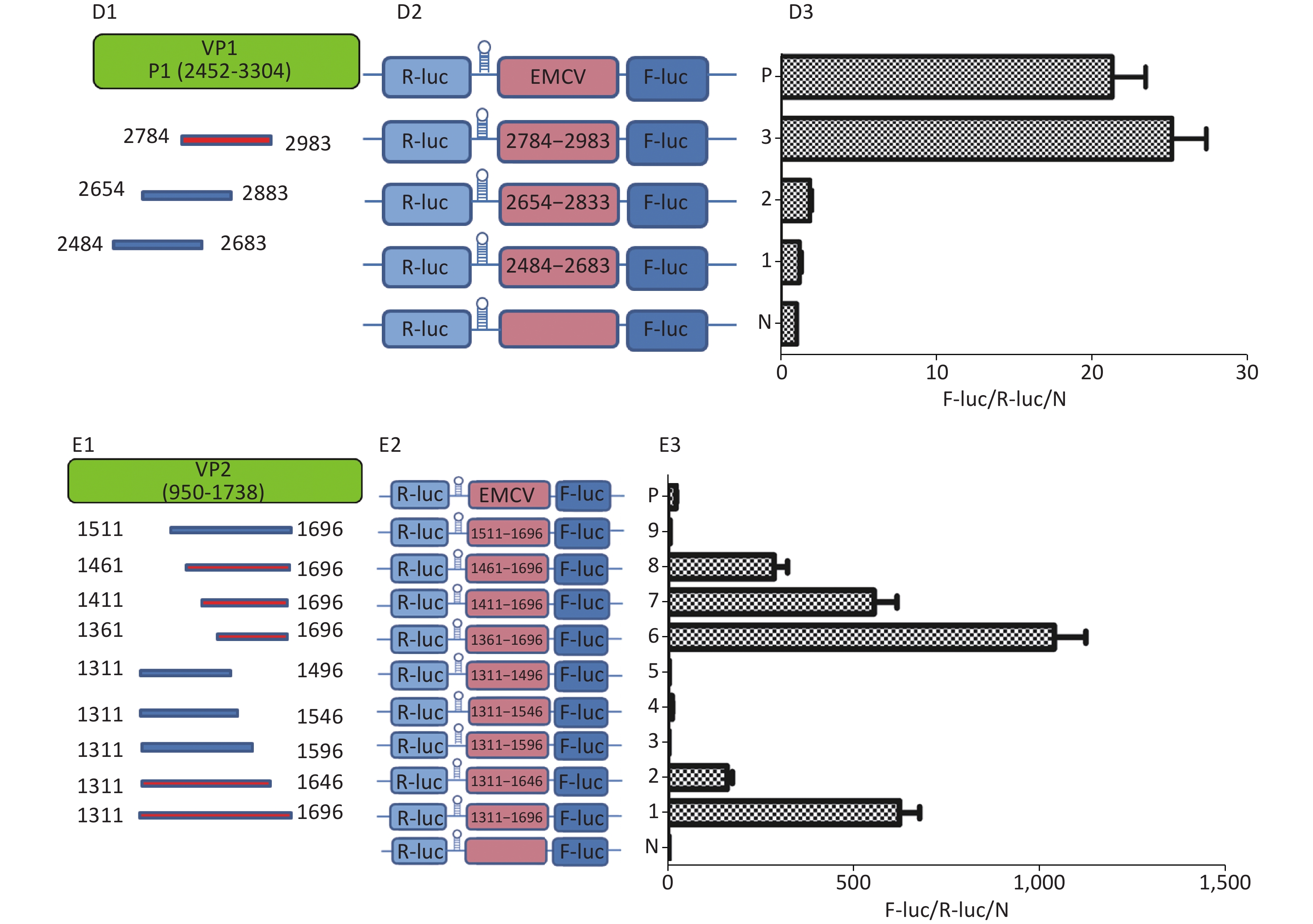
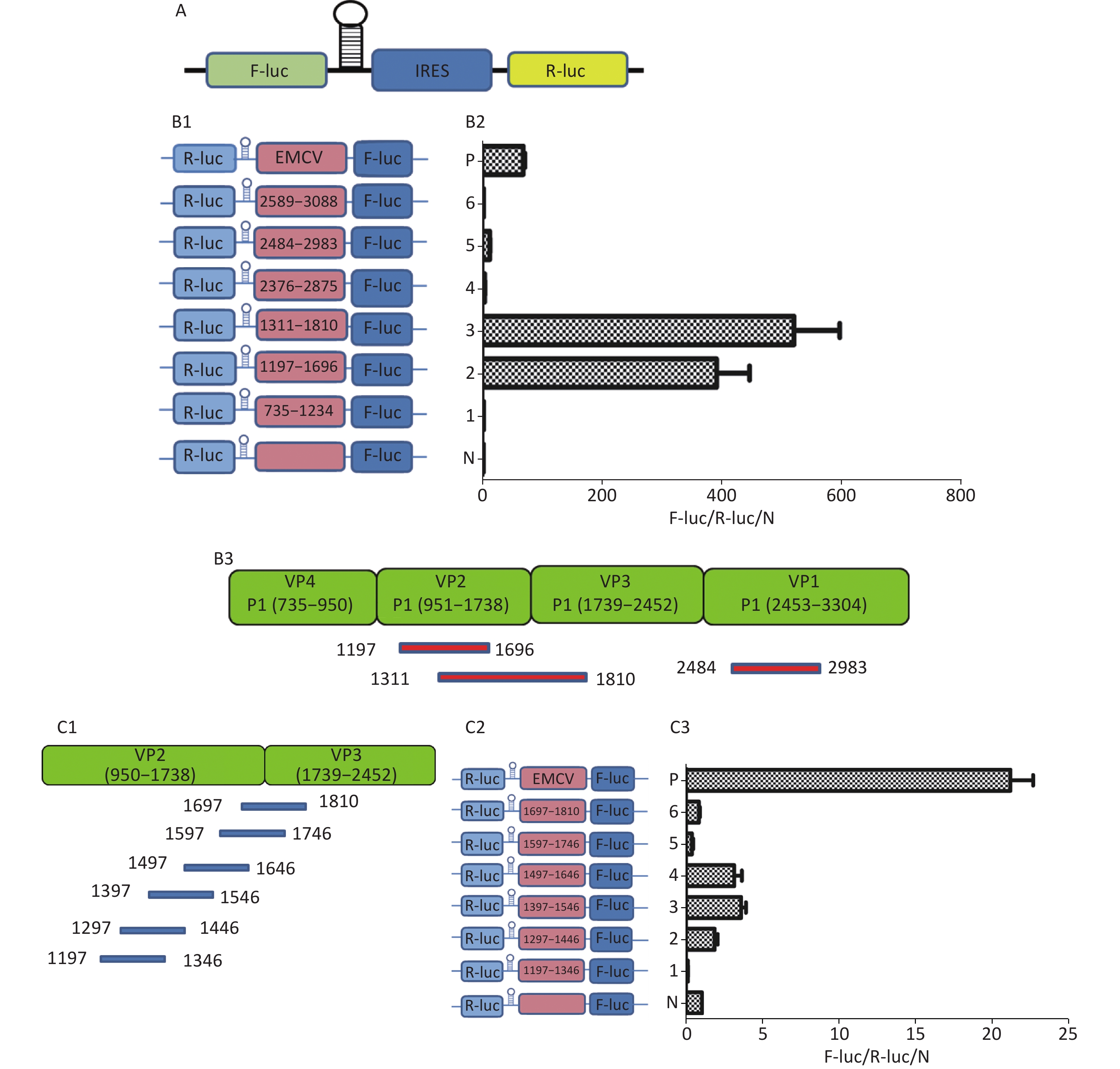
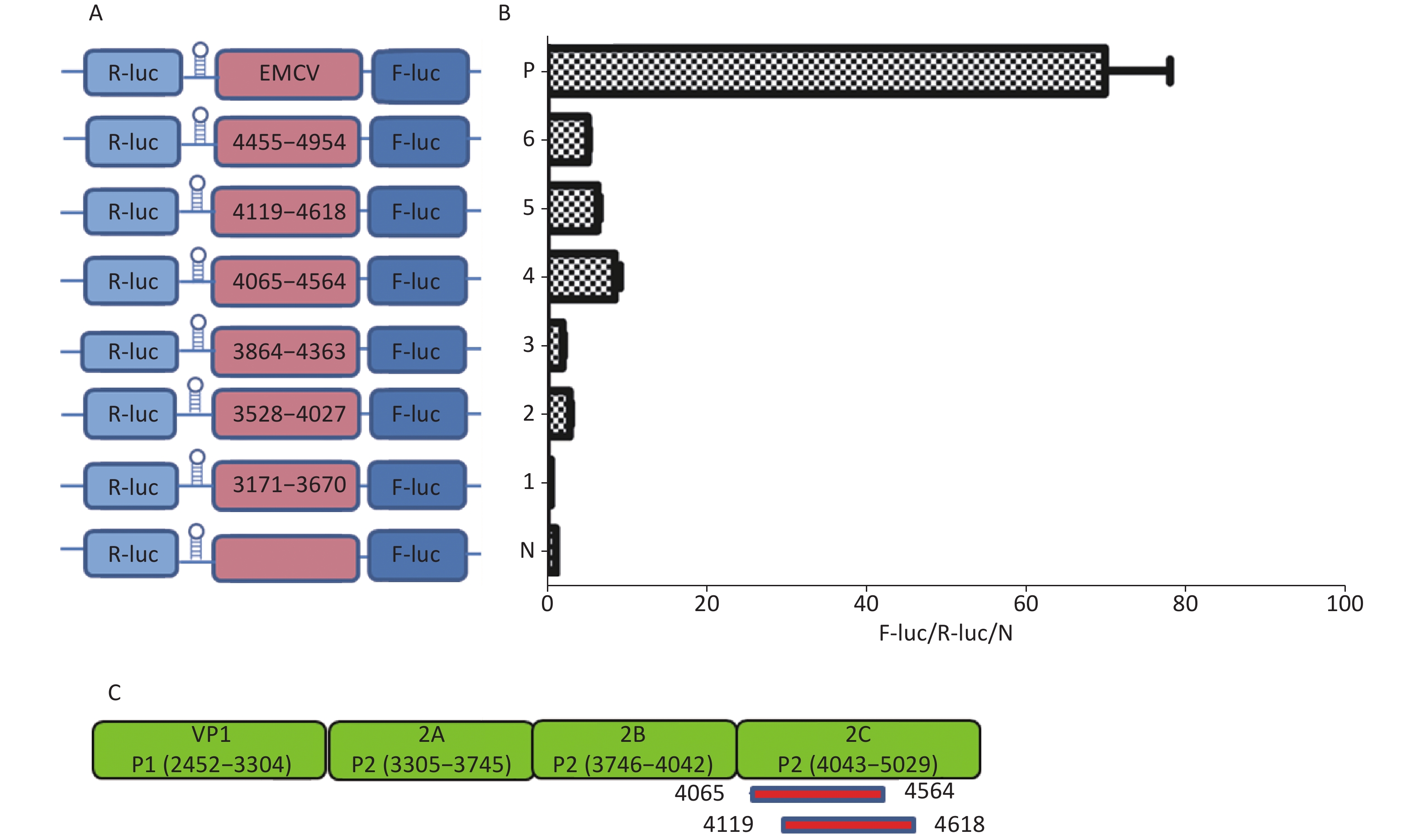
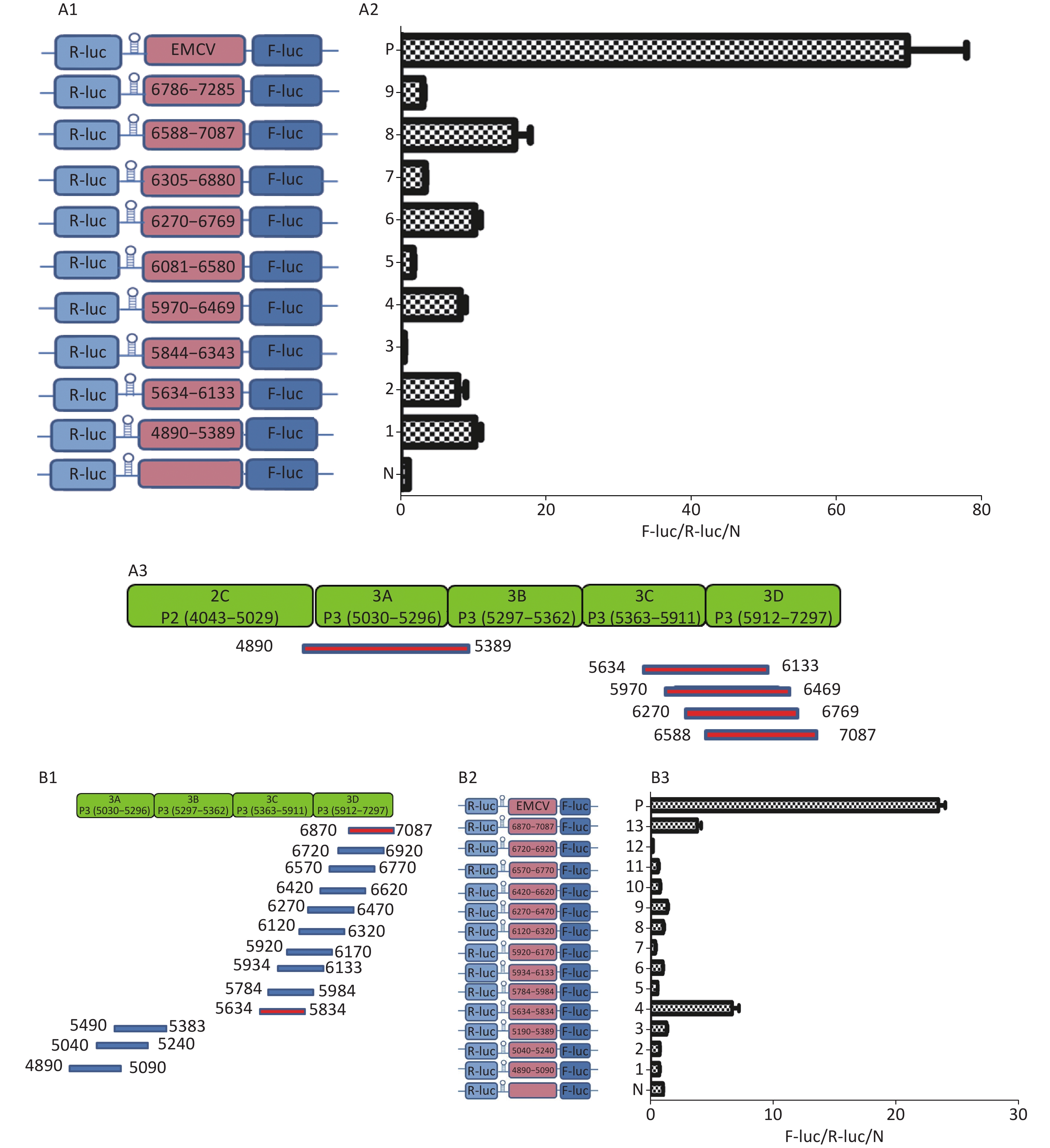
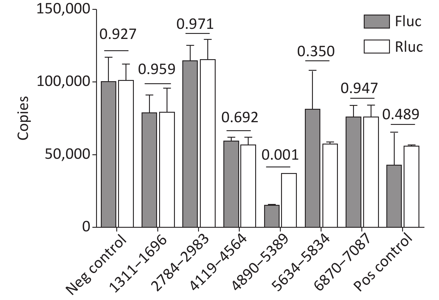
Objective This study aimed to identify internal ribosome entry sites (IRESs) in the open reading frame (ORF) of the Coxsackievirus B3 (CVB3) genome. Methods The sequences of P1, P2, or P3 of the CVB3 genome or the truncated sequences from each antithymocyte globulin (ATG) to the end of the P1, P2, or P3 gene were inserted into the pEGFP-N1 vector. After transfection, possible IRES-dependent green fluorescent protein (GFP)-fused proteins were detected by anti-GFP western blotting. The sequences of possible IRESs were inserted into specific Fluc/Rluc bicistronic vectors, in which the potential IRESs were determined according to the Fluc/Rluc activity ratio. Expression of Fluc and Rluc mRNA of the bicistronic vector was detected by RT-qPCR. Results After transfection of full length or truncated sequences of the P1, P2, or P3 plasmids, six GFP-fused protein bands in P1, six bands in P2 and nine bands in P3 were detected through western blotting. Two IRESs in VP2 (1461–1646 nt) and VP1 (2784–2983 nt) of P1; one IRES in 2C (4119–4564 nt) of P2; and two IRESs in 3C (5634–5834 nt) and 3D (6870–7087 nt) of P3 were identified according to Fluc/Rluc activity ratio. The cryptic promoter was also excluded by RT-qPCR. Conclusion Five IRESs are present in the CVB3 coding region.
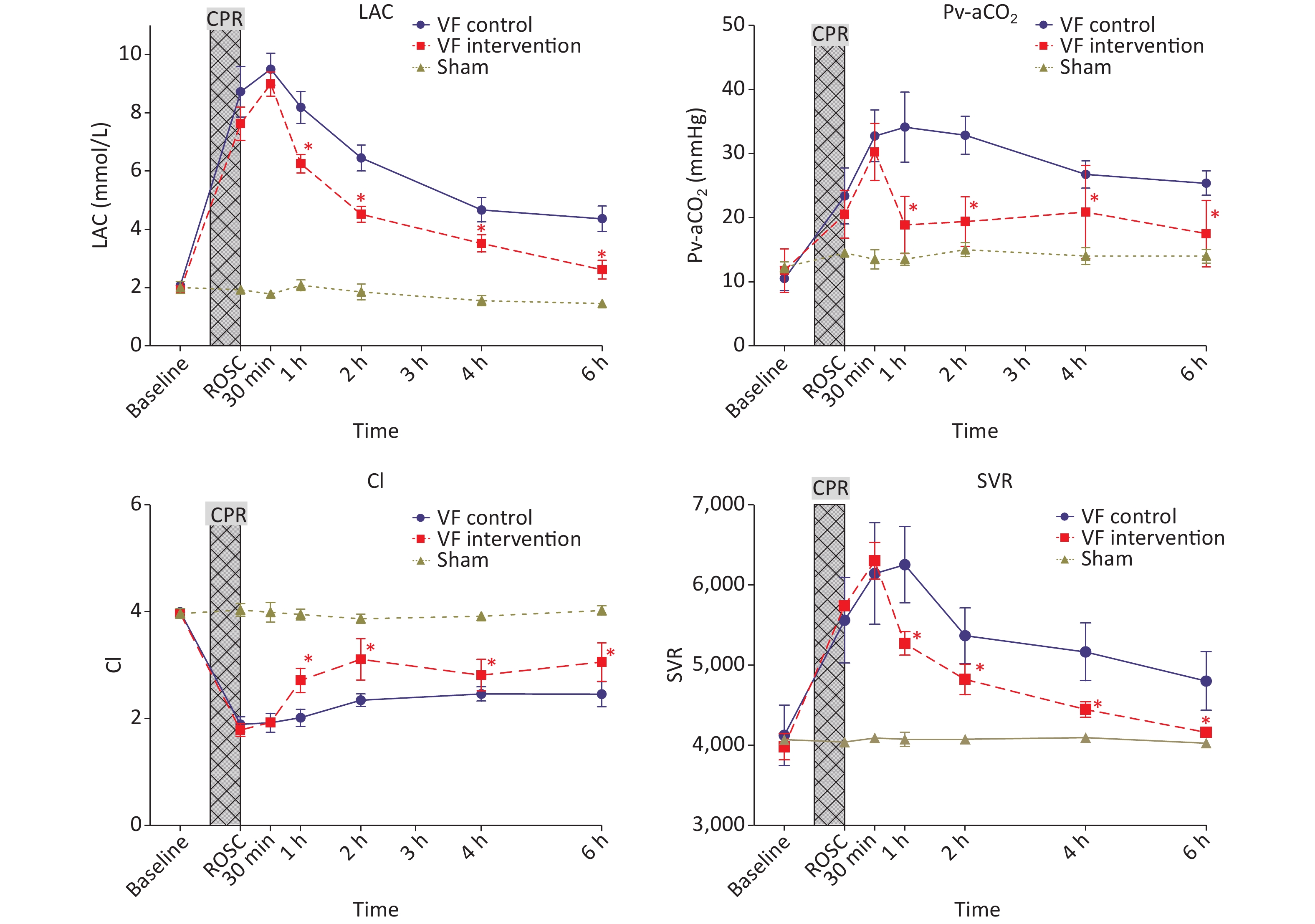
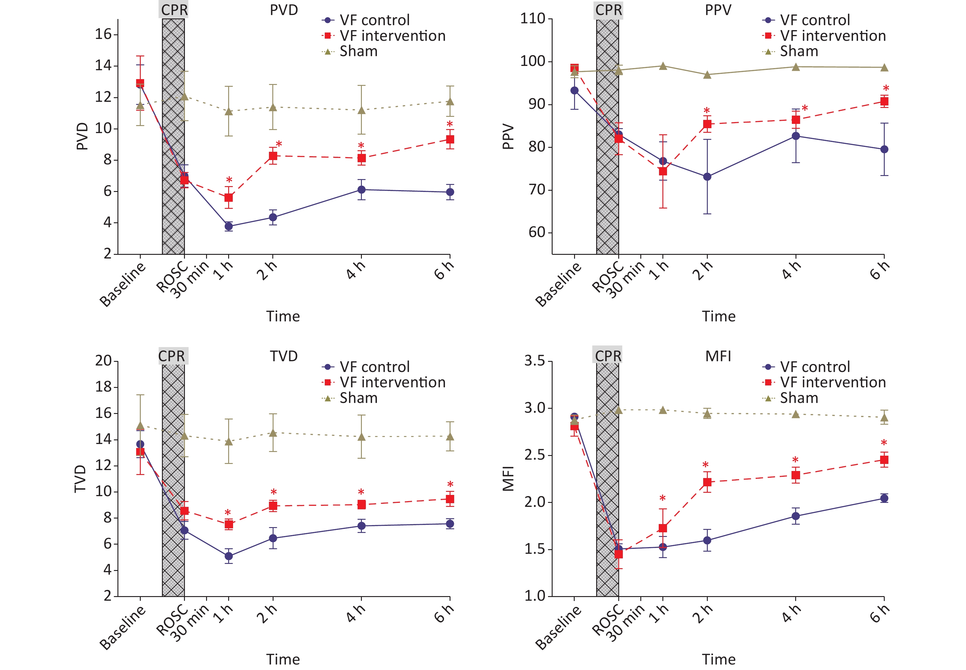
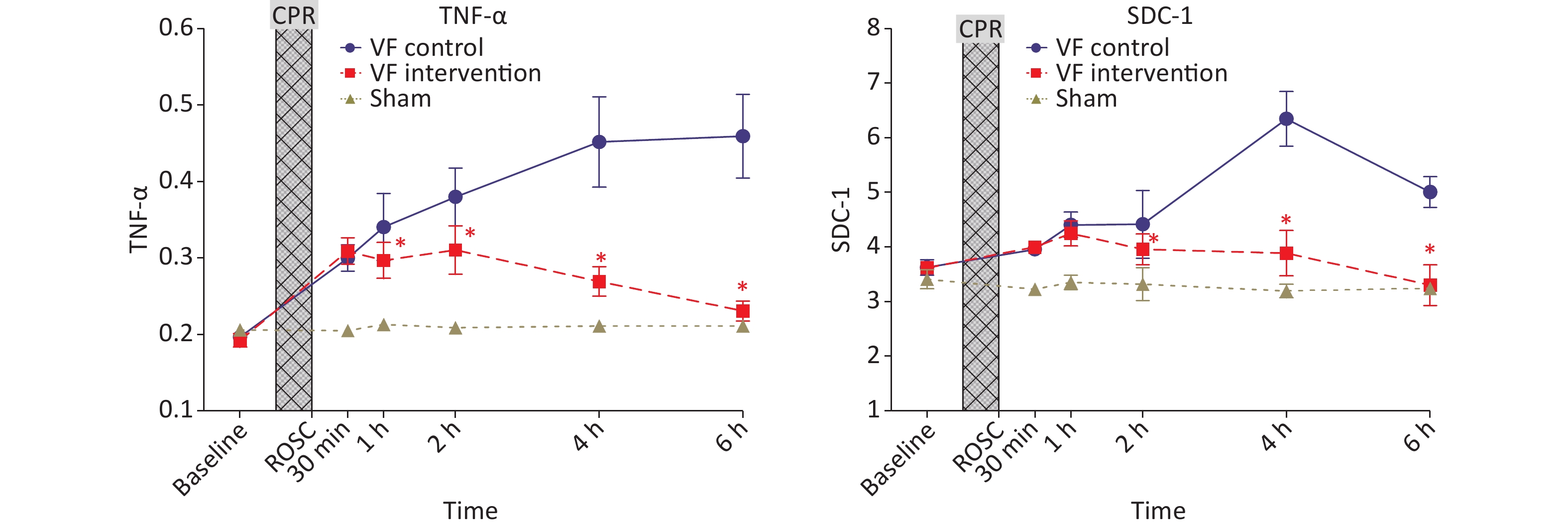
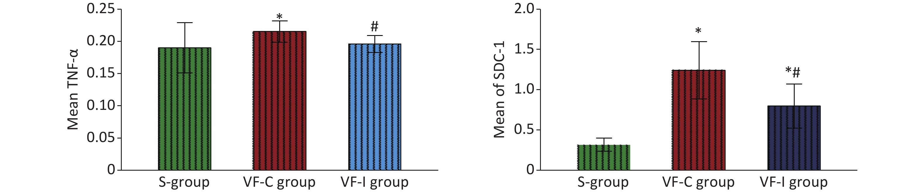
Objective This study aimed to examine the effects of microcirculatory dysfunction and 654-1 intervention after cardiopulmonary resuscitation on myocardial injury. Methods Landrace pigs were divided into a sham operation group (S group, n = 6), ventricular fibrillation control group (VF-C group, n = 8) and 654-1 intervention group (VF-I group, n = 8). Hemodynamics was recorded at baseline, at recovery of spontaneous circulation (ROSC), and 1 h, 2 h, 4 h and 6 h thereafter. Sidestream dark field (SDF) technology was used to evaluate and monitor the microcirculation flow index, total vessel density, perfusion vessel ratio, De-Backer score, and perfusion vessel density in animal viscera at various time points. Results After administration of 654-1 at 1.5 h post-ROSC, the hemodynamics in the VF-I group, as compared with the VF-C group, was significantly improved. The visceral microcirculation detected by SDF was also significantly improved in the VF-I group. As observed through electron microscopy, significantly less myocardial tissue injury was present in the VF-I group than the VF-C group. Conclusion Administration of 654-1 inhibited excessive inflammatory by improving the state of visceral microcirculation.


 Quick Links